

Breast Health Department
One-stop breast diagnosis and multidisciplinary breast cancer treatment, breast-conserving and nipple-sparing surgery with immediate reconstruction, planned by a dedicated breast council.
Three roads through breast cancer treatment
Which road is yours is decided by your tumor's size, position and tested biology at the breast council, never by a menu.
Breast-Conserving Surgery
The tumor removed with a safety margin, the breast kept, combined with oncoplastic techniques so safety and appearance are pursued together.
- Hospital stayTypically ~1 day
- Usually followed byRadiotherapy
- As effective asMastectomy, in suitable cases
Mastectomy + Reconstruction
When removing the breast is genuinely right: skin- and nipple-sparing techniques where suitable, with reconstruction planned before surgery, often in the same operation.
- TechniquesSkin- & nipple-sparing
- ReconstructionImmediate or delayed
- WithImplant or own tissue
Systemic & Radiation Therapy
Chemotherapy, sometimes before surgery to make conservation possible, hormone and anti-HER2 therapy chosen from your pathology, and precise radiotherapy.
- Chosen byReceptors & HER2 status
- Hormone therapy5+ years, at home
- RadiotherapyIncl. single-dose IORT
The sequence, surgery first, or drugs first, is itself a council decision made from your case. Ask what fits your diagnosis →
One clinic for the whole answer, diagnosis to reconstruction
Acibadem's dedicated breast clinics put examination, 3D tomosynthesis mammography, ultrasound and biopsy in one place, and plan every cancer case at a council where breast surgeons, plastic surgeons, oncologists, radiologists, pathologists and geneticists decide together.
- ✓One-stop diagnosis: mammography, ultrasound and biopsy in one visit
- ✓Breast conservation honestly pursued, including chemo-first strategies
- ✓Reconstructive surgeons in the room when removal is planned
- ✓Sentinel-node precision, no more armpit surgery than needed
- ✓Genetic counselling and high-risk screening built into the unit
- Prof. Dr. Fatih AydoğanBreast surgery
- Prof. Dr. Cihan UrasBreast surgery
- Assoc. Prof. Dr. A. Enes ArıkanBreast surgery
- Prof. Dr. Aykut SoyderBreast surgery
- Prof. Dr. Bülent SaçakPlastic & reconstructive surgery, breast reconstruction
- Assoc. Prof. Dr. Erdem GüvenPlastic & reconstructive surgery, breast reconstruction
From finding to answer, in days, not months
Everything a modern breast workup needs, inside one unit, so a worrying finding never waits weeks between appointments.
Which of these your case needs is decided at the council, and explained to you in plain language. Browse all medical technologies →
Six moments when a breast second opinion earns its keep
A written expert review of your diagnosis and plan, free, and with no obligation to travel. In breast cancer, verifying before deciding is diligence, not doubt.
Any of these sound familiar? Request a free written second opinion →
From sending your records to treatment complete
- Send recordsFree, prioritised review by the breast team
- Your planCouncil proposal + written estimate
- One-stop workupImaging, pathology re-review, confirmation
- Surgery weekOperation ± reconstruction, days, not weeks
- Completing treatmentRadiation & drug therapy, here or at home
- Home, connectedFull file + follow-up calendar your doctor can run
The support that carries you through treatment
Everything you want to know, answered below
Jump straight to the part of the guide you came for.
Quick answer
The Breast Health Department evaluates, diagnoses, and treats benign and malignant breast conditions using a coordinated, multidisciplinary approach. At Acibadem in Turkey, care may include imaging, biopsy, surgery, medical oncology, radiation oncology, genetic assessment, and follow-up planning tailored to each patient’s needs.
Breast health deserves its own front door in medicine, not a corner of a general department. At Acıbadem, breast care runs on a dedicated Breast Clinic model: one place where clinical examination, 3D tomosynthesis mammography, ultrasound and biopsy happen together, where every cancer case is planned by a multidisciplinary council of breast surgeons, plastic surgeons, oncologists, radiologists, pathologists and geneticists, and where protecting the breast, whenever oncologically safe, is treated as part of the cure rather than an afterthought. This guide explains the whole landscape: what a breast lump usually means, how screening and diagnosis work, every treatment from breast-conserving surgery to mastectomy with reconstruction, and what the journey looks like for a patient travelling from abroad.
Worldwide, breast cancer is the most common cancer in women, roughly one woman in eight will face it in her lifetime, and yet it is also among the cancers where early detection changes the story most. Most breast findings, meanwhile, turn out to be benign. Both truths matter, and this page takes each seriously.
Why women travel to Turkey for breast treatment
The reasons are practical. Speed: between a suspicious finding and a treatment plan, many health systems put weeks of waiting, for imaging, for biopsy, for results, for a surgical date. What patients come to us for is the treatment plan and the treatment itself, and there our diagnostic chain is designed to run in days rather than weeks. Completeness: modern breast care needs surgery, plastic and reconstructive surgery, medical oncology, radiotherapy, genetics and pathology working as one team, assembled here under one roof, with medical oncology and radiation oncology in the same hospital group. Cost: a written personalised estimate for your proposed plan comes before you travel. And underneath all three: a culture in which the question “can we save the breast, and if not, how do we rebuild it?” is asked from day one, in the same meeting that plans the cancer treatment itself.
The Breast Clinic model: one-stop diagnosis, one shared decision
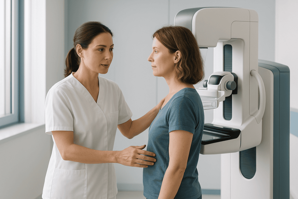
Acıbadem’s breast health units, with dedicated breast clinics at hospitals including Maslak, Altunizade and Ataşehir in Istanbul, are built on two principles. The first is one-stop diagnosis: clinical breast examination, 3D tomosynthesis digital mammography, high-resolution ultrasound and, where needed, image-guided needle biopsy are performed within the same unit, so a worrying finding moves from question to answer without weeks of appointments scattered across departments. Self-examination training and structured screening programmes, including personalised surveillance for high-risk women, run alongside.
The second principle is the multidisciplinary breast councilthe tumor board of breast care. Breast surgeons, plastic and reconstructive surgeons, medical oncologists, radiation oncologists, radiologists, pathologists and medical geneticists review each cancer case together and agree a single plan: whether surgery or drug therapy should come first, whether the breast can be conserved, how the armpit (axilla) will be assessed, and how reconstruction, if wanted, fits in. No single doctor, however senior, decides alone.
The surgeons: who will actually treat you
Breast treatment is a team sport, but its outcomes are carried by named people. The senior surgeons international patients most often meet at Acıbadem’s breast units:
- Prof. Dr. Fatih Aydoğanbreast surgery.
- Prof. Dr. Cihan Urasbreast surgery; among Turkey’s best-known names in breast cancer surgery.
- Assoc. Prof. Dr. A. Enes Arıkanbreast surgery.
- Prof. Dr. Aykut Soyderbreast surgery.
- Prof. Dr. Bülent Saçakplastic and reconstructive surgery; breast reconstruction.
- Assoc. Prof. Dr. Erdem Güvenplastic and reconstructive surgery; breast reconstruction.
That pairing is deliberate: breast surgeons and reconstructive surgeons work as one bench, because the decision to remove tissue and the plan to restore it belong in the same conversation. The full team, including the medical oncologists and radiation oncologists who complete the treatment, is listed further down this page, and every physician’s profile is public. When your records arrive, your case is matched to the surgeon whose practice fits it; you can also request a specific surgeon by name.
Found a breast lump? What it usually means
First, the statistics: most breast lumps are not cancer, cysts, fibroadenomas and other benign changes account for the majority of findings. But “usually benign” describes populations, not people, and two groups are wrongly reassured more often than any other: young women, and women who are pregnant or breastfeeding. A lump found during pregnancy or breastfeeding is not automatically a blocked duct; if it has not resolved within one to two weeks it needs an ultrasound, which is safe at every stage of pregnancy. Breast cancer often announces itself exactly this way: a firm, usually painless lump, noticed by the woman herself.
Beyond a lump, the changes that deserve a breast specialist’s attention are: skin dimpling or puckering; newly developing asymmetry; a nipple that has begun to pull inward; redness or swelling that does not settle; and spontaneous nipple discharge, particularly when it is bloody or clear and comes from a single duct. Pain alone is rarely how breast cancer presents, and cyclical breast pain linked to your period is common and usually harmless. Pain is not a green light, though: pain in one fixed spot that persists through a whole cycle, and any breast pain that comes with redness, swelling, skin change or a lump, is examined rather than explained away. None of these signs is a verdict.
Screening and early detection: the calendar that saves lives
Early-stage breast cancer is among the most treatable of all cancers, which turns screening from a bureaucratic chore into genuinely powerful medicine. Screening intervals differ legitimately between countries, many national programmes screen every two years, and some start at 50. The schedule Acıbadem’s own breast programmes follow, and the one we would discuss with you, is:
- From your 20sbreast awareness rather than a monthly ritual: know how your breasts normally look and feel, so that you notice a change and report it. A few days after your period ends is the easiest time to compare. Self-examination is not a screening test and has not been shown to reduce deaths on its own, so it never replaces mammography and a normal self-check never settles a symptom.
- From 40an annual clinical breast examination by a breast-experienced physician, and screening mammography at the interval you and your doctor agree; our own programmes work annually. No screening interval replaces reporting a symptom: a new lump between mammograms is assessed straight away, and a normal mammogram does not overrule a lump you can feel.
- Higher-risk womenwith a significant family history or a known genetic mutation, surveillance starts earlier and is personalised, often adding ultrasound or MRI to mammography on an individual schedule set with your doctor.
Modern 3D tomosynthesis mammography takes thin image “slices” through the breast, improving the detection of small cancers, including in dense breast tissue, over conventional two-dimensional images. For visitors, a complete breast check (examination, tomosynthesis mammography, ultrasound, and same-visit assessment of anything found) can be arranged as a single appointment, many international patients combine it with a wider health check-up.
Dense breasts, and when breast MRI is used
“Dense breast tissue” is a finding on your radiology report, not something you or your doctor can feel, and it has nothing to do with how firm your breasts are. It describes how much glandular and fibrous tissue shows on the mammogram compared with fat. Density tends to fall after the menopause, though many women stay dense for life.
Why density matters twice
Dense tissue is white on a mammogram, and so is a tumor, white on white, and a small cancer can hide in it. Separately from that imaging problem, dense tissue is itself associated with a higher risk of developing breast cancer. The two facts get blurred into one; both belong in your surveillance plan.
One consequence outranks the rest: a normal mammogram in dense breasts does not overrule a lump you can feel.
What tomosynthesis, ultrasound and MRI each add
Tomosynthesis takes thin slices through the breast, recovering part of what a flat image loses in dense tissue. Ultrasound adds further sensitivity, and also finds harmless things that then have to be explained. Breast MRI is the most sensitive of the three, and it is aimed at specific questions rather than used as a routine screen: high-risk surveillance, including known mutation carriers; mapping how far a proven cancer extends before surgery; judging a tumor’s response to chemotherapy given before surgery; and questions about implants. Density alone is a reason to discuss MRI with a breast specialist, not an automatic indication for it.
What the scan is like, and what it costs you
You lie face down with your breasts in a padded opening, arms over your head. The machine is loud and you must keep still. Contrast goes into a vein partway through, so kidney function and any previous contrast reaction are checked first. Say beforehand if you are or might be pregnant, and if you are claustrophobic: that is common and manageable. Before the menopause the scan is often booked to a particular week of your cycle, because normal tissue takes up contrast differently across the month. The downside is real: MRI finds shadows that turn out to be nothing, and each one costs an ultrasound, sometimes a biopsy, and weeks of worry.
From finding to answer: imaging, biopsy and pathology
When something needs explaining, the workup follows a disciplined sequence. Imaging first: tomosynthesis mammography and high-resolution ultrasound characterise the finding; breast MRI joins where it adds information, in dense tissue, in high-risk surveillance, or to map disease extent before surgery. Then tissue: if imaging cannot dismiss the finding, an image-guided needle biopsy, fine-needle or core, occasionally vacuum-assisted, takes a sample under local anesthesia, a short outpatient procedure rather than an operation. One rule outranks everything else here: normal imaging does not overrule a lump you can feel. If a lump is still there after a clear mammogram or ultrasound, it is re-examined and in almost all cases biopsied: a persistent palpable lump is followed to a tissue answer, never filed as reassured. Then pathology: the laboratory confirms what the tissue is and, if it is cancer, establishes the features that steer everything afterwards, hormone (estrogen/progesterone) receptor status, HER2 status and growth characteristics.
Findings that can be felt are one thing; for suspicious lesions that cannot be felt, the lesion is marked under mammography or ultrasound guidance, wire localisation or the ROLL technique, so the surgeon removes precisely the right tissue, verified by imaging during the operation itself. For international patients who arrive with a biopsy already taken at home, our pathologists re-review the slides as standard before any treatment is planned: the diagnosis underneath the plan must be verified, not assumed.
Benign breast conditions: the diagnoses that end with relief
A breast clinic’s daily work is mostly reassurance, delivered responsibly. The common benign findings:
- Cystsfluid-filled sacs, most frequent between 40 and 49; roughly one woman in fourteen develops them. Simple cysts are harmless; larger or uncomfortable ones can be drained with a fine needle in minutes.
- Fibroadenomasthe most common benign breast tumor, typically in women in their 20s and 30s. A fibroadenoma is a diagnosis made by imaging, and where there is any doubt by needle biopsy; it can never be diagnosed from your age or from how the lump feels, and being young does not exclude cancer. Once it has been confirmed, a simple fibroadenoma does not itself raise cancer risk: it is then monitored with imaging, and removed when large, growing or troubling. Any lump that changes, grows or feels different from the one that was checked is re-imaged rather than assumed to be the same lump.
- Intraductal papillomassmall growths inside a milk duct and the most frequent cause of bloody single-duct nipple discharge; investigated properly (including ductoscopy where useful) and usually removed, because a minority conceal something more serious.
- Mastitis and abscessbreast inflammation, treated with antibiotics and, for an abscess, drainage. An untreated breast abscess needs draining and can seed a bloodstream infection.
- Inflammation that is not an infectiona breast that becomes red, swollen, hot, heavy or orange-peel-textured over days to weeks, especially with no fever, outside breastfeeding, or after the menopause, is not assumed to be an infection. Inflammatory breast cancer imitates mastitis, often with no lump to feel, and it is the one breast presentation where weeks genuinely matter. Breast inflammation that has not clearly settled after a single course of antibiotics is investigated with imaging and a biopsy of the breast tissue and skin rather than treated with a second course.
- Phyllodes tumorsrare, fibroadenoma-like growths that are treated surgically with a margin of healthy tissue; the diagnosis is confirmed by pathology after removal.
The pattern to notice: benign diagnoses are made properly, not casually. Every “it’s nothing” at a good breast unit has imaging, and, where needed, tissue, behind it.
Breast cancer types and stages, explained simply
Breast cancer begins in the milk ducts or lobules. When abnormal cells are still confined there, carcinoma in situ, “stage 0”, the disease cannot yet spread, and treatment is at its most effective. From there, staging describes the tumor’s size and reach: stage 1, a small invasive tumor (under about 2 cm) without lymph-node spread; stage 2, a larger tumor and/or early spread to armpit lymph nodes; stage 3, locally advanced disease with more extensive node involvement; stage 4, disease that has travelled to distant organs, where treatment aims at control and quality of life over the long term.
Two things matter more than memorising numbers. First, stage is only half the story: the tumor’s biology (hormone receptors, HER2 status) shapes treatment just as strongly, which is why two women with “the same stage” can rightly receive different plans. Second, stage is measured, not guessed: it is established by imaging and pathology before your council fixes a plan, and it frames an honest conversation about what treatment aims to achieve.
Tumor biology: the subtypes that decide your treatment
Two women with identically sized tumors can get completely different treatment, and the reason sits in five lines of the pathology report: estrogen receptor (ER), progesterone receptor (PR), HER2 status, grade, and often Ki-67, a measure of how fast the cells divide. Ask for your report and find those lines.
- Hormone-receptor-positive, HER2-negativethe largest group. Endocrine therapy is the backbone; chemotherapy is used selectively. Risk of return is lower early but continues at a low level for many years, which is why treatment and follow-up are long.
- HER2-positivea subtype that behaved badly before targeted antibodies existed and behaves very differently now. Anti-HER2 drugs are given with chemotherapy, frequently before surgery so the response can be watched.
- Triple-negativeno hormone receptors and no HER2, so neither endocrine nor anti-HER2 drugs have a target. Chemotherapy is the main systemic tool, with immunotherapy added in some cases. It is more common in younger women and in BRCA1 carriers, which is why this diagnosis is itself a reason to be offered genetic testing.
- Grade and Ki-67these make no group of their own; they tell the council how aggressively a tumor within a group is behaving, and help decide whether chemotherapy is added.
Why triple-negative is usually treated with chemotherapy first
Giving chemotherapy before surgery does two jobs. It treats the disease immediately, and it shows the team in real time whether these drugs work on this tumor, information nobody has when chemotherapy follows the operation. If final pathology then finds no remaining invasive cancer in breast or nodes, that is a pathological complete response, one of the strongest signals available here. Where residual disease remains, that changes what is offered next.
Without quoting numbers that would not be yours: triple-negative disease carries its highest risk of return in the first years and that risk then falls: the opposite shape to hormone-receptor-positive disease, where the risk is lower but stretches over decades. Neither pattern predicts an individual.
DCIS and stage 0: cancer that has not learned to spread
Being told you have DCISductal carcinoma in situ, is a peculiar kind of frightening, because the word cancer arrives attached to a reassurance nobody explains. Plainly: DCIS is abnormal cells confined inside the milk ducts, behind an intact wall, and while they stay there they cannot spread to lymph nodes or other organs. Most of it is found on a screening mammogram, as microcalcifications, in a woman with nothing to feel.
Why you are offered surgery for something that cannot spread
Because some DCIS, left alone, eventually breaks through the duct wall and becomes invasive cancer, and medicine cannot yet reliably tell which. Treatment removes that possibility while it is still theoretical. Surgery is usually breast-conserving, taking the affected area with a margin, followed by radiotherapy to the remaining breast tissue. Mastectomy is recommended when DCIS is extensive or sits in more than one part of the breast, when clear margins cannot be achieved, or when radiotherapy is not possible for you, and there, immediate reconstruction is usually available.
What is not used, and what sometimes is
Chemotherapy has no role in pure DCIS: there is nothing circulating for it to hunt. Sentinel lymph node biopsy is usually unnecessary for the same reason, with one exception, if you are having a mastectomy, the sentinel node is checked during that operation, because the chance disappears once the breast is removed, and because final pathology sometimes finds an invasive focus the needle biopsy missed. Endocrine therapy is discussed afterwards in hormone-receptor-positive DCIS, to reduce the risk of a new cancer in either breast.
The overtreatment question
Some DCIS would never have caused harm in a woman’s lifetime, so treating all of it means treating some women who did not need it. Trials of monitoring low-grade DCIS instead of operating are running; outside them, surgery remains standard because no test safely identifies who could skip it. Ask for the detail that shapes your own decision, grade, extent on imaging, whether conservation is achievable.
Family history, BRCA and genetic counselling
About five percent of breast cancers are hereditary, driven by mutations passed through families, most famously in the BRCA1 and BRCA2 genes. Clues that genetics deserves a closer look include several affected relatives, breast cancer at young ages in the family, ovarian cancer, male breast cancer, or a known mutation in a relative. At Acıbadem, medical geneticists are part of the breast team: risk assessment, genetic counselling and, where indicated, testing are integrated into the workup, not outsourced as an afterthought.
The result changes real decisions. For a woman with cancer, a confirmed mutation can influence the choice of surgery and follow-up. For her healthy relatives, it opens a considered conversation about intensified surveillance or risk-reducing options, held with a counsellor, without pressure, and on your own timeline. And for women with a worrying family story and no mutation found, the result has to be read carefully rather than as an all-clear: most inherited risk is not explained by the genes we can currently test, so your surveillance plan is set by your family history as well as your test result. For some women that still means beginning mammography earlier, or adding MRI: a negative test lowers one specific worry, it does not cancel a family history.
If you carry a BRCA mutation: surveillance and risk-reducing surgery
A confirmed BRCA1 or BRCA2 result in a woman who does not have cancer is not a diagnosis and not an instruction to have surgery. It is a risk figure that earns you a plan.
What intensified surveillance involves
Carrier surveillance starts far earlier than population screening and leans on MRI, because young breast tissue is dense and hides cancers on mammograms. In practice that usually means annual breast MRI from the mid-to-late twenties, mammography added from around 30 to 35, the two often alternated so some imaging happens every six months, with clinical examination alongside; exact ages are set by your genetics team and your family history. Surveillance does not lower your risk of developing cancer. It is designed to find one early, and it only works if a new symptom is reported when it appears rather than saved for the next scan.
Risk-reducing mastectomy
Removing both breasts before any cancer appears substantially lowers the chance of ever developing breast cancer, and it is the most effective option available to a carrier. It does not reduce that risk to zero, a small amount of breast tissue always remains against skin and chest wall, and it does nothing for ovarian risk. Where a carrier chooses it, the operation is planned by the breast and reconstructive surgeons together, and in suitable anatomy it is performed with nipple-sparing technique and immediate reconstruction, which is why the reconstruction discussion belongs before the decision. The trade-offs are permanent: loss of nipple and breast sensation, the likelihood of further surgery over the years, a body that is different in the mirror. Choosing surveillance instead is a legitimate decision, not a refusal of care.
The ovaries, and the timing conversation
BRCA mutations also raise ovarian and fallopian tube risk, and there is no reliable ovarian screening test, which is why risk-reducing removal of tubes and ovaries is discussed separately, with a gynaecological team. Done before natural menopause it brings the menopause on immediately, with the bone, cardiovascular and sexual consequences that follow; whether any hormonal treatment is appropriate afterwards is a decision for your doctors, never a default. For women who have not completed their families the question becomes one of timing, best discussed years early rather than in a crisis.
One more duty comes with a positive result: siblings, children and parents each have a meaningful chance of carrying the same variant, and cascade testing lets them find out and act.
Planning your treatment: the multidisciplinary breast council
Once diagnosis and staging are complete, your case goes to the breast council, and this meeting is where modern breast medicine earns its results. The questions on the table are exactly the ones that matter: Should surgery come first, or would chemotherapy before surgery (neoadjuvant therapy) shrink the tumor enough to allow breast conservation where mastectomy once seemed inevitable? How will the armpit be assessed with the least possible surgery? If mastectomy is needed, can the skin, and the nipple, be preserved, and which reconstruction fits this patient’s anatomy and wishes? What do the receptors say about drug therapy, and where does radiotherapy belong in the sequence?
You receive the outcome as one coherent plan, in writing, explained in your language, with the reasoning, not just the conclusion. And a principle worth stating plainly: at a breast council, the reconstructive surgeon is present when removal is being planned. What is taken and what is rebuilt are designed together.
Breast-conserving surgery and oncoplastic breast surgery
For suitable tumors, the operation of choice removes the cancer with a margin of healthy tissue, lumpectomy, wide excision, quadrantectomy, and keeps the breast. Decades of evidence support a clear statement: in appropriate patients, breast-conserving surgery followed by radiotherapy treats the cancer as effectively as removing the whole breast. In appropriate patients that is why conservation is not a compromise: breast-conserving surgery with radiotherapy gives the same long-term survival as mastectomy, and the danger in breast cancer is distant spread, fought with systemic therapy rather than with larger operations than the tumor requires. Where a surgeon recommends mastectomy, it is because conservation would not achieve that same result in your particular tumour, ask for the reason, and expect a specific one.
Oncoplastic breast surgery is the craft that makes conservation look as good as it sounds: the surgeon combines cancer removal with plastic-surgery techniques, reshaping tissue, repositioning volume, adjusting the other breast for symmetry where wanted, so that oncological safety and a natural appearance are pursued in the same operation, not traded against each other. Non-palpable lesions are localised with wire or ROLL guidance beforehand and verified by imaging during surgery. Typical hospital stay is short, often a single night, and radiotherapy to the preserved breast usually follows to secure the result.
When pathology reports an involved margin: re-excision
Breast-conserving surgery removes the tumor with a rim of healthy tissue around it. That rim is the margin. The surgeon judges it in theatre by feel and by imaging the removed specimen, but the definitive answer comes from the laboratory afterwards, once the specimen has been sliced, inked and read under the microscope. Sometimes the report says cancer cells reach the inked edge. That is an involved, or positive, margin.
What happens next
You are called back for a second, smaller operation to take more tissue from that edge: a re-excision. It is common enough that a good unit plans for the possibility rather than treating it as a failure, and it does not mean the first operation went wrong or that the cancer has spread. Where disease turns out to be more extensive than the imaging suggested, or a clear margin cannot be reached while leaving a reasonable breast, mastectomy is discussed instead.
What it means for your travel
This is the reason not to book a tight return flight around your operation. Ask when your final pathology is expected, and stay reachable until it has been reported to you.
Mastectomy, including skin-sparing and nipple-sparing techniques
Sometimes removing the whole breast is genuinely the right operation: for large tumors relative to breast size, for disease in multiple areas of the breast, when radiotherapy is not possible, or by a well-counselled patient’s own choice. Two things have transformed what “mastectomy” means. First, technique: in suitable cases the breast envelope is preservedskin-sparing mastectomy, and for appropriate tumors positioned away from the nipple, nipple-sparing mastectomy, which keeps the skin and nipple-areola while removing the breast tissue beneath. Second, timing: reconstruction can very often begin in the same operation.
Equally important is what mastectomy is not: where breast-conserving surgery is oncologically appropriate, removing more does not add protection. And where mastectomy genuinely is needed, it is delivered with the reconstruction conversation already had, never as a fact presented on the morning of surgery.
Breast reconstruction: repair, not cosmetics
Reconstruction rebuilds a breast lost to cancer, and it deserves to be named correctly: this is reparative surgery, restoring what disease took, not a cosmetic extra, and it is planned by the plastic and reconstructive surgeons who sit on the breast council itself. Broadly, two roads exist. Implant-based reconstruction restores volume with a prosthesis, often beginning in the same operation as the mastectomy. Autologous reconstruction rebuilds the breast from your own tissue, most commonly from the abdomen, creating a warm, natural breast at the cost of a longer operation and recovery. Which fits you depends on your anatomy, planned radiotherapy, health and preferences; the options are laid out before your cancer operation, including the choice of reconstructing later, or, after full counselling, not at all. Both roads have real complications, and they belong in the conversation before you choose. Implant reconstruction can be complicated by infection, by capsular contracture, scar tissue tightening around the implant, by visible rippling, and by loss of the implant, which is more likely where radiotherapy is planned; implants are not lifetime devices, and further surgery over the years is common. Own-tissue reconstruction risks partial or, rarely, complete loss of the transferred tissue, areas of fat necrosis that feel like lumps and have to be checked, and weakness or bulging of the abdominal wall where the tissue was taken. Your surgeon will give you the figures that apply to your operation and your anatomy, not to an average.
Expectations, honestly: a reconstructed breast can look remarkably natural, in and out of clothes, and modern techniques include nipple reconstruction and adjustment of the other breast for symmetry. Normal sensation, however, does not return, some feeling may develop over time, and scars fade over one to two years without vanishing.
Breast reconstruction: the techniques, the timeline and the trade-offs
Reconstruction is the decision women research longest and hear explained latest. The two families of technique, and the factor that changes the answer most:
Implant-based reconstruction
An implant restores volume without a second surgical site, so recovery is shorter. In direct-to-implant reconstruction, where the preserved skin envelope and its blood supply allow it, the permanent implant is placed during the mastectomy itself. In expander-then-implant reconstruction, a tissue expander goes in first: an empty implant filled with saline through a small port at clinic visits, stretching the skin in stages until the permanent implant is fitted at a second operation.
Nobody describes the expander months. It is firm, sits high and does not move like a breast; after each fill there is a tight ache for a day or two; you are asymmetric in clothes until the exchange. It ends. The long-term issue is capsular contracturethe scar capsule the body forms around any implant tightening, so the breast becomes firm, rides higher and can ache. Correcting it means further surgery, and implants are not lifetime devices.
Reconstruction with your own tissue
Autologous reconstruction moves living tissue with its own blood supply. The DIEP flap takes skin and fat from the lower abdomen and joins its vessels to chest vessels under the microscope, leaving the abdominal muscle in place, which is why core strength recovers better than after the older TRAM flap, which takes muscle with the tissue. The latissimus dorsi flap brings muscle and skin from the back, often with a small implant for volume: the usual answer when abdominal tissue is insufficient, or when chest skin has been damaged by radiotherapy.
The cost is a second wound: DIEP and TRAM leave a long low abdominal scar and a repositioned navel, the latissimus flap a diagonal scar on the back. The operation is long, and the first days involve frequent checks on the flap’s circulation. What you get is a breast of your own tissue, warm, softening naturally, changing with your weight as the other one does.
Immediate, delayed, and what radiotherapy changes
This is the most consequential interaction in reconstruction. Radiotherapy delivered to a reconstructed breast makes capsular contracture, poor shape and implant loss more likely, and irradiated skin heals differently afterwards. So where radiotherapy is expected, an expander may hold the space through it with the definitive reconstruction done later, or own-tissue reconstruction may be delayed until the tissues have settled, months rather than weeks. That conversation belongs at the council table with the radiation oncologist present, before the mastectomy. One rule outranks the rest: reconstruction never delays cancer treatment. If a reconstructive step would postpone chemotherapy or radiotherapy, the cancer treatment goes first.
How many operations this really is
Almost nobody finishes in one. A complete reconstruction is usually a first operation, then revision stages, fat grafting to fill contour dips and soften the upper edge, often repeated; surgery on the other breast so the pair matches, which is part of the reconstruction and not vanity; nipple work last. The stages are spaced across months on purpose, because swelling has to resolve before a revision can improve anything.
The nipple and areola
Where the nipple could not be preserved, it can be rebuilt from small local skin flaps once the breast mound has settled, and the areola recreated by 3D tattooing, which can be strikingly convincing in a mirror. Two limits: a rebuilt nipple flattens over time and often needs touching up, and neither technique restores sensation.
Who is not a candidate
Smoking is the big one, nicotine constricts exactly the small vessels a flap and a preserved skin envelope depend on, and surgeons will ask you to stop well before the operation. Poorly controlled diabetes, significant vascular disease, previous abdominal surgery that divided the necessary vessels, or too little abdominal tissue can rule out particular flaps. Disease needing urgent systemic treatment postpones reconstruction rather than cancelling it. And if, fully informed, you do not want reconstruction, ask for a flat closure to be planned properly: a well-planned flat chest wall is a chosen outcome, not a failed one.
The armpit, sentinel nodes and lymphedema
Breast cancer’s first stop, when it travels, is usually the lymph nodes of the armpit, so assessing them is part of every cancer operation. The modern standard for clinically clear armpits is sentinel lymph node biopsy: the first node(s) draining the breast are identified with a tracer, removed and examined. If they are clear, the rest of the armpit is left alone, sparing most patients the fuller dissection of the past. When nodes are involved, a wider axillary dissection may be needed, and removing many nodes carries a real risk of lymphedemachronic arm swelling, which is uncommon after sentinel node biopsy and considerably more likely after a full dissection, particularly when the armpit is also irradiated.
That risk is exactly why the modern approach is precise rather than maximal, and why prevention is briefed, not hoped for. After axillary surgery you are taught the arm-care rules: begin gentle mobilisation early, build activity back in stages with your physiotherapist, including gradual, progressive strength work, which has been shown to be safe for the arm and is no longer restricted for life, keep the skin of that arm intact and treated, and report early tightness or swelling promptly, early lymphedema responds far better than established swelling. An arm on the operated side that becomes red, hot and painful, or swells over hours, particularly with a fever or shivering, is showing cellulitis, infection in an arm whose lymphatic drainage has been altered. It needs antibiotics started immediately, and every episode makes long-term swelling worse. Physiotherapy support, including a staged post-surgery exercise programme from first-day breathing and hand exercises to swimming and conditioning by week ten, is part of the care, coordinated with rehabilitation.
Who does what in your operation
Two different surgeons may work on you in the same operation, and it is worth knowing which does which part.
- The breast surgeon removes the cancer and assesses the armpit. In conserving surgery the reshaping of what remains, the oncoplastic part, is often done by the same surgeon.
- The plastic and reconstructive surgeon rebuilds: implant or expander, or a flap of your own tissue. A flap is microsurgery, joining vessels a few millimetres across under a microscope, and a different skill set from implant work.
- The radiologist marks a lesion that cannot be felt before you go to theatre. The pathologist reports the margins and receptors that decide everything afterwards.
Ask your surgeon which parts of your operation they will perform themselves, and who performs the rest. Every physician’s profile is public, and you can ask for a named surgeon.
Chemotherapy in breast cancer, including before surgery
Not every breast cancer needs chemotherapy, and when it is needed, its place in the sequence is a council decision. Given after surgery (adjuvant), it hunts microscopic cells beyond the surgical field to lower the risk of return. Given before surgery (neoadjuvant), it can shrink a tumor so effectively that breast-conserving surgery becomes possible where mastectomy first seemed unavoidable, one of the quiet revolutions of modern breast care, while also demonstrating, in real time, how the tumor responds to the drugs. In advanced disease, systemic therapy leads the plan.
Delivery follows the standards described on our medical oncology pages. A temperature of 38°C during chemotherapy is treated as an emergency, and antibiotics are started within the hour. Whether chemotherapy belongs in your plan is decided by tumor biology, stage and your overall health, not by routine.
Who actually needs chemotherapy: genomic tests on the tumor
“Not every breast cancer needs chemotherapy” is easy to say. The hard part is where the line falls for the women in the middle, hormone-receptor-positive, HER2-negative, with clear or few involved lymph nodes, because stage and grade alone do not settle it. For that group the decision is increasingly made with a genomic test performed on the tumor itself.
What the test does
Tissue already taken at biopsy or surgery is sent to a specialised laboratory. Assays such as Oncotype DX and MammaPrint measure the activity of a defined set of genes inside the tumor and return a recurrence score or a risk category. Nothing further is done to you: it is a test on tissue that has already left your body.
What the result changes
A low result identifies women whose likely benefit from chemotherapy is small enough that endocrine therapy alone is a reasonable plan, sparing them months of treatment they would gain little from. A high result identifies women who do stand to benefit, turning an anxious guess into a supported decision. Results in between are read together with age, nodes and tumor grade, exactly the question a multidisciplinary council exists to answer. These assays do not drive the decision in triple-negative or HER2-positive disease, where chemotherapy is planned on other grounds, and they never replace staging.
What to ask, and when
Before chemotherapy begins, ask in writing whether a genomic test applies to your tumor and whether it can be run on the tissue you already have. Ask alongside your existing plan, not instead of it: keep every appointment and treatment date you have been given while the answer is worked out. Do not delay treatment on your own initiative to wait for a test result.
Hormone therapy and targeted (anti-HER2) treatment
Two further pillars of systemic treatment are chosen directly from your pathology report. Hormone (endocrine) therapy applies when the tumor carries estrogen or progesterone receptors: the majority of breast cancers. Daily tablets block the signalling the cancer grows on, taken for five years or longer; unglamorous, and among the most effective long-term protections in oncology. One thing belongs with that: the side effects are managed rather than endured, hot flushes, joint pain and mood changes are common, and there are real options including changing to a different tablet, but this treatment is never stopped, paused or swapped on your own: stopping early measurably reduces the protection it gives, and the doctor who prescribed it decides any change together with you.
Targeted anti-HER2 therapy applies to the minority of breast cancers, around one in six to one in five, that overexpress the HER2 protein: antibody-based drugs directed at that target, typically given for about a year alongside or after other treatment in early disease, with periodic heart-function monitoring, because cardiac strain is the known side effect to watch. Its signs are breathlessness on mild effort or when lying flat, waking short of breath, swelling of the ankles, and a racing or irregular heartbeat; caught early, it is usually reversible once the drug is paused and treated.
Neither treatment is chosen on instinct. Receptor and HER2 testing is part of the standard pathology workup here precisely because these results decide who benefits, treatment matched to your tumor’s actual biology, the same principle that governs everything else on this page.
Endocrine therapy: which tablet, for how long, and what it does to you
For hormone-receptor-positive breast cancer, the tablet taken at home for years does more of the long-term protective work than anything that happened in theatre. It is also the treatment patients quietly abandon, because nobody warned them what it feels like.
Which drug, and why menopausal status decides
Tamoxifen blocks the estrogen receptor itself and works whether or not the ovaries are still producing estrogen, so it is the standard choice before the menopause. Aromatase inhibitorsanastrozole, letrozole, exemestane, stop estrogen being made in body tissues but cannot switch off the ovaries, so they are used after it. A premenopausal woman at higher risk may be offered ovarian suppression, injections that switch the ovaries off, allowing an aromatase inhibitor to be used instead. Men with breast cancer are treated with tamoxifen.
Five years or ten
Five years is the established minimum. Extending beyond it is offered when your individual balance, tumor size, node involvement, grade, tolerance, bone health, favours it: a real discussion with two defensible answers, not a formality.
The side effects, named
- Both families: hot flushes and night sweats, vaginal dryness and pain with sex, reduced libido, disturbed sleep, low mood, fatigue. These are what actually make women stop, and each has management worth asking for.
- Aromatase inhibitors: aching, stiff joints and muscles, worst in the morning and in the hands, common, real, and often improved by switching to a different aromatase inhibitor. They also accelerate bone-density loss, which is why DEXA scans are done at the start and repeated during treatment.
- Tamoxifen: a small increase in the risk of blood clots and of changes in the lining of the womb, which is why unusual vaginal bleeding is investigated rather than watched.
If it becomes intolerable
Say so early, to the doctor who prescribed it. The answer to unbearable side effects is almost never stopping: it is switching within the same family, changing to the other family, treating the specific symptom, or planning a supervised break. What must not happen is a decision taken alone in another country: stopping early measurably reduces the protection this treatment gives, and every change is decided by your prescriber together with you.
Radiotherapy, including single-dose intraoperative options
Radiotherapy secures the local result: after breast-conserving surgery it treats the preserved breast to prevent recurrence, and after mastectomy it treats the chest wall in higher-risk situations. Delivery is planned with radiation oncology on modern image-guided equipment, in short outpatient sessions over a planned course. A myth worth retiring: radiotherapy does not “burn the breast” in the way patients imagine. What it commonly causes is a sunburn-like reddening and soreness that builds through the course and settles over the weeks after it ends. A smaller number of women get skin that breaks down and needs dressings, which is why any raw or weeping skin is shown to the radiotherapy team rather than managed at home. The course itself, planning, the mask-free daily rhythm, breath-hold technique and the honest side-effect map, is told in full by the radiation oncology unit. Longer term, a treated breast can stay firmer, a little smaller or different in colour; those changes are usually permanent, and your radiation oncologist will tell you what to expect for your own plan.
For carefully selected patients, Acıbadem also offers single-dose intraoperative radiotherapy (IORT)radiation delivered directly to the tumor bed during the operation itself, in one dose, which in suitable cases can replace weeks of daily external sessions. Whether you are a candidate depends on tumor features and is a council decision; for international patients it can meaningfully change the length of stay, which is exactly why it is discussed at the planning stage rather than discovered afterwards.
Fertility, pregnancy and breast cancer under 40
For a young woman, the second question after survival is often whether she can still have children, and it is the part of breast cancer care most often raised too late to act on.
Before chemotherapy, not after
Chemotherapy can damage the ovaries permanently, and the older you are when you receive it the more likely that is. Preservation has to be arranged before the first cycle, and the window is short, commonly a couple of weeks. That delay is planned for and is not considered dangerous in most situations, but it only exists if someone opens the conversation. If nobody has raised fertility with you, raise it at the first consultation.
- Egg or embryo freezingthe established options, requiring a short course of ovarian stimulation. In hormone-receptor-positive disease, protocols designed to keep estrogen levels low are used.
- Ovarian tissue freezingan option where there is no time for stimulation, or before puberty.
- Ovarian suppression during chemotherapyinjections that quieten the ovaries through treatment, which may help preserve ovarian function. Not a substitute for freezing eggs or embryos, and not a guarantee.
Endocrine therapy, pregnancy and breastfeeding
Evidence now supports a planned, temporary interruption of endocrine therapy after a period of treatment in selected women trying to conceive: a pause scheduled with your oncologist, never one you take by yourself. Available evidence does not show that pregnancy after breast cancer makes the cancer more likely to return. Breastfeeding is often possible from an untreated breast; a breast that has had conservation and radiotherapy usually produces little or no milk. Effective non-hormonal contraception is used throughout treatment, because these drugs can harm a developing baby.
Breast cancer diagnosed during pregnancy
It happens, and it does not usually mean choosing between the pregnancy and the treatment. Breast surgery can be performed during pregnancy, and chemotherapy can be given in the second and third trimesters. Radiotherapy, endocrine therapy and anti-HER2 drugs are sequenced for after delivery. What is never appropriate is waiting until after the birth to investigate a lump: ultrasound is safe at every stage, and delay is the part of this that reliably makes the outcome worse.
Early menopause brought on by treatment
Chemotherapy, ovarian suppression and ovarian removal can all bring the menopause years or decades early, and arriving there abruptly at 34 is not the same as arriving gradually at 51. Expect hot flushes, disturbed sleep, vaginal dryness and pain with sex, mood changes and faster bone loss. All have management, most of it non-hormonal; whether any hormonal option is safe for you is an oncology decision.
Metastatic and recurrent breast cancer
Breast cancer that has spread to distant organs, bone, liver, lung or brain, is treated differently from early disease. The truthful frame first: metastatic breast cancer is generally not curable, and any centre implying otherwise is selling something. What has changed is duration and quality of life: in hormone-receptor-positive and HER2-positive disease especially, it is managed as a long-term illness, in lines of treatment, over years rather than months. That describes the disease as it is treated today; it is not a prediction about you.
What is done first
Spread is confirmed by imaging and, wherever a deposit is safely reachable, by biopsying it rather than assuming it matches the original tumor, receptor and HER2 status can change, and treating the old biology is a real and avoidable error. From there: hormone-receptor-positive disease is usually led by endocrine therapy combined with a CDK4/6 inhibitor rather than by chemotherapy; HER2-positive disease by anti-HER2 antibodies and antibody-drug conjugates; triple-negative disease by chemotherapy, with immunotherapy or a PARP inhibitor where the biology fits. Bone metastases additionally get bone-strengthening drugs, preceded by a dental check, and radiotherapy relieves painful bone deposits.
Complications treated as emergencies
Three complications of metastatic breast cancer are treated as hospital emergencies. Spinal cord compression shows itself as new or worsening back pain with weakness, numbness or tingling in the legs, unsteadiness, or a change in bladder or bowel control; a high blood calcium level as confusion, drowsiness, extreme thirst or persistent vomiting; brain metastases as a first seizure, sudden weakness or numbness down one side, sudden difficulty speaking, or a severe new headache with vomiting or drowsiness. Breathlessness at rest is assessed with the same urgency. Cord compression, high blood calcium and brain metastases are all treatable, and all are treated by the hour.
Recurrence in the treated breast, and whether to travel
A cancer returning in the same breast or on the mastectomy scar is a different situation from distant spread: it is usually treated with the intent to cure, often by mastectomy after previous conservation, and it is investigated urgently rather than watched. As for travelling, if you are mid-way through a line of treatment that is working, moving countries to start again usually costs more than it gains, and you will be told so in writing. A remote review runs alongside treatment, never instead of it.
Breast cancer in men
Men have breast tissue, and it can develop the same cancers. It is uncommon, and that is the difficulty: neither the man nor always the first doctor he sees is thinking of it, so it tends to be found later than it needed to be.
How it shows itself
Usually a firm, painless lump directly behind the nipple, on one side only. Also a nipple that pulls inward, crusting or an ulcer on the nipple, discharge, or a lump in the armpit. The workup is a woman’s workup: examination, mammography, ultrasound, and a needle biopsy where imaging does not settle it.
Treatment, and what it means for the family
Most male breast cancers carry hormone receptors, so endocrine treatment, commonly tamoxifen, is central, and it continues for years. Surgery is most often mastectomy with assessment of the armpit, because the tumor sits close to the nipple in a small volume of tissue. Radiotherapy, chemotherapy and anti-HER2 treatment follow the same logic as in women. Male breast cancer carries a strong association with BRCA2, so genetic counselling should be offered to every man diagnosed: the result changes the breast cancer risk of his daughters and sisters, and carries prostate and pancreatic implications for the men in the family.
Recovery: hospital days, drains and getting your arm back
Breast surgery recovers faster than most patients fear. Typical hospital stays: about a day after breast-conserving surgery, one to two days after mastectomy, two to five days when reconstruction is performed in the same operation. Surgical drains, thin tubes that prevent fluid collecting, usually stay from a few days up to one to two weeks depending on the operation; you can shower from about the second day with the drain protected. Pain is managed well with routine medication, eating is unrestricted from a few hours after surgery, and daily activities return progressively, from a few days after smaller operations to around four weeks after larger ones, longer where abdominal-tissue reconstruction is involved. Recovery also has warning signs: a fever or shivering, spreading redness or increasing pain around the wound, drain fluid that turns foul-smelling or suddenly heavy, and a breast or chest wall that swells rapidly or becomes tight and hard. After reconstruction with your own tissue, skin over the new breast that turns pale, dusky, cold or mottled is an emergency: the blood supply can usually be rescued, but only within hours. One-sided calf pain or swelling, and sudden breathlessness or chest pain, are the signs of a blood clot: a recognised risk of an operation followed by a flight.
The arm on the operated side gets its own programme: gentle hand, wrist and elbow movements from the first days; shoulder and posture work as healing allows; walking daily from the start; swimming and light resistance work typically from around six weeks, cleared by your team. The goals are practical, full shoulder motion, good posture, lymphedema prevention, and the programme is staged to your operation, not copied from a leaflet.
Recovery differs by operation, and the parts nobody warns you about
“Breast surgery” covers four quite different recoveries. Find yours.
- Breast-conserving surgerythe shortest. Soreness rather than severe pain, often no drain, daily life back first.
- Mastectomy alonea longer wound, usually a drain, and the first sight of the flat chest wall. That is its own event; have someone with you for it.
- Mastectomy with an implant or tissue expanderadd a band of tightness across the chest, and limits on lifting your arms until your surgeon lifts them.
- Reconstruction with your own tissuetwo healing sites, and the donor wound, not the breast, is what restricts you. Lifting, driving and work return latest here.
Seroma, numbness and the first bra
Once the drain is out, fluid often gathers under the wound: a seroma. A soft swelling that fills gradually is expected, and is drawn off with a needle in clinic if it becomes uncomfortable. A swelling that turns tight, hot or painful, or arrives with fever or spreading redness, is not part of expected recovery. Numbness of the chest wall, and of the inner upper arm after armpit surgery, is normal, often permanent, and rarely mentioned in advance. Wear a soft wire-free bra rather than nothing; support takes the drag off a healing wound. A prosthesis is fitted once the wound has healed and the swelling has settled.
Flying home
There is no fixed number of days. Your surgeon sets your flying date from your operation, your wound and your drains, and it belongs in writing on your discharge summary, get it before you book.
The breast cancer second opinion: before you decide anything
A remote second opinion is one of the most valuable steps available without boarding a plane, and in breast cancer there is usually time for it. A second opinion runs alongside existing care, never instead of it. Your imaging, pathology and proposed plan are reviewed by Acıbadem’s breast team, and a written assessment comes back, typically within about seven days, answering the questions that decide everything: Is the diagnosis complete, with receptors and HER2 tested? Is mastectomy genuinely necessary, or could neoadjuvant therapy open the door to breast conservation? Has reconstruction been offered and planned properly? Would a council have sequenced this differently?
Sometimes the answer is simple reassurance: your home plan is right, and we will say so in writing. Sometimes the review surfaces a real alternative worth discussing.
What breast cancer treatment costs in Turkey
No honest provider quotes a single price for “breast cancer treatment” before seeing your file, because the plan drives the cost: diagnosis and staging, the operation (conservation versus mastectomy, with or without reconstruction, implant or own tissue), radiotherapy, and systemic therapy, where modern targeted drugs are the world’s expensive ingredient, differ from patient to patient. What you receive instead is a written, itemised estimate for your specific proposed plan, prepared after your records are reviewed, before you travel, free and without obligation.
What can be said in general: because Acıbadem’s costs arise in the Turkish healthcare economy, comparable breast surgery, reconstruction and cancer treatment are generally substantially less expensive than private care in Western Europe or North America, commonly including travel and accommodation. If the plan changes mid-treatment, as cancer plans occasionally must, the financial picture is updated with the same transparency. Insurance documentation is prepared by the international patient office as standard.
Your journey from abroad, step by step
A breast cancer journey has enough unknowns; the logistics should not add more. The sequence in practice:
1. Send your records
Imaging (or reports), pathology if a biopsy exists, and a short history through a free consultation request. Breast cases are reviewed with priority.
2. Your plan and estimate
A proposed plan reflecting council input, surgery type, reconstruction options if relevant, expected systemic and radiation treatment, realistic durations, with the written cost estimate and a visa invitation letter if needed.
3. Arrival and one-stop workup
Airport transfer, interpreter, coordinator. In the first days: examination, any missing imaging, pathology re-review, receptor confirmation: the plan is confirmed on verified facts, and refined by the council if anything new emerges.
4. Surgery week
The operation as planned, with reconstruction where chosen. Hospital stay is measured in days, not weeks; final pathology returns within about a week and fine-tunes what follows.
5. The completing treatments
Radiotherapy and systemic therapy are scheduled around your life: some patients complete them here; many return home and continue with their own oncologist under a written plan agreed doctor-to-doctor; IORT, where suitable, compresses radiotherapy into the operation itself.
6. Home, with the thread unbroken
You leave with the complete file, operative notes, pathology, receptor status, treatment given and the follow-up calendar, and the team stays reachable for you and your home doctors.
Body, mind and family: the support around the medicine
Breast cancer strikes at more than tissue: it touches identity, femininity and the family around the patient, and pretending otherwise helps no one. Psychological support is offered as part of care, not as a confession of weakness: many patients move through shock, anger and low periods before finding their footing, and professional support measurably helps, as does contact with others who have walked the same road. Where body image and intimacy are affected, that too can be discussed with people who treat it as medicine, through psycho-oncology.
The practical layer runs alongside: nutrition counselling (weight control genuinely matters to recurrence risk), a staged exercise programme that rebuilds strength and protects the arm, and support for the companion who travels with you, because caregivers carry their own load, and our coordinators plan for two, not one.
After treatment: follow-up that actually follows
Breast cancer follow-up has a defined rhythm: examinations roughly every three months for the first three years, every six months for the following two, then annually. Follow-up is heaviest in the first years, when hormone-receptor-negative cancers are most likely to return, but for the hormone-receptor-positive cancers that make up the majority, a low, steady risk continues for twenty years and more. That is why follow-up continues annually for life, why endocrine therapy runs for years, and why a new symptom is reported whenever it appears rather than measured against how long ago you were treated. After breast-conserving surgery, the first mammogram of the treated breast comes about four to six months after radiotherapy ends, then annually; after mastectomy, the other breast is imaged every year. Additional scans and blood tests are ordered when something specific needs answering, not as an annual ritual for everyone.
For international patients, this calendar is written down and handed over: your home physician can run it, our team remains available for shared follow-up and imaging review, and hormone-therapy patients, on treatment for five years or more, get explicit long-term coordination. Survivorship also includes the unglamorous essentials: managing treatment side effects, protecting the arm, and having someone to call when a question will not wait for the next appointment. The findings that are investigated between scheduled visits rather than saved for them are a new lump or thickening in either breast, in the armpit or along the mastectomy scar; a rash or skin change on the chest wall; bone or back pain that is new, constant, worse at night or not eased by rest; a cough or breathlessness that persists; and unexplained weight loss.
Choosing where to be treated: the questions that reveal quality
Put these to every provider you consider, including us, and expect written answers:
- Will my case be planned by a multidisciplinary breast council? May I see the conclusion?
- Who is my named surgeon, and what is their breast-surgery profile?
- Will breast conservation be honestly evaluatedincluding chemotherapy before surgery if that could make it possible?
- If mastectomy is needed, will a reconstructive surgeon be involved before the operationand are skin-sparing and nipple-sparing techniques available to me?
- How will my armpit be assessed? Is sentinel lymph node biopsy the standard here?
- Will my pathology be re-reviewed, with receptor and HER2 testing?
- What exactly does the estimate includeand what would change it?
- How will follow-up work from my country?
What we will not promise
Breast cancer treated early is among modern medicine’s genuine success stories, and everything on this page reflects real, established practice. Here is what you will still not find us doing: quoting your personal cure probability in marketing text: that depends on your tumor’s stage and biology and belongs in a consultation; promising that conservation, or any operation, is certain before your imaging and pathology have been reviewed; or recommending travel when reviewing your file shows your home plan is already exactly right: you will simply be told so, in writing.
Why patients choose Acıbadem for breast health
A named team of senior breast and reconstructive surgeons who plan removal and restoration together, backed by medical oncology, radiation oncology, genetics and pathology at the same table. Techniques that respect both halves of the outcome, oncoplastic conservation, nipple-sparing mastectomy, immediate reconstruction, sentinel-node precision, single-dose intraoperative radiotherapy for suitable patients. Seven JCI-accredited hospitals. And an international patient organisation that turns a frightening diagnosis abroad into a written plan with a named coordinator, in your language, with your questions answered before you pack. Every element is verifiable before you decide, starting with a free review of your records.
Our Specialists Explain
 Breast Cancer Care at Acibadem Senology Institute
Frequently Asked Questions
I found a lump in my breast. How urgent is it?
A new breast lump is assessed within days rather than months. Most lumps prove benign, but that is a conclusion imaging (and sometimes biopsy) must reach, not a hope. At a one-stop breast clinic, examination, mammography, ultrasound and any needed biopsy can typically be arranged together, so the anxious waiting is kept as short as possible.
Is breast pain a sign of cancer?
Usually not. Breast pain, especially cyclical pain tied to your period, is very common and rarely how cancer presents; breast cancer is typically painless in its early stages. Pain that is new, persistent, one-sided or accompanied by other changes still deserves an examination, if only for the relief of a proper answer.
What does nipple discharge mean?
Discharge from both breasts or several ducts, milky or greenish, is usually benign. The pattern that needs prompt investigation is spontaneous discharge from a single duct, especially bloody or clear, most often caused by a benign intraductal papilloma, but properly investigated because a minority of cases involve something more serious.
At what age should I start mammograms?
For average-risk women: annual clinical breast examination and mammography from age 40, with monthly self-examination from your 20s. With a significant family history or known genetic mutation, surveillance starts earlier on a personalised schedule, discuss it with a breast specialist rather than guessing.
What is 3D (tomosynthesis) mammography?
A digital mammogram that captures thin slices through the breast rather than one flat image, improving detection of small cancers, particularly in dense breast tissue, and reducing recalls for overlapping-tissue shadows. It is the standard of breast imaging at Acıbadem’s breast units.
Can I have a full breast check as a visitor, without being a patient?
Yes. A complete breast screening, clinical examination, tomosynthesis mammography, ultrasound, and same-visit assessment of anything found, can be arranged as a standalone appointment or as part of a wider check-up programme during a stay in Istanbul.
How quickly can my case be reviewed if I already have a diagnosis?
Breast cancer cases are prioritised. A review is built on the imaging, the pathology report and a short history; the surgical team returns an assessment and proposed plan, and the formal written second-opinion service typically responds within about seven days.
Will you re-check my biopsy from home?
Yes, routinely. Our pathologists re-review your slides and confirm receptor and HER2 testing before any plan is fixed. The diagnosis underneath a treatment plan must be verified, discrepancies are uncommon but consequential, and this is how they are caught.
Can the breast really be saved, or is mastectomy safer?
For suitable tumors, breast-conserving surgery plus radiotherapy treats the cancer as effectively as mastectomy, decades of evidence support it, and removing more than the tumor requires adds no protection. Whether conservation fits your case depends on tumor size, location and biology, which is exactly what the council evaluates. Where mastectomy genuinely is needed, you will be told why, with reconstruction planned from the start.
What is nipple-sparing mastectomy, and am I a candidate?
A mastectomy that removes the breast tissue while preserving the skin envelope and the nipple-areola, allowing reconstruction with a remarkably natural result. It suits selected patients, generally earlier-stage tumors positioned away from the nipple. Candidacy is decided from your imaging and pathology by the council.
Can chemotherapy before surgery really save my breast?
In selected patients, yes. Neoadjuvant (pre-surgery) chemotherapy can shrink a tumor enough to make breast-conserving surgery possible where mastectomy first appeared unavoidable, and it shows the team, in real time, how your tumor responds to the drugs. Whether this sequence fits your case is a council decision made from your tumor’s size, biology and stage.
Will I lose my hair during treatment?
Only certain chemotherapy regimens cause hair loss, and not every breast cancer needs chemotherapy at all, hormone therapy and surgery alone are the whole plan for many patients. Before anything begins, your team walks you through your specific protocol’s realistic effects, so nothing about treatment arrives as a surprise.
What is a sentinel lymph node biopsy?
Instead of removing all armpit lymph nodes, the surgeon identifies the first node(s) draining the breast with a tracer, removes only those, and examines them. Clear sentinel nodes mean the rest of the armpit is left alone, sparing most patients the higher lymphedema risk of full dissection while answering the staging question precisely.
What is my risk of lymphedema, and can it be prevented?
After sentinel node biopsy alone the risk is low; after full axillary dissection, roughly one patient in four develops some degree of arm swelling. Prevention is taken seriously: staged arm exercises from the first days, avoiding heavy loads and shoulder-strap bags on the operated side, and reporting early tightness promptly, early lymphedema responds far better than established swelling.
Can reconstruction really happen in the same operation as mastectomy?
Very often, yes, immediate reconstruction with an implant or your own tissue is standard practice in suitable patients, and waking up with a reconstructed breast measurably softens the experience of mastectomy. Sometimes staging, planned radiotherapy or preference makes delayed reconstruction wiser; the options are laid out before your cancer operation.
Implant or my own tissue, which reconstruction is better?
Neither is universally better. Implant reconstruction means shorter surgery and recovery; autologous (own-tissue) reconstruction, typically from the abdomen, creates a warm, natural breast that ages with the rest of your body, at the cost of a longer operation, a second surgical site and a longer recovery. Anatomy, planned radiotherapy, health and your priorities decide; you will hear the trade-offs of each before choosing.
Will a reconstructed breast feel normal?
It can look remarkably natural, but normal sensation does not return, though some feeling may develop over time. Scars fade substantially over one to two years without disappearing. Patients told this before surgery are consistently the most satisfied afterwards.
What is single-dose intraoperative radiotherapy (IORT)?
Radiation delivered directly to the tumor bed during the operation itself, as a single dose. In carefully selected early-stage patients it can replace weeks of daily external radiotherapy: a meaningful difference for someone planning a stay abroad. Whether you are a candidate is a council decision based on your tumor’s features.
Do I need hormone therapy, and for how long?
If your tumor carries hormone receptors, as the majority do, endocrine tablets are usually recommended for at least five years, and they are among the most effective long-term protections in breast oncology. The tablets travel with you: this part of treatment continues at home, with coordination between our team and your own doctor.
What does HER2-positive mean for my treatment?
A minority of breast cancers, around one in six to one in five, overexpress the HER2 protein. For those patients, targeted antibody drugs directed at HER2, typically given for about a year in early disease, substantially improve outcomes, with periodic heart-function checks because cardiac strain is the known side effect to monitor. Your HER2 status comes from the standard pathology workup.
How long will I need to stay in Turkey?
For the surgical phase, commonly around one to two weeks, operation, recovery, final pathology, and the plan for what follows. Radiotherapy adds weeks if completed here (unless single-dose IORT suits your case), while chemotherapy and hormone therapy can often be delivered or continued at home under a written, doctor-to-doctor plan. Your proposal states the realistic timeline before you book anything.
How much does breast cancer surgery cost in Turkey?
It depends on the operation, conservation versus mastectomy, with or without reconstruction, implant versus own tissue, and on what systemic and radiation treatment your plan includes, so an honest number is personal. After your records are reviewed you receive a written, itemised estimate, free and without obligation; comparable care is generally substantially less expensive than private treatment in Western Europe or North America.
Is breast cancer hereditary? Should I be tested?
About five percent of cases are hereditary, most famously through BRCA1/BRCA2 mutations. Testing is worth discussing when the family story suggests it: several affected relatives, young ages at diagnosis, ovarian cancer, or male breast cancer in the family. At Acıbadem, genetic counselling and testing are part of the breast unit’s standard toolkit, for patients and, where relevant, for their relatives.
Can men get breast cancer?
Yes, rarely, but genuinely. A firm lump behind the nipple, nipple changes or discharge in a man deserve the same prompt assessment a woman’s symptoms would receive. Male breast cancer is treated by the same team with the same methods, and a family history of it is one of the clues that raises the question of genetic testing.
I was told mastectomy is my only option. Is a second opinion worth it?
Yes: this is among the most valuable second-opinion questions in breast cancer. Sometimes mastectomy genuinely is the right operation, and the review will confirm it with reasons. But in selected cases, neoadjuvant chemotherapy can shrink the tumor enough to open the door to breast conservation: an option worth exploring in writing before an irreversible decision.
What happens if cancer is found in my other breast later, or comes back?
This is exactly why structured follow-up exists: examinations every three months in the first three years, then six-monthly, then annual, with scheduled imaging of the treated and the other breast. It is aimed at what early detection genuinely changes: a recurrence in the treated breast, or a new cancer in the other breast, both very treatable when found early. It is not a hunt for spread elsewhere, because finding symptomless distant disease earlier has not been shown to help. What does help is telling your team promptly about any new symptom that persists, and your written follow-up calendar makes the schedule portable to your home country.
Will treatment affect my ability to have children?
Some breast cancer treatments can affect fertility, and for younger patients this is discussed before treatment begins, not after. Where family plans matter, fertility preservation options can be explored with the reproductive medicine team, and hormone-therapy timing is planned with your future in the conversation. Raise it at your very first consultation, before any treatment is agreed, and if nobody has raised it with you, ask directly. This is time-critical in a way that little else here is: egg or embryo freezing has to be arranged before chemotherapy starts, and once the first cycle has been given the opportunity has largely passed. Your team can tell you how short the delay would be: it is usually a matter of a couple of weeks, and it is a conversation worth having on day one rather than day thirty.
Can my companion stay with me, and will language be a problem?
Yes, and no. Your coordinator arranges companion-friendly accommodation and keeps your family informed throughout; interpreters cover more than 20 languages, consent conversations happen in a language you fully understand, and your medical documents are prepared for your home doctors.
Do you follow international treatment guidelines?
Treatment planning follows internationally recognised breast cancer guidelines and evidence, applied to your individual case by the multidisciplinary council. Where the evidence supports more than one legitimate path, as in breast cancer it often does, the options and their trade-offs are explained to you, and the choice is made with you.
How do I get started?
Send your imaging, pathology report (if a biopsy exists) and a short history through a free consultation requestor request a formal written second opinion on an existing diagnosis or plan. The breast team reviews your case with priority and returns an honest assessment, a proposed plan where treatment makes sense, and a written estimate. Free, confidential, and without obligation.
Conditions We Treat
Medically reviewed by the Acıbadem International Medical Board September 26, 2026
See our medical review board →
Update history
- PublishedJune 14, 2026
- Medical review approvedSeptember 26, 2026
- Last content updateSeptember 25, 2026
References4
- Breast Cancer: Screening uspreventiveservicestaskforce.org
- Breast Cancer Treatment (PDQ®) cancer.gov
- BRCA Gene Changes: Cancer Risk and Genetic Testing Fact Sheet cancer.gov
- Breast Reconstruction plasticsurgery.org
Add us as a Preferred Source to see more of our trusted health content across Google Search, AI Overviews and Discover.
Treatments in Breast Health Department
Specialists in this Unit


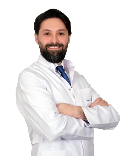


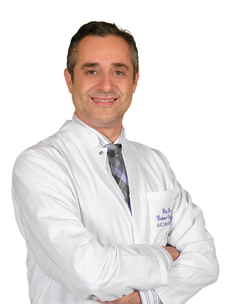
Available at these Hospitals







What patients say about this unit
★★★★★From 2,400+ verified patient reviews“From the first consultation to discharge, my breast reconstruction went smoothly. Dr. Özbebit was thorough, professional and genuinely caring. I would recommend this team to anyone.”
“I had been worried for months, but my breast cancer surgery was sorted out quickly and clearly. Dr. Parlakkılıç inspired complete confidence from the first meeting. I felt looked after at every turn.”
“I had been worried for months, but my breast reconstruction was sorted out quickly and clearly. The expertise of Dr. Öner put my mind completely at ease. The international patient office coordinated everything perfectly.”
“I flew in specifically for breast cancer surgery and was treated with the utmost professionalism. Dr. Alponat was thorough, professional and genuinely caring. I am deeply grateful for everything.”
Patient stories from this unit

Speak with our medical team
Share your case and our international patient team will guide you to the right specialist, free of charge.