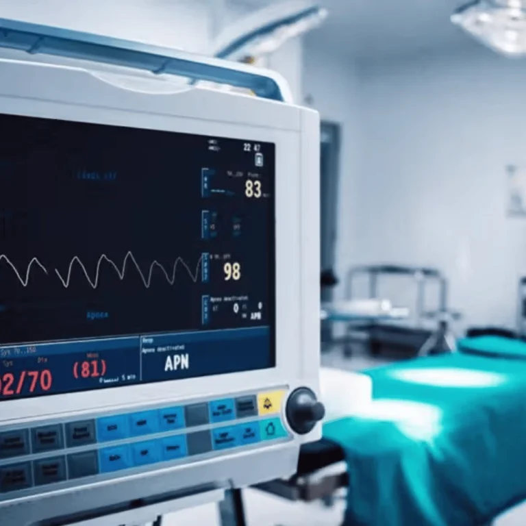
When someone gets diagnosed with pancreatic or gastric cancer, knowing the cancer’s stage is key. Endoscopic ultrasound (EUS) is a big help here. It’s a way to see how far the cancer has spread without a big surgery. EUS uses a special endoscope with an ultrasound probe. This probe makes clear pictures of the digestive tract and nearby tissues. Doctors can then check the tumor’s size, where it is, and if it’s touching other parts or lymph nodes. This info is vital for figuring out the cancer’s stage.
EUS helps doctors get a clear picture of the cancer. This leads to better treatment plans. It means patients have a better chance of getting the right treatment and living a better life.
What is Endoscopic Ultrasound (EUS)?
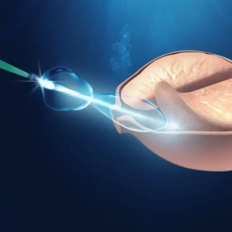
Endoscopic Ultrasound (EUS) is a cutting-edge medical procedure. It combines endoscopy and ultrasound to show detailed images of the digestive tract and nearby tissues. This tool has changed gastroenterology by giving clear views of the gastrointestinal wall and nearby areas.
The EUS procedure uses a flexible endoscope with an ultrasound transducer at the tip. This transducer sends high-frequency sound waves through the digestive wall. It creates detailed images of the tissues and organs around it. The procedure is done under sedation to keep the patient comfortable.
EUS Technology and Its Applications
EUS technology has improved a lot over time. It now includes Doppler technology. This lets doctors check blood flow through vessels. It’s key for looking at the blood flow of lesions and planning treatments.
| Key Features of EUS | Description | Clinical Significance |
|---|---|---|
| High-resolution imaging | Detailed visualization of the GI tract and surrounding tissues | Accurate diagnosis of lesions and staging of cancers |
| Doppler capability | Assessment of blood flow through vessels | Evaluation of vascularity and planning of interventions |
| Fine-needle aspiration (FNA) | Sampling of tissues or fluids for cytological examination | Diagnosis of malignancies and infections |
The EUS procedure is not just for looking at things. It’s also for treating them. It lets doctors take samples with fine-needle aspiration (FNA) for tests. This helps find cancers and infections. It also helps with treatments like draining pseudocysts and abscesses, and even managing pain in pancreatic cancer.
In summary, EUS is a big step forward in treating and diagnosing stomach and intestinal diseases. It’s a minimally invasive method that’s very accurate and can also treat some conditions.
How EUS Works
The EUS scan uses a special endoscope with an ultrasound probe. It looks at the layers of the stomach and nearby areas. This helps doctors see the digestive tract and tissues around it. It’s great for finding and understanding different problems, like cancer.
Step-by-Step Explanation of the EUS Procedure
The EUS procedure has a few main steps:
- Preparation: Patients get sedation to relax. The doctor explains what will happen and answers questions.
- Insertion: The EUS endoscope goes in through the mouth or rectum, depending on what’s being checked.
- Visualization: The ultrasound probe shows images of the stomach and nearby tissues on a screen. The doctor looks at these images.
- Sampling: If needed, the doctor might take a sample of tissue for more tests.
The EUS scan gives important details about tumors. It shows their size, where they are, and how far they’ve spread. This helps doctors figure out how serious the cancer is and what treatment to use.
| Step | Description | Significance |
|---|---|---|
| Preparation | Patient sedation and explanation of the procedure | Ensures patient comfort and understanding |
| Insertion | Careful insertion of the EUS endoscope | Access to the area of interest for imaging |
| Visualization | Generation of ultrasound images | Detailed examination of the GI tract and surrounding tissues |
| Sampling | Optional FNA for tissue collection | Provides tissue for histological examination |
Knowing how EUS works helps everyone understand its importance. It’s a key tool for managing stomach and intestinal cancers.
Clinical Applications of EUS
EUS has changed gastroenterology by making diagnosis and treatment easier. It’s a key tool for managing many conditions. This has greatly improved patient care and outcomes.
EUS is mainly used for finding and staging cancers in the gut. It helps see tumors clearly and check lymph nodes. This is vital for knowing how serious the cancer is and what treatment to use.
EUS also helps with other gut problems. It can find the cause of belly pain, check how bad pancreatitis is, and spot blockages in the bile duct.
Key Applications of EUS
- Cancer staging: EUS gives detailed images to see how far cancer has spread.
- Diagnostic sampling: EUS-guided fine-needle aspiration (FNA) gets tissue samples for tests.
- Treatment planning: EUS info helps decide if surgery or other treatments are best.
- Management of gastrointestinal disorders: EUS helps diagnose issues like pancreatitis and bile duct blockages.
EUS has many benefits in clinics. It’s a safe way to look inside the body without surgery. This lowers risks and speeds up recovery. EUS also lets doctors make quick decisions during procedures.
Using personal stories and examples shows EUS’s real impact. For example, a pancreatic cancer patient might get EUS to figure out the best treatment. This could greatly improve their life and chances of recovery.
Benefits of EUS in Cancer Staging
EUS is key in accurately staging cancers, like pancreatic and gastric ones. It gives clear images of tumors and the tissues around them. This makes it very useful for doctors.
One big plus of EUS is it gives detailed info without needing to do invasive tests. This makes patients more comfortable and lowers the chance of complications from other tests.
Advantages of EUS in Cancer Staging
EUS is great for cancer staging because it’s very good at finding tumors and seeing if they’ve spread. It can check if cancer has reached nearby lymph nodes or other parts of the body.
- Accurate T-staging and N-staging
- Guiding fine-needle aspiration for cytology
- Assessing the involvement of major blood vessels
| Imaging Technique | Resolution | Invasiveness | Diagnostic Accuracy |
|---|---|---|---|
| EUS | High | Minimally invasive | High |
| CT Scan | Moderate | Non-invasive | Moderate |
| MRI | High | Non-invasive | High |
The table shows how EUS compares to CT scans and MRI. EUS is best because it gives detailed images with little invasion.
In summary, EUS has many benefits in cancer staging. It helps a lot in making accurate diagnoses and treatment plans. Its role in cancer care is growing, bringing new ways to help patients.
EUS vs. Other Imaging Techniques
EUS diagnosis is key in oncology, but how does it compare to other imaging methods? To grasp its worth, we must look at EUS alongside other diagnostic tools used for pancreatic and gastric cancer staging.
Computed Tomography (CT) scans are a mainstay in cancer diagnosis. They give detailed views of internal structures, helping spot tumors and their spread. Yet, they might miss smaller lesions or accurately stage cancer. EUS, on the other hand, offers clear images of the GI wall and nearby tissues. It’s great for seeing how deep a tumor has invaded and finding nearby lymph nodes.
Magnetic Resonance Imaging (MRI) is also used for cancer staging. MRI excels in showing soft-tissue details and can check tumor spread. Though MRI is good for some cancers, EUS is preferred for its real-time images and ability to guide tissue sampling.
Positron Emission Tomography (PET) scans are used to see how active tumors are metabolically. They help spot distant metastases and disease burden. Yet, they don’t offer the detailed anatomy that EUS or CT scans do. A mix of imaging methods, including EUS, is often used for a full cancer stage understanding.
EUS stands out in imaging techniques for its strengths. It’s best for checking tumor spread and lymph node involvement. This info is key for deciding if a tumor can be removed and planning treatment. For instance, those with esophageal cancer can learn about treatment options on the Acıbadem International website.
| Imaging Technique | Strengths | Limitations |
|---|---|---|
| EUS | High-resolution images, real-time imaging, guides FNA | Limited depth penetration, operator-dependent |
| CT Scan | Detailed view of internal structures, widely available | May not detect smaller lesions, radiation exposure |
| MRI | Excellent soft-tissue resolution, no radiation | Not always suitable for all patients, more expensive |
| PET Scan | Assesses metabolic activity, detects distant metastases | Limited anatomical detail, radiation exposure |
In conclusion, EUS is a vital tool in cancer staging, with unique benefits for pancreatic and gastric cancers. While other methods have their own strengths, EUS’s high-resolution images and ability to guide tissue sampling make it essential in cancer care.
Risks and Limitations of EUS
EUS is very precise in cancer staging, but knowing its risks is key. It’s considered safe, but complications can happen.
Bleeding is a big risk, mainly if a biopsy is done. Infection is rare but can happen if the area isn’t kept clean. Pancreatitis is also a risk, mostly if the pancreas is touched during the procedure.
Common Risks and Complications
- Bleeding: More likely when biopsies are taken.
- Infection: Rare, but more common if sterile conditions are not maintained.
- Pancreatitis: Associated with procedures involving the pancreas.
Having an EUS expert do the procedure is key. They lower the risk of problems and make sure the results are right.
EUS can’t go through some areas because of blockages. It’s also hard to get enough tissue in some cases. Plus, it depends a lot on the doctor’s skill.
In short, EUS is a great tool, but knowing its risks and limits is important. By choosing experienced EUS experts, patients can make better choices. Doctors can also use EUS more effectively in treating cancer.
EUS in the Diagnostic Pathway
EUS is now key in diagnosing cancer. It’s a non-invasive way to check how serious a tumor is.
When someone is diagnosed with cancer, doctors use many tests to figure out what’s going on. EUS is important because it gives clear pictures of tumors and how they relate to other parts of the body.
Key Steps in the Diagnostic Pathway
- Initial assessment and screening
- Imaging tests such as CT scans and MRI
- EUS for detailed tumor staging
- Biopsy for tissue diagnosis
EUS is very helpful for pancreatic and gastric cancer. It helps doctors see how big the tumor is, where it is, and if it’s touching important areas. This info is key for deciding the best treatment.
Benefits of EUS in Cancer Staging
- Provides high-resolution images of tumors and surrounding tissues
- Enables accurate staging of cancer, which guides treatment decisions
- Helps avoid unnecessary surgery by identifying patients who are not candidates for curative resection
Using EUS in diagnosis helps doctors give better care to cancer patients. It helps create a treatment plan that fits each patient’s needs. This plan is made with info from EUS and other tests.
Advances in EUS Technology
Recent years have brought big changes to EUS technology. These changes are making it better for finding and understanding pancreatic and gastric cancers. EUS is now a key tool in diagnosing these cancers.
High-resolution ultrasound probes are a big part of these improvements. They give clear pictures of the GI tract and the tissues around it. This helps doctors make more accurate diagnoses and understand how far the cancer has spread.
Key Features of Advanced EUS Technology
- High-resolution imaging for better diagnostic accuracy
- Elastography for assessing tissue stiffness
- Contrast-enhanced EUS for improved visualization of lesions
- Advanced biopsy techniques for precise tissue sampling
Elastography is another important addition to EUS. It checks how stiff tissues are. This helps doctors tell if a lesion is cancerous or not. It’s been really helpful for looking at pancreatic lesions.
| Technology | Description | Clinical Benefit |
|---|---|---|
| High-Resolution EUS | Provides detailed images of the GI tract and surrounding tissues | More accurate diagnosis and staging of cancers |
| Elastography | Assesses tissue stiffness to differentiate between benign and malignant lesions | Improved diagnostic accuracy for pancreatic lesions |
| Contrast-Enhanced EUS | Enhances visualization of lesions using contrast agents | Better detection and characterization of lesions |
The future of EUS technology is exciting. Researchers are always finding new ways to use it. As EUS keeps getting better, it will play an even bigger role in fighting gastrointestinal cancers.
EUS Training and Expertise
The accuracy of Endoscopic Ultrasound (EUS) in diagnosing and staging cancers depends on the practitioner’s skills. As EUS becomes more important in oncology, the need for good training programs is clear.
EUS is a complex procedure that needs a deep understanding of the gut’s anatomy and diseases. It also requires the technical skills to do the exam safely and well. The benefits of EUS, like seeing tumors and tissues clearly, depend on well-trained practitioners.
Key Components of EUS Training
- Hands-on experience with EUS equipment and techniques
- Study of gastrointestinal anatomy and pathology
- Practice in interpreting EUS images and making accurate diagnoses
- Understanding of the clinical applications of EUS in cancer staging and treatment planning
Experienced practitioners make EUS exams more accurate. They also help patients by reducing risks. Plus, staying updated with EUS technology and techniques is key.
EUS Applications in Cancer Care
EUS is used in many ways in cancer care. It helps diagnose and stage pancreatic and gastric cancers. It also guides biopsies. Accurate staging with EUS leads to better treatment plans and outcomes.
- EUS-guided fine-needle aspiration biopsy for tissue diagnosis
- Staging of pancreatic and gastric cancers
- Evaluation of lymph nodes and other possible cancer sites
Highlighting the need for EUS training ensures top-notch care for patients. As oncology grows, EUS’s role will too. So, continuous education and training are vital.
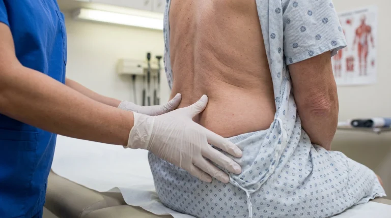
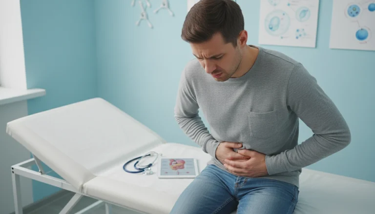
Patient Experience with EUS
Many patients find the idea of an EUS procedure scary. But knowing what to expect can really help. Understanding the steps before, during, and after can make a big difference.
Preparing for EUS
Getting ready for an EUS is important. You’ll likely be told not to eat or drink for a while before. Also, tell your doctor about any medicines you’re taking, like blood thinners. They might need to adjust or stop them.
- Follow the doctor’s instructions regarding fasting and medication.
- Arrange for someone to accompany you to the procedure and drive you home afterward.
- Wear comfortable, loose-fitting clothing.
On the day, you’ll get sedation to relax. The EUS uses an endoscope through your mouth or rectum. It uses ultrasound to see inside your body.
Post-Procedure Care
After the EUS, you’ll be watched for a bit to make sure everything is okay. You might feel a bit drowsy or sore. But most people can go back to normal in a day.
Here’s what to do after:
- Rest for the rest of the day.
- Avoid heavy meals and hard activities for 24 hours.
- Follow your doctor’s advice on medicine and when to come back.
Knowing how EUS uses and EUS diagnosis work can make you appreciate it more. Being ready and knowing what to expect can make the EUS process easier and less scary.
Conclusion: The Future of EUS in Oncology
Endoscopic Ultrasound (EUS) is playing a bigger role in cancer staging. This is thanks to new technology and the skills of EUS experts. They say EUS is key for diagnosing and planning treatment for pancreatic and gastric cancers.
The future of EUS looks bright. It will work better with other tools and treatments. This means better care for patients with more accurate and less invasive treatments.
EUS will keep being important in fighting cancer. It gives clear pictures of tumors and the areas around them. This helps doctors plan the best treatment for patients, leading to better care.