Nasopharyngeal cancer is a form of cancer that starts in the nasopharynx — the upper part of the throat behind the nose — and is considered a distinct type within head and neck cancers because of its unique causes, behavior, and care. While it’s less common worldwide than some other head neck cancer types, nasopharyngeal cancer is more frequent in certain regions (for example, parts of East and Southeast Asia) and is strongly linked to the Epstein‑Barr virus (EBV) in many cases.
This article explains what nasopharyngeal cancer is, common symptoms to watch for, how doctors diagnose and stage the disease, and the main treatment options (including when radiotherapy and systemic treatment are used). If you or someone you know has persistent one‑sided ear problems, a painless neck lump, or unexplained nosebleeds, see an Ear, Nose, and Throat (ENT) specialist promptly — early evaluation can make a difference.
What is Nasopharyngeal Cancer?
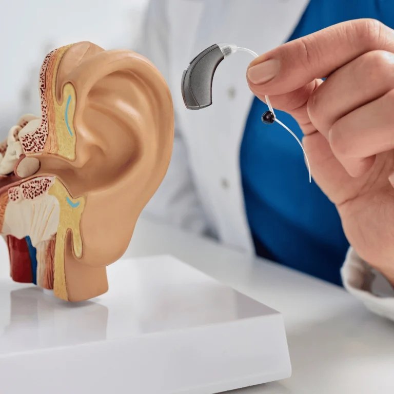
The nasopharynx is the space behind the nose and above the back of the throat at the base of the skull. It connects the nose, the back of the throat, and the upper airway; air passes through the nasopharynx on its way to the larynx and lungs. Nasopharyngeal cancer occurs when cells in this area begin to grow uncontrollably, forming tumor tissue.
Several different types of tumors can arise in the nasopharynx. Key types to know about include:
- Nasopharyngeal carcinoma: This is the most common form of nasopharyngeal cancer and develops from the epithelial cells (the surface cells that line the nasopharynx). Pathologists typically classify carcinoma into subtypes — non‑keratinizing undifferentiated, non‑keratinizing differentiated, and keratinizing squamous cell — with the non‑keratinizing undifferentiated subtype most often linked to Epstein‑Barr virus (EBV) infection and generally more radiosensitive.
- Lymphoma: Lymphoma arises from lymphocytes (immune system cells) and can involve the nasopharynx. Lymphoma often presents differently from carcinoma — for example, rapidly growing, softer masses and systemic symptoms can point toward lymphoma rather than carcinoma.
- Minor salivary gland cancers: These include adenocarcinoma and adenoid cystic carcinoma and originate from small salivary gland tissue in the nasal and oral regions. Their behavior and treatment can differ from epithelial nasopharyngeal carcinoma.
Clinical note: a firm, painless lump in the neck (enlarged lymph nodes) is a common presenting sign of nasopharyngeal carcinoma — when you see a neck node that’s unexplained, especially in people from higher‑risk regions, an evaluation of the nasopharynx is important.
What are the Risk Factors?
Nasopharyngeal cancer develops from a mix of viral, genetic, and environmental influences. Infection with the Epstein‑Barr virus (EBV) is a major risk factor for many cases, but EBV exposure alone does not mean a person will develop cancer — the risk rises when viral infection combines with other factors.
Risk factors can be grouped as non‑modifiable and potentially modifiable:
- Non‑modifiable: Infection with Epstein‑Barr virus (EBV) (strongly linked to non‑keratinizing nasopharyngeal carcinoma), family history of nasopharyngeal cancer, and ancestry or race — the disease is more common in certain populations, particularly in parts of East and Southeast Asia.
- Potentially modifiable: Long‑term dietary exposures (early‑life consumption of salt‑preserved foods such as salted fish, which can contain carcinogenic nitrosamines), heavy alcohol use, and certain environmental or occupational exposures (smoke from wood fires, air pollution, and chemical vapors) may increase risk.
Epidemiology notes: nasopharyngeal cancer shows distinct age patterns — in some regions there’s a peak in adolescence/young adulthood and another in middle age — and it is seen more often in men than in women. These patterns vary by population and geography.
If you have a family history of nasopharyngeal cancer, come from a higher‑risk region, or have persistent ENT symptoms, talk to your healthcare provider about evaluation and whether surveillance (for example, clinical exams or EBV‑related testing in specialist settings) is appropriate for you.
What are the Symptoms of Nasopharyngeal Cancer?
Early nasopharyngeal cancer often causes few or nonspecific symptoms and can be mistaken for common problems such as sinus infections or allergies. Because of this, diagnosis may be delayed until more specific signs appear or the disease spreads to nearby lymph nodes in the neck.
Common symptoms can be grouped by area to make them easier to spot:
- Neck: A painless lump or swelling in the neck from enlarged lymph nodes is the most common warning sign — unexplained neck lumps should be evaluated, especially in people from higher‑risk regions.
- Nasal/oral: Persistent nasal congestion, nosebleeds, or blood in saliva can occur when the tumor affects the nasal cavity or nasopharynx.
- Ear and hearing: One‑sided ear symptoms such as ringing (tinnitus), a feeling of fullness, persistent ear infections, or unilateral hearing loss — especially if they don’t respond to usual treatment — are important red flags.
- Throat and head: Sore throat, persistent headache, or symptoms from tumor spread to nearby structures (for example, facial numbness) may be present.
When to see a doctor: See an ENT specialist if you have a painless neck lump lasting more than two weeks, persistent one‑sided ear problems, repeated ear infections that don’t improve, or unexplained nosebleeds. Early evaluation — including examination of the nasopharynx and imaging or biopsy when indicated — helps detect nasopharyngeal cancer sooner.
What Causes Nasopharyngeal Cancer?
Like many cancers, nasopharyngeal cancer develops after a series of genetic changes that let cells divide uncontrollably, invade nearby tissue, and sometimes spread to other parts of the body. In nasopharyngeal carcinoma, these changes most often affect epithelial cells — the surface cells that line the nasopharynx. Infection with the Epstein‑Barr virus (EBV) is the single most important known risk factor for many cases, but EBV infection by itself is common and only a small proportion of people with EBV go on to develop cancer. Risk reflects an interaction between the virus, genetics, and environmental exposures.
Below are common factors that increase the likelihood of developing nasopharyngeal cancer; these are grouped as non‑modifiable and potentially modifiable:
- Non‑modifiable factors: Infection with the Epstein‑Barr virus (EBV) — strongly associated with the non‑keratinizing subtype of nasopharyngeal carcinoma; family history of nasopharyngeal cancer; and ancestry/race (the disease is more common in parts of East and Southeast Asia and in some immigrant communities).
- Potentially modifiable factors: Long‑term dietary exposures such as early‑life and frequent consumption of salt‑preserved foods (for example, salted fish), which may contain nitrosamine carcinogens; heavy alcohol use; and environmental or occupational exposures (smoke from wood fires, air pollution, and certain chemical vapors) that may increase risk.
Additional considerations: Men are affected more often than women, and age patterns vary by region — some populations show a peak in young adulthood and a separate peak in middle age. The exact reasons for these patterns are complex and involve genetic and environmental interactions.
Takeaway: EBV is a key causal factor but not the sole cause; nasopharyngeal cancer may result from a combination of viral exposure, inherited susceptibility, and environmental or dietary factors. If you have a family history of nasopharyngeal cancer, come from a higher‑risk population, or have ongoing ENT symptoms, discuss evaluation and possible surveillance with your healthcare provider.
Is Early Diagnosis Possible?
Early diagnosis is possible, especially when people or clinicians act on persistent warning signs. Routine annual health checkups can pick up suspicious findings, but targeted tests are often needed to detect nasopharyngeal cancer at an earlier stage.
Which signs should prompt further testing? Persistent one‑sided ear problems, an unexplained painless lump in the neck, repeated nosebleeds, or ongoing nasal congestion warrant evaluation. For people at higher risk (family history or from endemic regions), clinicians may consider additional surveillance.
Common tests used to investigate suspected disease include nasoendoscopy (direct visualization of the nasopharynx), biopsy of suspicious tissue for pathology, and imaging such as MRI or PET/CT to assess extent. In specialist centers and some high‑risk screening programs, plasma EBV DNA testing is also used as an adjunctive tool to detect early disease or monitor for recurrence.
How is the Diagnosis Made?
Step 1 — History and physical exam: The clinician begins with a detailed medical history (symptoms, risk factors such as family history or origin from a high‑risk region) and a focused head and neck exam. Palpation of the neck for enlarged lymph nodes (nodes) is important because an unexplained, painless neck lump is a common presenting sign.
Step 2 — Direct visualization and biopsy: If suspicion is raised, the nasopharynx is examined directly using nasoendoscopy (a thin, flexible tube with a camera). If a suspicious lesion is seen, an endoscopic biopsy is taken — the tissue sample is the definitive test to tell whether abnormal cells are benign or malignant and to determine the tumor subtype. Biopsy and pathology also help define tumor grade and guide staging.
Step 3 — Imaging and staging: Once cancer is confirmed, imaging tests evaluate the extent of local disease and search for spread (cancer spread) to lymph nodes or distant sites. Common imaging includes MRI for detailed local and regional assessment and PET/CT to detect nodal involvement and distant metastases. The combination of pathology and imaging determines the tumour (T), node (N) and metastasis (M) categories and the overall stage — staging is essential to choose the right treatment.
Step 4 — Additional tests and monitoring: Ancillary tests may include blood work and, in specialist centers, plasma EBV DNA testing to support diagnosis or for baseline monitoring. In some cases, ultrasound of the neck and fine‑needle aspiration of suspicious nodes are used. Your care team may also order dental, nutritional, and audiologic assessments prior to treatment planning.
Practical notes: an endoscopic biopsy is usually done under local anesthesia or light sedation; imaging exams like MRI or PET/CT are noninvasive but can take time and may require contrast. If you have persistent red‑flag symptoms (a painless neck lump, unilateral hearing loss or ongoing ear infections, repeated nosebleeds), seek ENT evaluation promptly — early diagnosis improves treatment options.
What Are the Stages of the Disease?
Accurate staging is essential to decide whether treatment aims to cure the patient or to control symptoms (palliative care). Staging describes three core components: the size and local extent of the primary tumor (T), whether cancer has spread to regional lymph nodes in the neck (N), and whether distant metastasis (M) is present. Together these give an overall stage group (I–IV) that guides treatment choices.
Key practical points:
- Early stages (stage I–II) typically involve a small primary tumor with no or limited spread to lymph nodes and are often treated with curative radiotherapy (often combined with chemotherapy depending on stage).
- Advanced local or regional disease (for example, large primary tumors or extensive nodal involvement such as N3 — very large or bilateral nodes) increases the risk of cancer spread and usually requires combined modality treatment and careful screening for distant metastases.
- If distant metastasis (M1) is present at diagnosis — reported in roughly a minority of patients (about 5–10% in some series) — treatment objectives typically shift toward systemic therapy to control cancer spread and symptoms.
Because nodes in the neck are a common pathway for spread, thorough evaluation of lymph nodes neck (clinical exam, ultrasound, and cross‑sectional imaging such as MRI or PET/CT) is important during staging. In patients with advanced nodal disease, clinicians often perform more extensive distant metastasis screening to guide appropriate therapy.
How is Nasopharyngeal Cancer Treated?
Overview: Treatment is tailored to the stage, subtype, and overall health of the person. For many patients with nasopharyngeal carcinoma, the main curative approach involves radiotherapy combined with systemic therapy; surgery has a more limited role. Multidisciplinary care — involving radiation oncology, medical oncology, ENT (ear, nose & throat), nutrition, dental, and speech/swallow teams — optimizes outcomes and manages side effects.
Radiotherapy (radiation therapy)
- What it is: External beam radiotherapy is the primary local treatment for nasopharyngeal cancer; highly focused beams deliver radiation to the tumor and involved lymph nodes in the neck.
- When it’s used: Radiotherapy alone may be sufficient for very early-stage disease. For most locally advanced cases, radiotherapy is combined with systemic treatment to improve control.
- Technical notes: Modern techniques (intensity‑modulated radiotherapy, IMRT) allow precise targeting to spare nearby tissues (salivary glands, spinal cord), reducing long‑term toxicity.
- Side effects: Common acute effects include skin irritation in the treatment areas, dry mouth (xerostomia), mucositis (mouth and throat soreness), difficulty swallowing, and temporary hearing changes. Late effects can include persistent dry mouth, dental problems, neck stiffness, and occasionally long‑term hearing loss; supportive care and rehabilitation (dental care, swallowing therapy) are important.
Systemic therapy (chemotherapy and other cancer treatments)
- Role: Chemotherapy is used alongside radiotherapy to increase the chance of cure in more advanced local/regional disease and to treat distant spread. Common approaches include concurrent chemoradiation (chemotherapy given during radiotherapy) and, for some patients, induction (neoadjuvant) chemotherapy given before radiotherapy to shrink large tumors.
- Administration: Drugs are usually given intravenously; regimens are selected by medical oncologists based on the stage and patient fitness. Systemic therapies can cause fatigue, nausea, low blood counts, and increased infection risk — your care team will provide supportive medications and monitoring.
- Emerging options: In selected situations, targeted therapies and immunotherapy are being studied or used, particularly for recurrent or metastatic disease; discussion with a medical oncologist about clinical trials may be appropriate.
Surgery
- Primary role: Surgery is not usually the first‑line treatment for primary nasopharyngeal tumors because of the anatomic location. However, it has a role for selected recurrences and for salvage of persistent disease.
- Neck management: For cancer that has spread to lymph nodes, a therapeutic or salvage neck dissection (surgical removal of cancerous lymph nodes) may be performed when residual or recurrent nodal disease persists after radiotherapy.
Treatment planning and what patients can expect
- Multidisciplinary planning: Your team will review pathology, imaging (MRI, PET/CT), and overall health to recommend a personalized plan. Treatment timing and sequence (induction chemo → chemoradiation, or concurrent chemoradiation) depend on stage and fitness.
- Supportive care: Expect baseline dental checks, nutrition counseling, and hearing evaluation as part of preparation. During treatment, you will have frequent monitoring for side effects and supportive therapies to manage symptoms.
- Rehabilitation and survivorship: After treatment, many patients need rehabilitation such as swallowing therapy, dental follow‑up, and ongoing surveillance for recurrence. Emotional and social support resources are also important.
Treatment complications and monitoring
Complications are typically divided into acute (during or shortly after treatment) and late effects (months to years later). Acute toxicities often involve rapidly dividing tissues in the radiation field (mucosa, skin, bone marrow). Late complications depend on treatment dose and volume and individual factors (age, prior surgery). Regular follow‑up visits, imaging as indicated, and, where available, plasma EBV DNA testing help detect recurrence early and guide further care.
When to seek care: If cancer comes back or new symptoms develop (for example, a new neck lump, increasing pain, or breathing/swallowing changes), contact your oncology/ENT team promptly — early evaluation improves the chance of effective salvage treatment.
Is it Possible to Prevent?
There is no guaranteed way to prevent nasopharyngeal cancer, but people can take steps that may reduce their risk. Because Epstein‑Barr virus (EBV) infection, genetic background, and environmental or dietary exposures all interact to influence risk, the most practical approach focuses on risk reduction and early detection rather than absolute prevention.
Practical risk‑reduction measures:
- Limit consumption of salt‑preserved foods (for example, salted fish) and processed preserved foods that may contain carcinogenic compounds.
- Avoid heavy alcohol use and reduce exposure to cigarette smoke and indoor air pollution (including prolonged exposure to wood smoke where possible).
- Adopt a healthy lifestyle—balanced diet, regular physical activity, and maintaining overall health—which supports the immune system and general well‑being.
People at higher risk (those with a family history of nasopharyngeal cancer or from regions where the disease is more common) should discuss personalized surveillance with their healthcare provider. In specialist centers, targeted approaches such as clinical examinations, nasoendoscopy, imaging, or plasma EBV DNA testing are sometimes used for earlier detection or monitoring.
If you are concerned about your risk or have persistent symptoms, talk with your clinician about steps you can take and whether referral to an ENT specialist is appropriate.