The Science Behind Tomography
Medical imaging has transformed how healthcare professionals diagnose and treat patients. Tomography, a key technology, enables detailed cross-sectional imaging of the body. This has revolutionized the field. Tomography provides a clear view of internal structures. This has significantly improved diagnostic capabilities. Healthcare providers can now identify conditions more effectively and develop targeted treatment plans.
The impact of tomography on healthcare technology has been profound. It has enhanced patient care and outcomes. As medical imaging evolves, tomography’s role is expected to expand. This will drive further innovations in diagnostic techniques and treatment options. Tomography’s science lies in its ability to create detailed cross-sectional images of the body’s internal structures. This is key to its role in diagnostic medicine.
Definition and Basic Principles
Tomography is a diagnostic imaging technique that produces images of specific sections or slices of the body. It uses a tomographic device to capture data, which is then reconstructed into an image. This method allows for the visualization of internal structures without surgical intervention.
Cross-Sectional Imaging Explained
Cross-sectional imaging is a core aspect of tomography, enabling healthcare professionals to view the body’s internal structures in detail. It captures images of thin slices or sections of the body, which are then reconstructed into 2D or 3D images. This technique is invaluable for diagnosing a wide range of medical conditions.
Difference Between Tomography and Conventional Radiography
Unlike conventional radiography, which provides a 2D representation of a 3D object, tomography offers a more detailed view by capturing images of specific sections. Conventional radiography superimposes all structures within the beam’s path, potentially obscuring important details. Tomography, by contrast, eliminates this overlap, providing clearer images of internal structures.
Understanding the science behind tomography, including its definition, principles, and the advantages of cross-sectional imaging, helps healthcare professionals use this technology effectively for diagnostic purposes.
Historical Development of Tomographic Imaging
The history of tomography is a rich tapestry of innovation and discovery. It has transformed the field of medical imaging over the decades.
Early Tomographic Techniques
Early tomographic techniques began in the 1930s. The first practical tomographic device was developed by Alessandro Vallebona, an Italian radiologist. His work laid the foundation for modern tomographic imaging.
Key Milestones in Tomography Evolution
The evolution of tomography has seen several key milestones. The introduction of computed tomography (CT) in the 1970s by Godfrey Hounsfield and Allan McLeod Cormack was a major breakthrough. Their work revolutionized diagnostic imaging.
Nobel Prize-Winning Contributions
The development of CT scans was recognized with the Nobel Prize in Physiology or Medicine in 1979. Hounsfield and Cormack were awarded for their groundbreaking work. This highlights the significant impact of their contributions on medical imaging history.
Their innovation continues to influence contemporary medical imaging techniques. It shapes the future of diagnostic medicine.
Types of Tomography in Modern Medicine
The field of tomography has grown significantly, introducing various imaging modalities for different diagnostic needs. This growth enables healthcare professionals to choose the best imaging technique for each patient’s case. This tailored approach improves patient care across multiple medical specialties.
Computed Tomography (CT)
Computed Tomography (CT) scans employ X-rays to generate detailed cross-sectional images of the body. They are invaluable for:
- Emergency room diagnostics, such as assessing injuries from accidents
- Detecting cancers and monitoring their progression
- Guiding interventional procedures like biopsies
Positron Emission Tomography (PET)
Positron Emission Tomography (PET) uses a radioactive drug (tracer) to display both metabolism and chemical flow within the body. PET scans are essential for:
- Oncology, to assess the spread of cancer
- Cardiology, to evaluate heart function
- Neurology, to diagnose brain disorders
Single-Photon Emission Computed Tomography (SPECT)
Single-Photon Emission Computed Tomography (SPECT) is a nuclear medicine tomographic imaging technique. It is widely applied for:
- Bone and gallium scans
- Cardiac stress tests
- Brain perfusion studies
Magnetic Resonance Imaging (MRI)
Magnetic Resonance Imaging (MRI) employs powerful magnetic fields and radio waves to create detailed images of internal structures without X-rays. It excels in soft tissue imaging.
Functional MRI Applications
Functional MRI (fMRI) is a specialized MRI application that measures brain activity by detecting blood flow changes. It is used for:
- Mapping brain function before surgery
- Research into neurological disorders
- Cognitive neuroscience studies
In conclusion, the diverse range of tomographic techniques in modern medicine supports a personalized approach to diagnosis and treatment. This enhances patient care across various medical fields.
How Computed Tomography Works
To grasp the essence of Computed Tomography, we must explore its fundamental elements: X-ray generation and image reconstruction. CT scans are vital in today’s medical field, delivering detailed views of the body’s internal parts.
X-ray Generation and Detection
The journey starts with X-ray creation. A CT scanner employs an X-ray tube to generate X-rays, which then traverse the patient’s body. Sensors on the scanner’s opposite side capture these X-rays.
The X-ray tube and detectors are positioned on a gantry that circles the patient. This movement is essential for assembling a complete image.
Image Reconstruction Algorithms
The data from the detectors is analyzed through advanced image reconstruction algorithms. These algorithms transform the X-ray attenuation data into detailed cross-sectional images of the body.
Modern algorithms, like iterative reconstruction, refine image quality. They minimize noise and artifacts, boosting diagnostic precision.
Hounsfield Units and Image Interpretation
The images are displayed in Hounsfield Units (HU), a scale that measures X-ray attenuation. Tissues are differentiated by their HU values, aiding in the identification of various structures and abnormalities.
Contrast Media in CT Imaging
Contrast media are used to highlight specific structures or lesions. These substances are given orally or intravenously, depending on the examination type.
Contrast media significantly enhance CT scan diagnostics, most noticeably in vascular and oncological studies.
Tomography in Diagnostic Medicine
Tomography is a cornerstone in modern diagnostic medicine, providing deep insights into the human body. It offers detailed cross-sectional images, transforming how medical professionals diagnose and treat conditions.
Neurological Applications
In neurology, tomography is key for diagnosing and monitoring brain and nervous system conditions. Techniques like CT and MRI are essential for spotting stroke, tumors, and diseases like Alzheimer’s. These methods give high-resolution images, allowing for precise lesion localization and disease tracking.
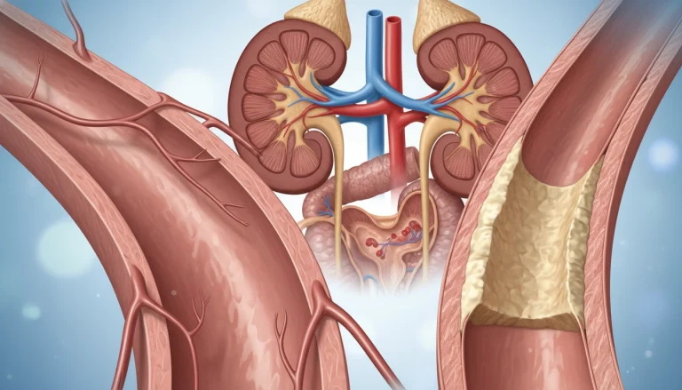
Cardiovascular Imaging
Tomography is vital in cardiovascular imaging, showing the heart and its blood vessels. CT angiography and cardiac MRI are critical for spotting coronary artery disease, assessing heart function, and planning surgeries. They help identify plaque buildup, aneurysms, and other heart issues.
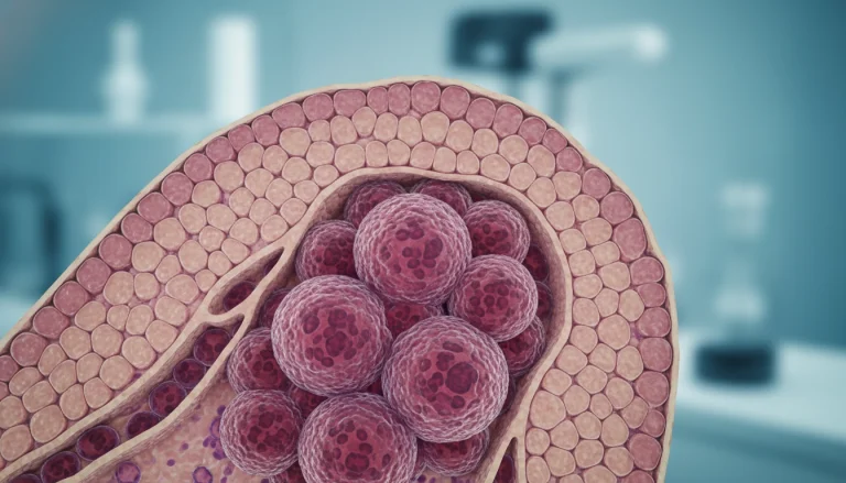
Oncology Diagnostics
In oncology, tomography is essential for cancer diagnosis and staging. PET-CT combines metabolic and anatomical data, aiding in tumor detection, aggressiveness assessment, and treatment monitoring. This approach is vital for tailored cancer care.
Musculoskeletal Evaluation
Tomography is also key in musculoskeletal evaluations. CT scans are great for complex fractures, bone infections, and tumors. MRI, with its superior soft tissue contrast, is used for ligament and tendon injuries, as well as spinal disorders. Detailed images aid in accurate diagnosis and treatment planning.
In conclusion, tomography’s versatility and diagnostic precision make it essential in modern medicine. It significantly improves patient care across various specialties.
Patient Experience During Tomographic Procedures
The patient experience during tomographic procedures encompasses preparation, the scan itself, and post-procedure care. Grasping these elements can ease concerns and enhance patient outcomes.
Preparation Requirements
Preparation for tomographic procedures typically involves removing any metal objects, such as jewelry or glasses, that could interfere with the imaging. Patients may also be required to change into a hospital gown.
For certain types of tomography, like CT scans, patients might need to fast for a few hours before the procedure or drink a contrast agent to enhance image quality.
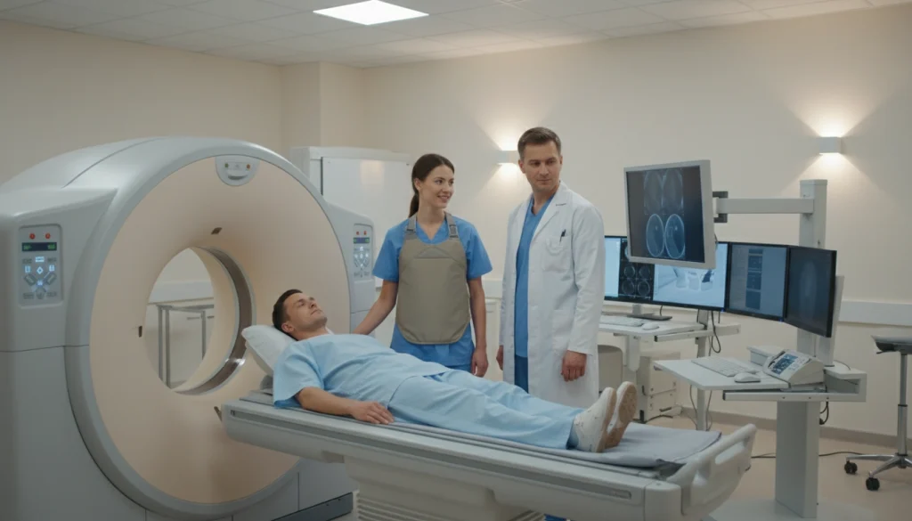
What to Expect During the Scan
During the scan, patients are positioned on a table that slides into the tomographic machine. They are required to remain as motionless as possible to ensure clear images.
The scan’s duration can vary, but most procedures are relatively quick, lasting anywhere from a few minutes to about an hour.
Post-Procedure Care
After the scan, patients can typically resume their normal activities unless specified by their healthcare provider.
If a contrast agent was used, patients might be monitored for a short period to check for any adverse reactions.
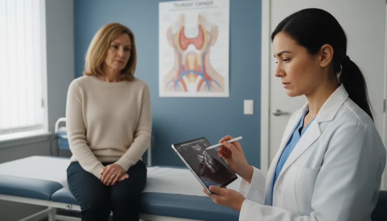
Addressing Patient Anxiety
Patient anxiety is a common concern for tomographic procedures. Healthcare providers often address this by explaining the procedure in detail and answering any questions the patient may have.
Some facilities also offer relaxation techniques or sedation to help patients feel more comfortable during the procedure.
Interventional Tomography Techniques
Tomography has revolutionized interventional techniques, allowing for more precise procedures. These techniques are now vital in modern medicine. They provide accurate guidance and real-time images during various treatments.
CT-Guided Procedures
CT-guided procedures use computed tomography for detailed images. These images guide surgeons during interventions. This method is key in biopsies, drainages, and tumor treatments, where accuracy is essential.
- Enhanced precision in targeting lesions
- Real-time monitoring of the procedure
- Reduced risk of complications
Real-Time Tomographic Imaging
Real-time tomographic imaging enables continuous monitoring during procedures. This is critical for adjusting techniques as needed. It ensures the success of the intervention.
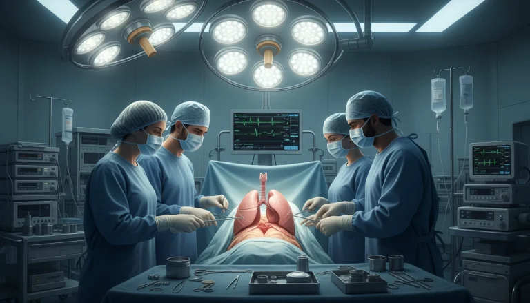
Minimally Invasive Surgical Applications
Interventional tomography in minimally invasive surgery has greatly improved patient care. It reduces recovery times and minimizes tissue damage. These procedures are less traumatic for patients and often lead to fewer complications.
- Reduced hospital stay
- Less post-operative pain
- Faster return to normal activities
Interventional tomography techniques are continually evolving. Advances in technology promise even greater precision and safety in medical interventions.
Tomography in Dental and Maxillofacial Imaging
Advanced tomographic imaging is transforming dental and maxillofacial diagnosis and treatment planning. It offers detailed, three-dimensional views of oral structures, previously unattainable with traditional two-dimensional radiography. This technology has become essential in dental and maxillofacial imaging.
Cone Beam Computed Tomography
Cone Beam Computed Tomography (CBCT) is a specialized version of traditional CT, ideal for dental and maxillofacial imaging. It uses a cone-shaped X-ray beam for data capture in a single rotation. This results in faster scanning times and lower radiation doses compared to conventional CT scans. Cosmetic dentistry procedures frequently rely on CBCT for precise planning.
Applications in Dental Surgery and Orthodontics
CBCT’s ability to provide accurate 3D images has transformed dental surgery and orthodontics. It offers precise assessments of bone density, volume, and morphology. This is critical for implant placement, orthognathic surgery, and orthodontic treatment planning.
The detailed images help identify anatomical structures and possible complications. This enhances treatment outcomes significantly.
3D Reconstruction for Treatment Planning
The 3D reconstruction capabilities of tomographic imaging are invaluable for treatment planning in dental and maxillofacial surgery. Clinicians can create detailed 3D models from tomographic data. This allows for better visualization of complex anatomy and simulation of surgical procedures.
It also aids in developing more effective treatment plans. This is highly beneficial for complex cases, such as impacted teeth or significant facial asymmetry.
Industrial Applications of Tomography
Tomography extends its reach beyond medical imaging, profoundly influencing numerous industrial sectors. Its capability to deliver detailed cross-sectional images makes it a critical asset in various fields.
Non-Destructive Testing
In the realm of non-destructive testing (NDT), tomography stands out. It enables the inspection of materials and structures without causing harm. This is essential for evaluating the integrity of components in sectors like aerospace, automotive, and construction.
- Detection of internal defects or flaws
- Analysis of material composition
- Examination of complex geometries
Quality Control and Material Analysis
Tomography is also widely applied in quality control and material analysis. It offers detailed images of materials’ internal structures. This aids in identifying defects, analyzing material properties, and ensuring adherence to manufacturing standards.
- Inspection of welds and joints
- Analysis of porosity and density in materials
- Verification of assembly integrity
Archaeological and Cultural Heritage Preservation
Tomography also plays a vital role in preserving archaeological and cultural heritage. It allows for the non-invasive study of artifacts, providing insights into historical objects without causing damage.
The application of tomography in these fields highlights its versatility. It offers significant advantages in terms of detailed imaging and analysis.
Radiation Safety and Dose Considerations
Radiation safety is a critical concern in tomographic imaging. The benefits of accurate diagnosis must be weighed against the risks of radiation exposure. As tomography evolves, understanding and mitigating radiation exposure is a top priority.
Radiation Exposure in Different Tomographic Techniques
Different tomographic techniques involve varying levels of radiation exposure. For example, Computed Tomography (CT) scans generally involve higher doses of radiation than conventional X-rays. The dose can vary significantly based on the protocol, the body part imaged, and the patient’s size.
Dose Reduction Strategies
Several strategies have been developed to reduce radiation doses in tomographic imaging. These include optimizing scan protocols, using dose modulation techniques, and implementing advanced image reconstruction algorithms. These allow for lower dose scans without compromising image quality.
Risk-Benefit Analysis
A thorough risk-benefit analysis is essential for every tomographic examination. It involves weighing the diagnostic benefits against the risks of radiation exposure. For most patients, the benefits of accurate diagnosis outweigh the risks.
Special Considerations for Pediatric Patients
Pediatric patients require special consideration due to their increased sensitivity to radiation and longer life expectancy. This increases the risk of long-term effects. Protocols for pediatric tomographic imaging are tailored to minimize dose while maintaining diagnostic image quality.
By understanding the factors that influence radiation exposure and implementing strategies to minimize dose, healthcare providers can ensure that tomographic imaging is used safely and effectively.
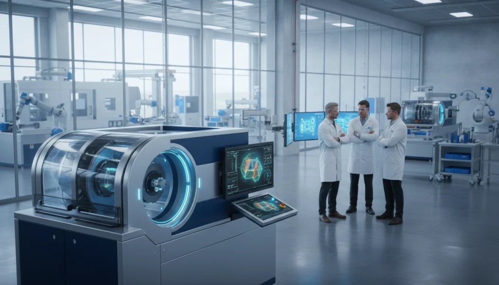
Technological Advancements in Modern Tomography
Technological breakthroughs are reshaping tomographic imaging, boosting diagnostic precision and patient care. Recent innovations have significantly enhanced image quality, reduced scanning times, and broadened application areas.
Dual-Energy CT
Dual-Energy CT scanners employ two distinct X-ray energy spectra to deliver richer tissue composition details. This advancement improves material characterization and enhances visibility of specific lesions or structures.
Spectral Imaging
Spectral imaging extends CT technology’s capabilities, enabling differentiation of various body materials. It’s highly beneficial in oncology and cardiovascular imaging.
Ultra-High Resolution Systems
Ultra-high resolution CT systems provide unmatched detail, allowing clinicians to spot smaller structures and minor abnormalities. These systems are invaluable in neurological and musculoskeletal imaging.
Portable Tomographic Devices
The emergence of portable tomographic devices is introducing advanced imaging to underserved areas. These devices are ideal for emergency use and for patients who cannot be moved easily.
The fusion of these technological advancements in tomography is revolutionizing diagnostic medicine. It’s leading to more precise diagnoses and tailored treatment plans. As technology advances, we can anticipate even better image quality, patient comfort, and clinical results.
Artificial Intelligence and Tomography
The integration of artificial intelligence (AI) in tomography is transforming medical imaging. It’s making tomographic techniques more accurate, faster, and better at diagnosing. This is a significant leap forward in healthcare technology.
AI-Assisted Image Interpretation
AI algorithms are now helping interpret tomographic images. They can spot patterns and anomalies that humans might miss. This boosts diagnostic accuracy.
For example, AI can detect early cancer signs by analyzing tissue changes. This is a game-changer in disease detection.

Deep Learning in Image Reconstruction
Deep learning is being used to enhance image reconstruction in tomography. These models learn from vast datasets, producing clearer images with less noise.
This improvement not only makes images clearer but also reduces radiation doses. It makes tomography safer for patients.
Automated Diagnosis Systems
AI-powered automated diagnosis systems are emerging. They provide initial diagnoses from tomographic images. This streamlines clinical workflows and eases healthcare professionals’ workloads.
Predictive Analytics in Patient Care
Predictive analytics, another AI application, forecasts patient outcomes from tomographic data. It personalizes treatment plans and enhances patient care.
By analyzing historical and real-time data, predictive models can identify high-risk patients. They suggest preventive measures.
The field of AI in tomography is rapidly advancing. Ongoing research aims to improve its capabilities and healthcare applications.
Economic and Accessibility Aspects of Tomographic Imaging
Tomographic imaging, a groundbreaking field, faces major economic hurdles that limit its accessibility. The high costs of equipment, maintenance, and operation for modalities like CT and MRI scanners are significant barriers.
Cost-Effectiveness Analysis
Assessing the cost-effectiveness of tomographic imaging requires examining both direct and indirect costs. Direct costs include equipment and personnel, while indirect costs involve patient preparation and post-procedure care. Research indicates that despite initial expenses, the diagnostic precision of tomographic imaging can lead to improved patient outcomes. This could potentially lower overall healthcare costs.
- Reduced need for exploratory surgeries
- Improved diagnostic accuracy
- Better patient management plans
Healthcare Reimbursement Policies
Reimbursement policies are vital in determining the accessibility of tomographic imaging. These policies vary widely across different healthcare systems. This variation impacts how frequently and under what conditions tomographic imaging is employed.
Global Disparities in Access to Advanced Imaging
There’s a notable global disparity in access to tomographic imaging technologies. Countries with higher incomes have more access to these technologies than those with lower incomes.
The economic aspects of tomographic imaging are complex. They involve not just the cost of the technology but also the broader healthcare infrastructure and policies that support its use.
The Transformative Impact of Tomography on Modern Healthcare
Tomography has transformed Modern Healthcare, bringing unmatched diagnostic capabilities. Its effects are clear in more accurate diagnoses, better patient care, and the advancement of healthcare technology. It provides detailed cross-sectional images of the body. This allows healthcare professionals to detect and treat medical conditions more effectively.
The Transformative Impact of Tomography is evident in its diverse applications. It ranges from neurological and cardiovascular imaging to oncology diagnostics and musculoskeletal evaluation. As technology advances, Tomography’s role in shaping healthcare’s future will grow.
Advancements in Artificial Intelligence and image reconstruction are making Tomography more sophisticated. This leads to faster and more accurate diagnoses. As Modern Healthcare adopts these innovations, Tomography’s benefits will expand. This will result in better patient outcomes and enhanced healthcare delivery.
FAQ
Q: What is tomography?
A: Tomography is a medical imaging technique. It creates detailed cross-sectional images of the body’s internal structures. This allows for accurate diagnoses and treatment planning.
Q: How does computed tomography (CT) differ from conventional radiography?
A: CT scans combine X-rays and computer technology for detailed cross-sectional images. In contrast, conventional radiography shows a two-dimensional view of internal structures.
Q: What are the different types of tomography used in modern medicine?
A: Modern medicine employs several tomography types. These include computed tomography (CT), positron emission tomography (PET), single-photon emission computed tomography (SPECT), and magnetic resonance imaging (MRI).
Q: How does tomography contribute to diagnostic medicine?
A: Tomography is vital in diagnostic medicine. It offers detailed images of internal structures. This enables healthcare professionals to diagnose and manage various conditions, from neurological disorders to cancer.
Q: What is the role of artificial intelligence in tomography?
A: Artificial intelligence (AI) enhances tomography. It improves image interpretation and reconstruction. AI also aids in developing automated diagnosis systems, leading to more accurate patient care.
Q: How is radiation safety ensured during tomographic procedures?
A: Radiation safety is ensured through dose reduction strategies and careful patient selection. Adherence to established protocols and guidelines is also key. This minimizes radiation exposure while maintaining image quality.
Q: What are the benefits of using tomography in dental and maxillofacial imaging?
A: Tomography, like cone beam computed tomography, offers detailed 3D images. These images are essential for accurate diagnosis and treatment planning in dental surgery and orthodontics.
Q: How is tomography used in industrial applications?
A: Tomography is applied in industrial settings for non-destructive testing and quality control. It’s also used for material analysis and preserving cultural heritage. This highlights its versatility beyond medical imaging.