

What Cancers Can an Abdominal CT Scan Detect: Preparation, Procedure and Results
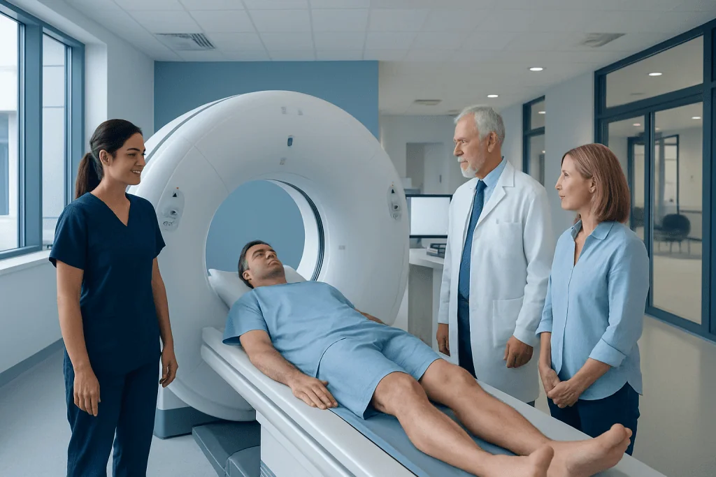
Abdominal CT can detect suspicious findings in organs such as the liver, pancreas, kidneys, bowel, bladder, ovaries and adrenal glands. A CT image may suggest cancer, but a radiologist usually cannot confirm a diagnosis without clinical information and often a biopsy.
Key Takeaways
- Abdominal CT can detect suspicious findings in organs such as the liver, pancreas, kidneys, bowel, bladder, ovaries and adrenal glands.
- A CT image may suggest cancer, but a radiologist usually cannot confirm a diagnosis without clinical information and often a biopsy.
- CT is commonly used to investigate symptoms, assess the extent of a known cancer and monitor response to treatment.
- Some small, early or biologically subtle cancers may not be visible on CT, and other tests may be more appropriate.
- Most people return to normal activities immediately after an abdominal CT scan; contrast dye requires a few additional safety considerations.
An abdominal CT scan can reveal suspicious masses, enlarged lymph nodes, organ changes and possible cancer spread in the abdomen and pelvis. It is a valuable diagnostic tool, but it cannot confirm cancer on its own; further imaging, blood tests or a biopsy may be needed.
Overview: What cancers can an abdominal CT scan detect?
What cancers can an abdominal CT scan detect? An abdominal computed tomography (CT) scan can show suspicious masses or abnormalities in abdominal and pelvic organs, including the liver, pancreas, kidneys, bowel, bladder, ovaries, uterus, adrenal glands and lymph nodes. It can also help identify whether a known cancer may have spread to the liver, lymph nodes, abdominal lining or other nearby structures.
CT uses a series of X-ray images taken from different angles to create detailed cross-sectional views of the body. A contrast dye is often used to make blood vessels and organs easier to distinguish. Although CT is very useful for finding abnormalities, it does not diagnose cancer with certainty. Many non-cancerous conditions can look similar on a scan, so doctors may recommend blood tests, another imaging examination, follow-up scanning or a tissue biopsy.
An abdominal CT is not a routine screening test for every type of cancer. It is usually requested because of symptoms, abnormal laboratory findings, an existing cancer diagnosis or a need to investigate a finding seen on another test. The best test depends on the organ involved and the individual’s medical history.
How abdominal CT works and when it may be recommended
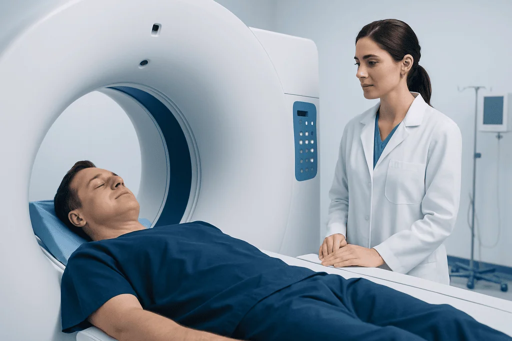
During CT imaging, an X-ray tube rotates around the body while computer software combines the images into detailed slices. These slices allow radiologists to assess the size, shape, density and blood supply of organs and any abnormal areas. Images may include the abdomen alone or the abdomen and pelvis, depending on the clinical question.
Doctors may recommend an abdominal CT when a person has persistent or unexplained symptoms such as abdominal pain, unintentional weight loss, jaundice, changes in bowel habits, blood in the urine or stool, abdominal swelling, or abnormal liver, kidney or blood test results. It may also be used after an ultrasound or MRI identifies a lesion that needs closer evaluation.
For people with known cancer, CT can help stage the disease by showing the primary tumor and possible spread. It can also guide treatment planning, assess response to chemotherapy or other therapies, and monitor for recurrence. A clinician weighs the likely benefit of imaging against radiation exposure and any contrast-related risks.
What cancer shows up on an abdominal CT scan?
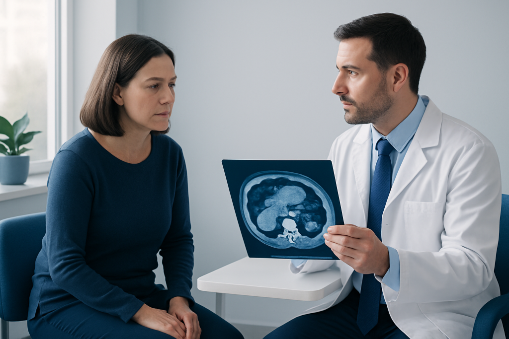
An abdominal CT scan may show cancers that begin in, or spread to, organs within the abdomen and pelvis. Examples include cancers of the liver, pancreas, kidneys, colon, rectum, stomach, bladder, ovaries and adrenal glands. It may also show enlarged lymph nodes, fluid in the abdomen, thickening of the bowel wall or deposits on the abdominal lining that warrant further assessment.
CT is particularly useful for evaluating the liver because it can identify lesions that may represent primary liver cancer or spread from a cancer elsewhere. It is also frequently used to assess suspected pancreatic, kidney and colorectal cancers. For more information on cancer that begins in the liver, see liver cancer.
A CT scan can detect lesions of different sizes, but detection is affected by the lesion’s location, size, contrast enhancement pattern and the quality of the images. Small tumors and subtle early disease can be more difficult to see. In some cases, ultrasound, MRI, endoscopy, colonoscopy or PET-CT provides complementary information.
CT can also help doctors identify a safe route for sampling a suspicious area. When appropriate, CT-guided biopsy allows a specialist to obtain tissue for laboratory testing, which can establish the diagnosis and help determine the most suitable treatment.
Can a radiologist tell if it is cancer?
A radiologist can often describe whether a finding has features that are suspicious for cancer, such as an irregular mass, invasion of nearby tissues, enlarged lymph nodes or a pattern of spread. The report may classify an abnormality as likely benign, indeterminate or concerning. However, imaging appearances alone do not always distinguish cancer from infection, inflammation, cysts, scar tissue or other non-cancerous changes.
Radiologists interpret CT images alongside the reason for the scan, previous imaging, laboratory results and relevant medical history. Comparing a new scan with earlier images can be especially helpful because growth or change over time may influence the level of concern.
When a CT finding is uncertain or suspicious, the next step may be targeted MRI, ultrasound, endoscopy, repeat imaging after a defined interval, or biopsy. A pathology assessment of tissue is generally the most reliable way to confirm the type of cancer. Results should be discussed with the doctor who ordered the scan, as they can explain what the finding means in the context of the individual’s health.
Preparing for and having an abdominal CT scan
Preparation depends on whether contrast dye is planned. The imaging team may ask the person not to eat for several hours beforehand, while clear fluids may be allowed. Instructions vary by facility and clinical situation, so patients should follow the directions they receive. They should tell the team about pregnancy or a possible pregnancy, kidney disease, diabetes, thyroid conditions, asthma, previous contrast reactions and all medicines or supplements.
Before the scan, metal items may need to be removed and the person may change into a gown. If intravenous contrast is used, a small cannula is placed in a vein. Some abdominal studies also use an oral contrast drink. During injection, a brief warm sensation, metallic taste or feeling of needing to urinate can occur; these sensations usually pass quickly.
The person lies on a table that moves through the doughnut-shaped CT scanner. The examination itself is painless and commonly takes only a few minutes, though preparation can take longer. The radiographer may ask the person to hold their breath briefly to reduce movement and improve image clarity. Staff can see, hear and communicate with the patient throughout the scan.
Results, recovery, benefits and possible risks
Most people can return to usual activities immediately after a CT scan. Those who receive contrast are often advised to drink fluids if this is medically appropriate, helping the body clear the dye through the kidneys. If sedation is needed for a special circumstance, the person may require someone to accompany them home and should follow the specific aftercare instructions provided.
A radiologist reviews the images and sends a report to the referring clinician. Timing varies according to the setting and urgency. Results may be described as normal, benign-appearing, indeterminate or suspicious. An abnormal result does not automatically mean cancer; it indicates that the doctor may need to investigate further.
The principal benefits of CT are its speed, detailed anatomical information and ability to assess multiple organs in one examination. Risks include exposure to a low dose of ionizing radiation and, when contrast is used, an uncommon allergic-like reaction or kidney-related complication in people with significant kidney impairment. The healthcare team uses contrast and radiation only when the anticipated diagnostic benefit is greater than the risk.
People should seek urgent medical advice after contrast if they develop breathing difficulty, facial or throat swelling, widespread hives, severe dizziness or other signs of a serious reaction. Mild warmth or a transient odd taste during the injection is common and is not usually a sign of allergy.
How common is cancer after a CT scan?
A CT scan does not cause cancer to appear immediately afterward, and an abnormal result after a scan does not mean the scan created a cancer. Often, the scan is performed because symptoms or other tests already raised a concern, and it may reveal a condition that was present before the examination.
CT uses ionizing radiation, which carries a very small potential long-term risk that increases with repeated or unnecessary exposure. For an individual medically indicated scan, this possible risk is generally considered low compared with the benefit of identifying an important condition. Imaging teams use protocols designed to keep radiation as low as reasonably achievable while maintaining diagnostic image quality.
People who are worried about previous scans can ask their clinician why each test was needed and whether future follow-up could use an alternative such as ultrasound or MRI. It is important not to avoid recommended imaging when it could clarify a serious diagnosis or guide timely treatment.
What cancer cannot be detected by CT scan?
No CT scan can reliably detect every cancer. Very small tumors, early-stage disease and cancers that do not create a distinct mass or visible structural change may be missed. CT is also less suitable than other methods for certain tissues and organs, including many brain, breast, skin, cervical and prostate cancers.
For example, mammography and breast MRI are designed for breast assessment, while cervical screening detects precancerous cervical changes that CT cannot identify. Endoscopy and colonoscopy can directly inspect the lining of the digestive tract and identify small polyps or early surface cancers that may not be apparent on CT. MRI may provide greater detail for some liver, pelvic, prostate and soft-tissue findings.
CT may also be limited in identifying small lesions in a moving bowel, early stomach cancer, very small peritoneal deposits and some low-contrast liver lesions. A normal CT result is reassuring for many significant structural abnormalities, but it does not rule out every possible cause of symptoms. Persistent or worsening symptoms should be reviewed by a qualified doctor.
When to seek medical care
Medical assessment is appropriate for persistent or unexplained abdominal pain, ongoing bloating, yellowing of the skin or eyes, unintended weight loss, loss of appetite, a new persistent change in bowel habits, blood in the stool or urine, or a new abdominal lump. These symptoms have many possible causes, most of which are not cancer, but they deserve timely evaluation when they continue or worsen.
Urgent care is important for severe or sudden abdominal pain, vomiting blood, black tar-like stools, large amounts of rectal bleeding, fainting, high fever with severe pain, or jaundice accompanied by significant illness. A clinician can decide whether CT is the right test or whether other investigations should come first.
Where cancer is suspected or confirmed, care may involve radiology, gastroenterology, surgery, medical oncology, pathology and other specialties. Oncology care may include further diagnostic assessment and a personalized treatment plan. Acibadem International’s multidisciplinary specialists and JCI-accredited hospitals diagnose and treat cancer for international patients.
Frequently asked questions
What cancers can an abdominal CT scan detect?
An abdominal CT scan can identify suspicious changes involving the liver, pancreas, kidneys, bowel, bladder, ovaries, adrenal glands and abdominal lymph nodes. It can also show possible spread of cancer to the liver, lymph nodes or lining of the abdomen. A suspicious CT result requires further clinical assessment and may require biopsy for confirmation.
Can a radiologist tell if it is cancer?
A radiologist can identify imaging features that raise or lower concern for cancer, but CT images alone often cannot confirm the diagnosis. Benign cysts, inflammation and infections can sometimes resemble tumors. Tissue testing through a biopsy is commonly needed to determine whether cancer is present and, if so, what type it is.
How common is cancer after a CT scan?
A CT scan does not make cancer develop immediately afterward. It may find a cancer that was already present, particularly when it is ordered to investigate symptoms or an abnormal test. The radiation from a medically necessary individual CT scan carries a small potential long-term risk, but the diagnostic benefit usually outweighs this risk.
What cancer cannot be detected by CT scan?
CT may miss very small, early or subtle tumors, especially those that do not change the structure of an organ. It is not the main test for many breast, skin, cervical, prostate or brain cancers. Other examinations, such as mammography, MRI, screening tests, endoscopy or biopsy, may be better suited to these conditions.
Do I need contrast dye for an abdominal CT scan?
Many abdominal CT scans use intravenous contrast because it helps show blood vessels and differences between normal and abnormal tissue. Some scans can be performed without contrast, depending on the clinical question. The doctor and radiology team consider kidney function, past reactions and other health factors before using contrast.
How long does it take to get abdominal CT scan results?
The scan images are reviewed by a radiologist, who prepares a report for the doctor who requested the examination. Results may be available quickly in urgent situations, while routine reports can take longer depending on the facility. The referring clinician should explain the findings, their significance and whether follow-up tests are needed.
References
- American Cancer Society
- Radiological Society of North America
- National Cancer Institute
- U.S. Food and Drug Administration
- World Health Organization
This article is for general information only and is not a substitute for professional medical advice. Please consult a qualified doctor about your individual situation.
Persistent digestive symptoms? Get evaluated in Turkey
JCI-accredited · board-certified surgeons · reply within 24h
Add us as a Preferred Source to see more of our trusted health content across Google Search, AI Overviews and Discover.

More from the Health Library



Related Specialists

Asst. Prof. Dr. Mustafa Sungur
Orthopedic Surgery & Traumatology
Dr. Sema Tekin Koşargelir
Cosmetic Dermatology
Dr. Sercan Başböyük
Anesthesiology
Prof. Dr. Bülent Kisacikoğlu
Cardiovascular Surgery