

Cardiovascular Surgery
Coronary bypass, valve repair and replacement, aortic aneurysm and dissection, carotid and peripheral arterial surgery, varicose veins and venous disease, congenital correction and mechanical support.

Two specialties that share a name
Cardiac surgery stops or works around a beating heart. Vascular surgery treats arteries and veins everywhere else. Someone coming for a bypass and someone coming for varicose veins are both in the right department and will otherwise have nothing in common.
The heart
Coronary bypass, valve repair and replacement, and the catheter alternatives the heart team weighs against them.
The aorta and arteries
Aneurysm and dissection, carotid disease, and the arteries of the legs, open and endovascular.
The veins
The largest everyday workload in the unit, and the one most often dismissed as cosmetic when it is not.
The heart team decides, not the specialty you happened to reach first
Almost every significant decision here can reasonably be made in more than one way, and each option is performed by a different person. That is precisely the situation in which a formal review, surgeon, interventional cardiologist and imaging specialist on the same images, stops the referral route from determining the treatment.
It matters most where the evidence is close. Bypass against stenting in multivessel disease. Surgery against TAVR in a patient who could have either. Operating on an aneurysm now against watching it for another year. In each of those a confident single answer, offered without alternatives, is worth a second opinion.
What we will not do
- Offer surgery without the alternative having been weighed. If a catheter procedure is reasonable, it is discussed.
- Replace a mitral valve that could have been repaired to make the operation shorter.
- Choose a smaller incision at the cost of what is achieved inside. A perfect repair through a sternotomy beats an imperfect one through a port.
- Treat varicose veins without a duplex map, or treat surface veins while leaving the reflux that feeds them.
- Quote a survival figure as though it described you. Risk in cardiac surgery depends far more on the patient than on the operation.
Cardiovascular surgeons who lead this work
What actually happens, in order
Send the images, not the reports
Angiogram, echocardiogram and CT in their original digital form. In this field the images decide the plan and a written report frequently does not carry what a surgeon needs to see.
Heart team review
Surgeon, interventional cardiologist and imaging specialist on the same images before a recommendation exists. A proportion of reviews conclude that a catheter procedure, or medical treatment and surveillance, is the better course.
Assessment on arrival
What cannot be done remotely: repeat imaging where the outside study is inadequate, lung function, carotid imaging where indicated, and a dental assessment before any valve is implanted.
Surgery, intensive care, ward
Mobilisation and breathing work from the first day, because chest infection and clots are the commonest early problems and both are prevented by moving. Length of stay is decided at the time.
Before the flight home
The operative note, the prosthesis details for any valve or graft, the anticoagulation plan and target, and the rehabilitation referral. Anticoagulation monitoring is arranged at home before departure, not after.
Six things worth knowing first
Most heart disease never reaches a surgeon
Medication, angioplasty and stenting handle the great majority of it under cardiology. Surgery is for the situations where the evidence puts it ahead, and those situations are specific.
Who you saw first should not decide the treatment
Bypass or stent, surgery or TAVR, repair or replace, each is performed by a different person. That is exactly why a heart team reviews the images together before anything is offered.
Neither surgery treats the disease
A graft and a stent are both mechanical answers to one consequence of atherosclerosis. The medication afterwards is what slows the disease, and stopping it is the commonest reversible mistake.
Repair beats replacement where it is achievable
A repaired mitral valve preserves function and avoids a prosthesis and anticoagulation. Repair rates vary substantially between surgeons, which makes it a fair question to ask directly.
Varicose veins are frequently not cosmetic
Aching, swelling, skin staining, hardening above the ankle and ulceration are all venous. A duplex scan distinguishes a cosmetic problem from a failed circuit, and comes before that judgement.
For claudication, walking comes before stenting
Supervised exercise improves walking distance substantially and approaches what revascularisation achieves for this symptom, without its risks. It is first-line, not a delaying tactic.
Jump to what you came for
Quick answer
Cardiovascular surgery covers the surgical treatment of heart and blood vessel diseases, including coronary artery disease, valve disorders, aortic conditions, congenital heart defects, and peripheral vascular problems. At Acibadem in Turkey, this unit evaluates each patient with advanced cardiac imaging and planning, then provides procedures ranging from minimally invasive and endovascular techniques to open surgery, followed by intensive postoperative monitoring…
What our cardiovascular surgery unit covers, and who it is for
Cardiovascular surgery is the surgical treatment of the heart, its valves, the aorta and the blood vessels of the body. It is two disciplines that share a name: cardiac surgery, which stops or works around a beating heart, and vascular surgery, which treats arteries and veins everywhere else. A person coming for a bypass and a person coming for varicose veins are both in the right department and will otherwise have nothing in common.
At Acıbadem International the work is organised into five strands.
- Coronary surgerybypass grafting for coronary disease that stenting cannot adequately treat, on or off the heart-lung machine.
- Valve surgeryrepair and replacement of the aortic and mitral valves by open, minimally invasive and catheter routes, with the choice made by a heart team rather than by a single operator.
- Aortic surgeryaneurysm and dissection of the thoracic and abdominal aorta, treated open or endovascularly, including root replacement in connective tissue disease.
- Vascular and venous surgerycarotid disease, peripheral arterial disease and limb salvage, dialysis access, and the large everyday workload of varicose veins and chronic venous disease.
- Congenital and advanced heart failurecorrection of structural defects, and mechanical support and transplantation where the heart itself has failed.
Two borders are worth stating. Everything treated without an operation, medication, angioplasty and stenting, pacemakers and ablation for rhythm disorders, echocardiography and stress testing, belongs to cardiology, and the great majority of heart disease is managed there and never reaches a surgeon. Stroke that has already happened is neurological rather than surgical, though the carotid artery that caused it is dealt with here, alongside neurology.
Who is this page for? People told they need a bypass who want to know why a stent will not do. People with a valve problem being watched, wondering what decides the moment to operate. People with an aneurysm found by accident on a scan for something else. People with legs that ache when they walk, or veins they have been told are cosmetic and are not. And relatives trying to understand what an operation involves before agreeing to it on someone else’s behalf.
Heart bypass surgery: coronary artery bypass graft (CABG)
Heart bypass surgeryCABG, or coronary artery bypass graft surgery, does not clear a blocked coronary artery. It routes blood around the blockage using a new vessel taken from elsewhere in the body and joined beyond the narrowing. The distinction matters, because it explains why bypass suits diffuse disease that stenting handles badly: a graft is placed beyond the entire diseased segment rather than propping open one point within it.
The operation is usually performed through the breastbone, with the heart stopped and the circulation maintained by a heart-lung machine, though it can be done on a beating heart. The grafts are the substance of it: the internal mammary artery from inside the chest wall, the radial artery from the forearm, and the saphenous vein from the leg. Which and how many depends on which arteries are diseased and on the patient.
The reason bypass has not been displaced by stenting, forty years into the stent era, is that it keeps outperforming it in specific groups, disease in all three coronary territories, disease of the left main artery, reduced pump function, and diabetes. Those are the situations a heart team is convened for, and they are described under bypass versus stenting.
Which grafts are used, and why one of them matters more than the rest
Not all grafts are equal, and the difference is measured in decades rather than months. The left internal mammary arterythe LIMA graft, is left attached to its origin under the collarbone and joined to the left anterior descending artery, the vessel supplying the largest territory of heart muscle. It stays open in the great majority of patients at ten years and beyond, far better than any alternative, and using it to the left anterior descending is close to a rule in modern practice rather than a preference. A bypass operation that does not use it needs a specific anatomical reason.
The saphenous vein from the leg is quick to harvest and versatile, and it is what most additional grafts are made from; veins were never designed for arterial pressure, and a proportion narrow over the years, which is the main reason people need further treatment a decade later. The radial artery from the forearm performs better than vein in the long run and is used for targets with severe narrowing, after checking that the hand’s other artery can supply it alone. Some units use both mammary arteries in younger patients, a technically more demanding operation with better long-term patency and, in some groups, more wound healing difficulty.
The practical consequence for a patient is a question worth asking before surgery: which grafts are planned, and is the left internal mammary artery going to the left anterior descending.
Off pump bypass, the heart lung machine and cardiopulmonary bypass
The heart lung machinecardiopulmonary bypasstakes over the work of the heart and lungs, oxygenating the blood and returning it to the body, so the heart can be stopped and held still while the grafts are sewn. It is the invention that made cardiac surgery possible. It is also the reason for a set of effects specific to this kind of operation: an inflammatory response, fluid shifts, and a period of cloudy thinking afterwards that patients often describe more vividly than the surgery itself.
Off pump bypass is performed on a beating heart, with the target segment stabilised mechanically, avoiding the machine altogether. It appeals for exactly the reasons above and it has been studied extensively. The honest summary of that evidence is that off-pump reduces some short-term effects, particularly in patients at high risk from the machine, badly diseased aortas, poor kidney function, previous stroke, but has not shown superior long-term results in general, and in less experienced hands the grafts can be fewer or less well placed. It is a tool for particular patients and particular surgeons rather than a better operation.
Bypass vs stent: how the choice is actually made
The bypass vs stent question is decided by anatomy and by the patient, and in a well-run hospital it is decided by a heart team: a surgeon and an interventional cardiologist looking at the same angiogram together, rather than by whoever the patient happened to see first. That structure exists because the incentive to treat with whatever the person in the room performs is real and well documented.
Broadly: stenting suits disease confined to one or two discrete points, and it is far less invasive with a much shorter recovery. Bypass suits disease in all three vessel territories, significant left main disease, long diffuse narrowings, chronic total occlusions, reduced left ventricular function, and diabetes, where surgery has repeatedly shown better long-term survival and freedom from further procedures. Complexity is scored formally on the angiogram to inform this, and the score is a guide rather than the decision.
Two things belong alongside that. Neither operation treats the disease itself, which is atherosclerosis affecting the whole arterial tree; both are mechanical solutions to one consequence of it, and the medical treatment continues afterwards regardless. And urgency changes everything: an unstable presentation is treated by whatever restores blood flow fastest, and the elegant comparison above applies to planned decisions.
How long does bypass surgery take, and what happens on the day
The operation itself typically runs to several hours and varies with the number of grafts, the quality of the target vessels and whether anything else is being done at the same time. Around it sits a longer day: anaesthesia and line placement beforehand, and a period in intensive care afterwards. Most people are ventilated for the first hours and wake in intensive care with drains, a urinary catheter and monitoring lines, all of which come out over the following days.
Time in theatre is a poor thing to compare between hospitals, and a shorter operation is not a better one. The variables worth asking about are how many grafts are planned, whether the left internal mammary artery is being used, and what the plan is if the anatomy found is worse than the angiogram suggested.
Life expectancy after bypass surgery
This page does not quote survival figures, and the reason is that they depend far more on the patient than on the operation. Age, kidney function, pump function, diabetes, lung disease and how far the atherosclerosis has progressed elsewhere all move the number more than any surgical variable, and a figure drawn from a published series describes that series rather than an individual.
What can be said usefully is this. Bypass is durable: the mammary graft in particular commonly outlasts a decade and often much longer. It relieves angina reliably and, in the groups where it is indicated over stenting, it improves survival compared with the alternative. It does not stop coronary artery disease itself, which is atherosclerosis of the whole coronary tree, so a proportion of people need further treatment years later, and vein grafts are usually what fails first. And the single largest determinant of the years after surgery is not the surgery: it is what happens to the disease afterwards, which is decided by medication taken as prescribed, blood pressure, glucose, lipids and smoking. Surgeons say this so often that it sounds like a disclaimer; it is the most evidence-backed sentence in this section.
Open heart surgery: what the term covers
Open heart surgery means an operation performed on the heart itself, usually with the chest opened and the circulation supported by a machine. It is a category rather than a procedure: bypass grafting, valve repair and replacement, aortic root surgery, correction of congenital defects and transplantation are all open heart operations. People are frequently told they need one without being told which, and the difference in what it involves is considerable.
What the operations share is the access and the physiology. The chest is opened, the pericardium around the heart is entered, the circulation is taken over so the heart can be stopped, and at the end the heart is restarted and the machine withdrawn. What differs is what happens in between, and how long the heart has to be still.
Sternotomy, mini sternotomy and how the chest is closed
A sternotomy divides the breastbone down the midline to reach the heart, and the bone is brought back together at the end with stainless steel wires that stay there permanently. The wires set off no alarms and are not felt; they are visible on every chest X-ray afterwards and are frequently a surprise to patients who were not told.
The consequence that matters is that the breastbone is a healing bone, and bone takes weeks to knit. That is the entire basis of the restrictions after cardiac surgery, described under sternal precautions. It is also the reason wound problems are taken seriously in this population: diabetes, obesity, smoking, and the use of both mammary arteries all raise the risk of the sternum healing badly, which is a far more serious problem than a superficial wound.
A mini sternotomy divides only the upper portion of the breastbone, leaving the lower part intact. It is used mainly for aortic valve surgery. The bone is more stable afterwards and recovery is quicker; the exposure is narrower, so it is not suitable for every case and not for every surgeon.
Minimally invasive heart surgery and robotic heart surgery
Minimally invasive heart surgery reaches the heart between the ribs rather than through the breastbone, most commonly on the right side for mitral valve work, with the heart-lung machine connected through the groin vessels. Because the sternum is untouched, the restrictions afterwards are far lighter and return to normal activity is faster. Robotic heart surgery performs the same operations through smaller ports with instruments held by a robotic platform under the surgeon’s direct control, and is used principally for mitral valve repair and for some coronary and rhythm procedures.
The honest framing is that these are different routes to the same operation, not better operations. What must not change is what is achieved inside: a mitral valve repaired through a small incision but repaired imperfectly is a worse outcome than a perfect repair through a sternotomy. They suit isolated, well-selected problems: a single valve, a suitable body habitus, no previous surgery in that space, acceptable groin vessels, and they are unsuitable for multi-procedure operations and for many redo cases. A unit that offers a minimally invasive route for everything is describing a marketing position; a unit that offers it for nothing is behind.
Open heart surgery recovery time
Recovery after a sternotomy runs on the bone’s timetable, and the honest version has three phases people are rarely given in advance.
Hospital, roughly the first week. Intensive care for the first day or so, then a ward. Sitting out of bed and walking begin early and deliberately. Drains and lines come out over the first days. Breathing exercises are given for a reason: the chest hurts, people breathe shallowly, and chest infection is the commonest early complication.
Weeks two to eight, at home. Fatigue dominates and it is more profound than most people expect, not soreness but a whole-body tiredness that comes in waves. Appetite is poor for a while, taste changes are common, and sleep is disturbed. Low mood in the second to sixth week is common enough to be considered part of the normal course rather than a complication; it usually lifts. Walking distance is built up daily and is the main measure of progress.
Two to three months onward. The breastbone is largely healed by around eight to twelve weeks, restrictions lift, and driving and heavier activity resume on the surgeon’s timetable. Full recovery of energy takes longer than the bone does, three to six months is usual, and longer in older patients. Recovery after minimally invasive surgery follows a considerably shorter version of this because the sternum was never divided.
Sternal precautions
Sternal precautions are the restrictions that protect a divided breastbone while it knits, and they are almost always given as a list of prohibitions without the reason, which makes them easy to disregard. The reason is simple: the two halves of the sternum are held by wires, and movements that pull them apart, or push them apart through the arms, delay healing and, rarely, cause the closure to fail.
The pattern is consistent across units. No lifting beyond a light weight, commonly set around four to five kilograms, for the first weeks. No pushing or pulling with the arms, which includes getting out of bed or a chair by pushing on the armrests: the “hug a pillow and use your legs” instruction exists for this. No reaching both arms behind or overhead in a way that spreads the chest. No driving until cleared, because an emergency stop and a seatbelt both load the sternum. Coughing and sneezing while hugging a pillow against the chest.
The specific weights, the duration and when each restriction lifts are set by the operating team, and modern practice has moved toward less restrictive advice in many centres. What has not changed is the principle, and the point in recovery when people breach it is predictable: around week three, when they feel well enough to forget.
Heart valve surgery and heart valve replacement
Heart valve surgery treats valves that have become too tight to open properly, too leaky to close properly, or both. Two questions decide everything about it: whether the valve can be repaired or must be replaced, and when the moment to intervene has arrived. Neither is answered by symptoms alone.
The timing question is the one patients find hardest, because they are frequently told they need surgery “eventually” and sent away with a scan interval. The reasoning behind that is real. Operating too early exposes someone to the risks of surgery and, if a valve is replaced, to a prosthesis they must live with for decades. Operating too late allows the heart muscle to sustain damage that does not fully reverse after the valve is fixed. The window in between is what the surveillance scans exist to find, and the measurements that move the decision, chamber size, pump function, pressures, and the gradient across the valve, are followed with cardiology.
Aortic stenosis
Aortic stenosis is narrowing of the valve between the left ventricle and the aorta, and it is the commonest valve disease requiring treatment in adults. Most cases are degenerative, developing over decades as the valve calcifies with age; a substantial minority arise in a bicuspid valve and appear earlier.
The natural history is what makes it important. Severe aortic stenosis can be entirely silent for years, and then the classical symptoms appear, breathlessness on exertion, chest tightness, and light-headedness or blackouts on effort. The appearance of symptoms marks a sharp change in outlook, and it is the reason a valve that has been watched for years is suddenly treated with urgency. That is also why anyone under surveillance is asked repeatedly about exertion, and why people who have quietly reduced what they do, taking the lift, walking more slowly, stopping activities they blame on age, need to say so, because a symptom avoided is easily reported as absent.
Medication does not treat it. There is no drug that opens a calcified valve, and the treatment is mechanical: surgical replacement or the catheter route described below.
Aortic valve replacement
Aortic valve replacement removes the diseased valve and sews in a prosthesis, through a full sternotomy or a mini sternotomy. It is one of the most reliably beneficial operations in cardiac surgery: relief of the obstruction is immediate and complete, and the ventricle that thickened to overcome it remodels over the following months.
The decision that accompanies it is the type of valve, and it is genuinely the patient’s to make with guidance rather than a purely technical matter. A mechanical valve is made of pyrolytic carbon and does not wear out, so a second operation for valve failure is unlikely, at the cost of lifelong anticoagulation with warfarin, regular blood monitoring, and the bleeding and dietary constraints that come with it. Direct oral anticoagulants are not suitable for mechanical valves. A tissue valve, made from bovine or porcine tissue, needs no long-term anticoagulation, and it degenerates over time, faster in younger patients, so a further procedure becomes likely, though that is increasingly a catheter procedure inside the old valve rather than another operation.
The rule of thumb is age, with the crossover usually discussed somewhere around the sixth decade, but the real inputs are wider: whether pregnancy is planned, since warfarin is unsuitable in pregnancy; bleeding risk and occupation; whether reliable anticoagulation monitoring is available where the person lives, which matters a great deal for international patients; and how the individual feels about the two different kinds of future. Both are correct answers.
TAVR and TAVI: replacing the aortic valve by catheter
TAVRtranscatheter aortic valve replacement, called TAVI in Europe and the United Kingdom for implantation rather than replacement, delivers a new valve mounted on a collapsible frame through a catheter, usually from the femoral artery in the groin, and deploys it inside the diseased valve. The old valve is pushed aside rather than removed. There is no sternotomy, no heart-lung machine and no cardiac arrest, and many patients are walking the same day.
It began as a treatment for people too high-risk for surgery, and it has moved steadily down the risk scale as the evidence accumulated; in many countries it is now the default for older patients regardless of surgical risk. That migration is the reason the heart team exists, because the choice is no longer obvious.
What still favours surgery: younger age, where the durability of a surgical valve over decades matters and the long-term performance of a transcatheter valve is less established; a bicuspid valve with unfavourable anatomy; disease of the aortic root or ascending aorta needing treatment at the same time; another valve or coronary disease requiring an operation anyway; and access vessels too small or too diseased for the catheter route. What favours TAVR: age and frailty, previous cardiac surgery, a hostile chest, lung disease, and the general balance in older patients where recovery from a sternotomy is itself the risk.
Two considerations belong in the conversation rather than in the small print. Conduction disturbance is more common after TAVR than after surgery, and a proportion of patients need a permanent pacemaker afterwards. And a valve implanted inside a valve constrains what can be done later, which is precisely the argument that matters most in someone with decades ahead of them.
Mitral regurgitation: mitral valve repair before mitral valve replacement
Mitral regurgitation is leakage backwards through the mitral valve, and it divides into two conditions that share a name. Primary regurgitation is a fault of the valve itself, usually degenerative disease in which the leaflets become floppy and prolapse. Secondary regurgitation is a valve pulled out of shape by a dilated, failing ventricle; the valve is structurally normal and the disease is the muscle. The distinction governs treatment entirely: the first is a surgical problem, the second is largely a heart failure problem in which fixing the valve treats a symptom of the disease.
Mitral valve repair preserves the patient’s own valve, reshaping the leaflets and supporting the ring with an annuloplasty device. Where it is achievable, it is clearly superior to replacement: better preservation of ventricular function, no prosthesis, and no lifelong anticoagulation. In degenerative disease, repair is achievable in the great majority of cases in experienced hands, and the phrase “in experienced hands” is doing real work in that sentence. Repair rates vary substantially between surgeons, and the likelihood of leaving with your own valve depends on who operates.
That gives a patient one question that is more useful than any other: what proportion of degenerative mitral valves does this surgeon repair rather than replace, and what is the plan if repair is not possible once the valve is seen. Mitral valve replacement is the answer where the valve is destroyed by rheumatic disease, endocarditis or heavy calcification, and the mechanical-versus-tissue choice is the same as described for the aortic valve.
Bicuspid aortic valve
A bicuspid aortic valve has two leaflets instead of three. It is the commonest congenital cardiac abnormality, present in a small percentage of people, and most live for decades without knowing. It matters here for two reasons.
The first is that two leaflets take more mechanical punishment than three, so the valve calcifies earlier, bicuspid patients typically present with aortic stenosis a decade or two before those with a normal valve, and often need treatment in middle age rather than old age.
The second is less well known and more important. A bicuspid valve is frequently accompanied by weakness of the wall of the ascending aorta, which dilates independently of how the valve is behaving. That is why anyone with a bicuspid valve is imaged for aortic size as well as valve function, why the threshold for replacing the aorta is lower in this group than in degenerative disease, and why the aorta is often replaced at the same operation as the valve even when the valve was the reason for surgery. It is also why first-degree relatives are commonly offered a screening echocardiogram.
Aortic aneurysm and dissection
The aorta is the main artery leaving the heart, and it fails in two ways: it widens gradually into an aortic aneurysm, or it tears along its wall as an aortic dissection. The first is usually silent and found by accident; the second is sudden and is one of the true emergencies in medicine. They are related, because an aneurysmal aorta is more likely to dissect, and both are treated by the same surgeons.
Aortic dissection
An aortic dissection is a tear in the inner lining of the aorta that allows blood to force its way into the wall itself, splitting the layers apart along the length of the vessel. The false channel that results can compress the true one, cut off the branches supplying the brain, kidneys, gut or legs, rupture into the chest, or tear back into the heart and disable the aortic valve.
The classical description is sudden severe chest or back pain, often described as tearing or ripping and at its worst from the very first moment, which distinguishes it from the crescendo of a heart attack. It is uncommon enough to be missed and dangerous enough that missing it matters, and the presentations that mislead are well documented: pain that migrates as the tear extends, a stroke, a cold painless leg, or abdominal pain.
Classification governs treatment and is simple. A dissection involving the ascending aorta is a surgical emergency operated without delay, because untreated mortality rises by the hour. A dissection confined to the descending aorta is usually managed medically at first, with strict blood pressure control, and treated by stent graft if a complication develops. The predisposing factors are long-standing hypertension above all, connective tissue disorders such as Marfan syndrome, a bicuspid aortic valve, and pre-existing aneurysm.
Abdominal aortic aneurysm
An abdominal aortic aneurysm is a permanent dilatation of the abdominal aorta beyond one and a half times its normal diameter. It is far more common in men, in smokers, in those over sixty-five and where a first-degree relative has had one, which is why several countries screen men in that age group with a single ultrasound.
Aortic aneurysm symptoms are, in the great majority, absent. Most are found incidentally on a scan done for something else, and that silence is the whole clinical problem: the first symptom in an unmonitored aneurysm can be rupture, which is frequently fatal before help arrives. A minority cause deep abdominal or back pain, or a pulsation the person notices.
Management is surveillance until a threshold, then repair. Small aneurysms are watched with interval ultrasound and treated by controlling blood pressure and stopping smoking, which slows expansion measurably. Repair is considered when the diameter crosses a threshold generally around five and a half centimetres in men and somewhat lower in women, when expansion is rapid, or when it becomes symptomatic, because below that threshold the risk of the operation exceeds the risk of the aneurysm, and above it the balance reverses. The threshold is lower in connective tissue disease. This section is the only place on this page where the size logic is set out; other sections refer to it rather than restating it.
EVAR: endovascular aneurysm repair
EVARendovascular aneurysm repairlines the aneurysm from the inside with a stent graft delivered through the femoral arteries, excluding the weakened wall from the circulation without opening the abdomen. Compared with open repair it has markedly lower early mortality and a much shorter recovery, and it has become the default where the anatomy allows.
The anatomy is the constraint, and it is specific: an adequate length of healthy aorta below the kidney arteries to seal against, suitable angulation, and access vessels large and straight enough to deliver the device. Where those are not met, open repair remains the operation, and branched or fenestrated devices extend the endovascular option to aneurysms involving the visceral arteries in specialist centres.
The trade-off is surveillance. A stent graft can leak around or through its seals, and the aneurysm sac can continue to grow, so EVAR commits the patient to imaging follow-up indefinitely and to a higher rate of further procedures than open repair. Open repair, once healed, generally needs no such surveillance. That is the genuine comparison, a lower upfront risk against a longer tail of monitoring, and it is why open repair is still chosen in younger patients with good anatomy and long life expectancy.
Aortic root replacement and the Bentall procedure
The aortic root is the segment where the aorta meets the heart, containing the valve and the origins of the coronary arteries. When it dilates, in Marfan syndrome and related connective tissue disorders, with a bicuspid valve, or degeneratively: the valve leaks because its supporting structure has stretched, and the aorta is at risk of dissection.
Aortic root replacement replaces that whole segment. The Bentall procedure is the classical version: a composite graft carrying a mechanical or tissue valve replaces root and valve together, and the coronary arteries are re-implanted into it. It is durable and reliable, and with a mechanical valve it commits the patient to lifelong anticoagulation.
The alternative is a valve-sparing root replacement, in which the patient’s own aortic valve is preserved and re-suspended inside a graft. It avoids a prosthetic valve and anticoagulation entirely, which matters most in the young patients this operation is often needed for. It requires leaflets that are structurally healthy and it is technically demanding, so it is concentrated in centres that do it regularly, and a patient facing root surgery in their thirties or forties should ask directly whether valve preservation is possible in their case, because it is the difference between a life with warfarin and a life without it.
Carotid artery stenosis, carotid endarterectomy and carotid artery stenting
Carotid artery stenosis is narrowing of the arteries in the neck that supply the brain, caused by atherosclerotic plaque. Its importance is not the narrowing itself but what breaks off it: fragments of plaque and clot that travel to the brain and cause a stroke or a transient ischaemic attack.
Carotid endarterectomy opens the artery and removes the plaque from inside it, restoring the lumen and, more to the point, removing the source of the debris. It is one of the best-studied operations in surgery, and the evidence is unusually specific about who benefits.
The essential distinction is between symptomatic and asymptomatic disease. Where a severe stenosis has already caused a stroke or transient ischaemic attack on that side, surgery clearly reduces the risk of a further stroke, and the benefit is greatest when it is done soon after the event rather than months later, timing is part of the treatment. Where the stenosis is found incidentally in someone who has had no symptoms, the balance is much closer, because modern medical treatment has substantially reduced the stroke risk of asymptomatic disease, and the decision weighs the individual’s stroke risk against the operative risk in that unit.
Carotid artery stenting is the catheter alternative, placing a stent across the narrowing with a protection device to catch debris. It avoids a neck incision and the risk of cranial nerve injury, and it carries a somewhat higher risk of stroke around the procedure in most comparisons, particularly in older patients. It is generally preferred where the neck is hostile, previous surgery or radiotherapy, or where the lesion sits high or low beyond comfortable surgical reach. Whichever is chosen, the medical treatment for atherosclerosis continues afterwards; neither procedure treats the disease.
Peripheral artery disease
Peripheral artery disease is atherosclerosis of the arteries supplying the legs. It is common, it is under-diagnosed, and it is a marker of disease elsewhere: someone with narrowed leg arteries very often has narrowed coronary and carotid arteries too, which is why finding it changes the whole medical plan and not just the leg.
Claudication and intermittent claudication
Claudicationproperly intermittent claudicationis cramping pain in the calf, thigh or buttock brought on by walking a predictable distance and relieved within minutes of stopping. The pattern is the diagnosis: reproducible with exertion, relieved by rest without needing to sit or bend, and returning at the same distance. Muscle demand outstrips a supply that is adequate at rest.
What surprises people is the treatment. For claudication alone, the first-line management is not surgery: it is a supervised exercise programme, stopping smoking, and medical treatment of the atherosclerosis. Structured walking, walking to near-maximal pain, resting, repeating, improves walking distance substantially, and in several comparisons matches or approaches what revascularisation achieves for this symptom, without its risks. It works by developing collateral supply and improving muscle efficiency, and it requires the thing patients least want to hear, which is walking into the pain rather than avoiding it.
Revascularisation, angioplasty and stenting, or bypass, is considered where claudication is genuinely lifestyle-limiting despite that programme, and where the anatomy is suitable. It is not offered to prevent progression, because most claudication does not progress to limb threat.
Diabetic foot and the vascular contribution to it
The diabetic foot is where three problems meet, and treating any one alone fails. Neuropathy removes the pain that would have warned of a pressure point, so damage is often advanced before it is noticed. Arterial disease impairs the supply needed to heal it, and in diabetes that disease characteristically affects the smaller arteries below the knee, which are harder to treat than the larger vessels. Infection then progresses in tissue that cannot mount a normal response.
The vascular contribution is assessed in every diabetic foot ulcer rather than assumed absent, because a foot that will not heal on antibiotics and dressings very often has an arterial problem nobody has looked for. Restoring blood flow, where it is inadequate, is what converts an unhealing ulcer into a healing one. The rest of the care, glucose control, offloading pressure from the ulcer, debridement and infection management, runs alongside it with endocrinology, and the outcome depends on all of it happening at once rather than in sequence.
Critical limb ischaemia (critical limb ischemia)
Critical limb ischemia is a different condition from claudication and is treated with an entirely different urgency. Here the blood supply is inadequate even at rest: pain in the foot at night that is relieved by hanging the leg out of bed, ulceration that will not heal, or gangrene. The limb is threatened, and without restoration of blood flow a substantial proportion of these legs are lost.
It is the situation in which revascularisation is unequivocally indicated, by whichever route the anatomy favours, and in which speed matters. It is also where diabetic foot disease and vascular disease meet: neuropathy removes the pain that would have warned, so ulceration is often advanced when it is first seen, and infection, pressure and poor supply compound each other. Care in that situation is shared with endocrinology and, where infection is established, with the specialties managing it.
Femoral popliteal bypass and bypass surgery in the leg
Femoral popliteal bypass routes blood from the femoral artery in the groin to the popliteal artery behind the knee, around a diseased segment: the same principle as coronary bypass, in a larger vessel. The conduit is either the patient’s own saphenous vein, which performs considerably better in the long run, or a synthetic graft where no suitable vein exists.
The choice between bypass and endovascular treatment follows the pattern of the disease: short, focal narrowings are treated by angioplasty and stenting first, while long occlusions and disease below the knee in a threatened limb often do better with a vein bypass. Distal bypasses to the arteries of the calf and foot are performed for limb salvage and are technically demanding. Follow-up surveillance of a vein graft is part of the treatment, because a narrowing found in a still-open graft can be corrected, and a graft that has already occluded frequently cannot.
Varicose veins
Varicose veins are the visible end of a mechanical problem. Veins in the leg carry blood upward against gravity, assisted by one-way valves and by the calf muscles squeezing them at every step. When those valves fail, blood falls back down the vein, reflux, and the pressure below rises. The bulging veins are the consequence; the failed valves above them are the cause, and this is why treating the visible vein without addressing the source is the commonest reason varicose veins come back.
They are not automatically a cosmetic matter, and being told they are is a frequent source of poor care. Symptoms attributable to venous disease include aching and heaviness that worsen through the day and with standing, swelling of the ankle, night cramps, itching and restlessness of the legs. Beyond symptoms sits the progression described under chronic venous insufficiency: skin discolouration, hardening of the tissues above the ankle and, eventually, ulceration. Bleeding from a superficial varicosity and superficial thrombophlebitis are the acute complications.
Assessment is by duplex ultrasound, and it is not optional. The scan maps which veins are refluxing, where the incompetence begins and whether the deep system is patent, information that determines which treatment is appropriate and which would fail. Any provider proposing treatment without a duplex map is treating the appearance rather than the circuit.
Varicose vein treatment: the modern options
Varicose vein treatment has changed completely in twenty years, and the change is worth understanding because the operation many people have heard about from a relative is no longer the standard.
Modern treatment closes the refluxing trunk vein from the inside, through a needle puncture, under local anaesthetic, with the patient walking out afterwards:
- Endovenous laser ablation and radiofrequency ablation both heat the vein wall from within a catheter until it seals and is gradually absorbed. They require a ring of local anaesthetic around the vein for comfort and protection. These are the most established endovenous methods and the benchmark the others are measured against.
- Cyanoacrylate closure, known by the trade name VenaSeal, seals the vein with a medical adhesive. No heat means no tumescent anaesthetic and no thermal injury risk, at the cost of leaving a foreign material in place; a minority develop a local inflammatory reaction to it.
- Foam sclerotherapy, including the standardised preparation sold as Varithena, injects a foamed detergent that irritates the lining so the vein scars closed. It handles tortuous veins that a catheter cannot pass and is often used alongside another method.
- Phlebectomy removes the bulging surface tributaries through tiny incisions, usually at the same sitting as trunk ablation, because closing the source does not always empty veins that have already stretched.
Compression stockings after treatment, and as a treatment in their own right for people who do not want or cannot have intervention, relieve symptoms and support healing; they do not correct reflux and the veins return when they are removed.
Endovenous laser ablation and radiofrequency ablation
Endovenous laser ablation passes a laser fibre inside the refluxing trunk vein and withdraws it slowly, heating the wall until the vein seals and is absorbed over the following months. Radiofrequency ablation does the same with a heating element rather than a laser, at a controlled lower temperature along segments of the vein.
Both need a ring of dilute local anaesthetic placed around the vein under ultrasound, tumescent anaesthesia, which both numbs it and pushes surrounding tissue away from the heat. That injection is the least comfortable part of the procedure and the reason it works safely. Results between laser and radiofrequency are close enough that the choice is largely operator preference; both close the vein reliably and both leave the patient walking out the same day, with tightness and bruising along the treated segment for a few weeks.
Sclerotherapy and spider veins
Sclerotherapy injects an irritant solution into a vein so that its walls stick together and it is absorbed. In fine surface vesselsspider veins, the red and blue threads visible in the skin: it is the standard treatment, given as a series of sessions with compression afterwards.
One point matters more than the technique. Spider veins are frequently fed by an underlying incompetent vein that is invisible from the surface, and treating them without checking for that source produces a good early result and rapid recurrence. Duplex assessment before treating what looks like a purely cosmetic problem is the difference between a lasting result and a subscription. Where the source is treated first, the surface work that follows is both easier and more durable.
Vein stripping: what it was and where it remains
Vein strippingsurgically tying off the vein at the groin and pulling the trunk out through the leg, was the standard operation for decades and is what most people picture when varicose vein surgery is mentioned. It works, and it is done under general anaesthetic with a longer recovery, more bruising and more discomfort than the endovenous methods above, which is why guidelines in most countries now place it below them.
It has not disappeared. It remains appropriate for very large or extremely tortuous veins unsuitable for a catheter, for some recurrent disease after previous surgery, and where endovenous equipment is not available. What it should not be is the default offer, and a patient told that stripping is the only option is entitled to ask why the endovenous alternatives do not apply to them.
Chronic venous insufficiency
Chronic venous insufficiency is the long-term consequence of venous hypertension in the leg: the pressure that should fall when you walk stays high, and over years the tissues above the ankle change because of it. Venous insufficiency is the same condition described more loosely, and the two terms are used interchangeably.
It arises from two different starting points, and telling them apart matters. Superficial reflux, failed valves in the visible veins, the situation described under varicose veinsis correctable, and correcting it can halt and partly reverse the skin changes. Deep venous disease, most often the aftermath of a previous deep vein thrombosis that scarred and destroyed the valves, is far harder to correct, and management is then aimed at controlling pressure rather than curing the cause. Many patients have both.
The progression is recognisable and follows a well-described sequence: aching and swelling; then brown staining of the skin above the ankle as iron leaks from congested capillaries; then a hard, tight, inverted-champagne-bottle appearance to the lower calf as the fat beneath the skin becomes fibrotic; then eczema; and finally ulceration. Recognising the middle of that sequence is the point, because that is when intervention still changes the ending.
Treatment rests on compression, graduated stockings or bandaging, which is the single most effective measure and the one most poorly complied with, on correcting superficial reflux where the duplex scan shows it, on leg elevation and calf exercise, and on skin care. Weight, immobility and prolonged standing all load the system and are part of the plan whether or not anyone wants them to be.
Venous ulcer
A venous ulcer is a break in the skin, characteristically above the inner ankle, that will not heal because the underlying venous pressure prevents it. It accounts for the majority of leg ulcers, and it is distinguished from an arterial ulcer, which is painful, punched-out and found on the toes or pressure points, by its shallow irregular shape, its surrounding skin changes, and by the fact that the pulses are present.
That distinction is not academic: compression is the treatment for a venous ulcer and is dangerous in a limb whose arterial supply is inadequate, which is why the arterial circulation is assessed before compression is applied. That single check is the most important step in leg ulcer care.
Healing takes months and relapse is common, which is the honest position. Compression is the mainstay; correcting superficial reflux, where the scan shows it, both speeds healing and substantially reduces recurrence, and that finding changed practice: an ulcer is a reason to assess the veins for treatment, not a reason to postpone it. Ulcers that fail to progress despite adequate treatment are reassessed rather than continued, because malignancy and other causes present this way.
Deep vein thrombosis
Deep vein thrombosis is a clot in the deep veins, most often of the calf or thigh, presenting with swelling, pain, warmth and sometimes redness, usually in one leg, and that asymmetry is a large part of the suspicion. Its immediate danger is that part of the clot travels to the lungs, and its long-term consequence is the damage to the vein valves that produces the post-thrombotic form of chronic venous insufficiency years later.
Diagnosis is by duplex ultrasound, guided by clinical probability and a D-dimer test, and treatment is anticoagulation prescribed and monitored by the treating doctors: the duration depends on whether the clot was provoked by an identifiable event, and that decision belongs to them. Surgical and catheter treatments have a narrow role: clot removal or thrombolysis is considered in extensive iliofemoral thrombosis in younger patients, and a filter in the vena cava is reserved for those who genuinely cannot be anticoagulated.
What belongs in a surgical page is the prevention. Every operation on this page carries a risk of venous thrombosis, and it is managed deliberately, early mobilisation, calf compression during surgery, and pharmacological prophylaxis where indicated. It is also why long-haul travel after surgery is timed by the operating surgeon rather than by the return booking.
Thoracic outlet syndrome
Thoracic outlet syndrome is compression of the nerves or blood vessels as they pass through the narrow space between the collarbone, the first rib and the scalene muscles on their way to the arm. It is one condition anatomically and three conditions clinically, and conflating them is why it has a reputation for being over- and under-diagnosed at the same time.
Neurogenic thoracic outlet syndrome is by far the commonest, accounting for the great majority of cases: compression of the brachial plexus producing pain, numbness and tingling in the arm and hand, weakness, and symptoms worsened by working with the arms raised. It has no single confirmatory test, which is the source of most of the controversy around it, and the diagnosis is clinical with imaging used to exclude other causes. First-line treatment is physiotherapy directed at posture and the shoulder girdle, and it is genuinely effective in a large proportion; surgery is reserved for well-selected patients who have not responded.
Venous thoracic outlet syndrome presents abruptly with a swollen, blue, heavy arm, typically in a young active person after repetitive overhead activity, effort thrombosis of the subclavian vein. It is treated urgently, with anticoagulation and often catheter-directed clot removal, followed by decompression of the space to prevent recurrence.
Arterial thoracic outlet syndrome is the rarest and the most dangerous: compression of the subclavian artery, usually by a cervical rib, causing damage to the vessel wall, aneurysm and embolisation of clot into the hand. It presents with a cold, painful or discoloured hand and requires surgery.
Decompression, where indicated, means removing the first rib or a cervical rib and dividing the scalene muscle, sometimes with reconstruction of the vessel. The reason to set out the three types plainly is that they carry three different thresholds for operating, and the surgical results in the vascular forms are considerably more predictable than in the neurogenic one.
Other vascular work: kidney, gut and dialysis access
Three areas complete the vascular workload and are frequently overlooked in descriptions of this specialty.
Renal artery stenosis
Renal artery stenosis is narrowing of an artery to a kidney, usually atherosclerotic, presenting as hypertension that is difficult to control, deteriorating kidney function, or fluid on the lungs without an obvious cardiac cause. Its treatment has moved decisively toward medical management: trials of stenting in atherosclerotic disease largely failed to show benefit over good medical treatment, and intervention is now reserved for specific situations, rapidly deteriorating function, recurrent flash pulmonary oedema, or fibromuscular dysplasia in younger patients, which responds well to angioplasty. Care is shared with internal medicine and nephrology.
Mesenteric ischaemia (mesenteric ischemia)
Mesenteric ischaemia is inadequate blood supply to the intestine. In its acute form it is a surgical emergency in which the diagnosis is frequently delayed because the pain is out of proportion to the abdominal findings. In its chronic form it produces pain after eating, fear of eating and weight loss, a triad that is often attributed to something else for a long time, and is treated by stenting or bypass of the mesenteric arteries.
Dialysis access is the third, and it is a substantial part of vascular surgery that is almost never described in accounts of the specialty: it has its own section under AV fistula and dialysis access.
AV fistula: the dialysis fistula and vascular access
An AV fistulaan arteriovenous fistula, joins an artery directly to a vein in the arm. Under arterial pressure the vein enlarges and its wall thickens over several weeks, a process called maturation, producing a vessel that can be needled repeatedly for haemodialysis. A native dialysis fistula outlasts a synthetic graft and a tunnelled catheter, and carries far less infection risk than either.
Two consequences follow, and both are frequently missed. Because maturation takes weeks to months, a fistula is created in advance of dialysis being needed rather than when it becomes urgent, and the commonest reason someone starts dialysis on a catheter is that nobody referred them in time. And because the fistula will be built from a forearm vein, those veins need protecting in anyone approaching kidney failure: cannulas and blood tests go elsewhere, which is a small instruction with a large effect on what is possible later.
Atrial fibrillation surgery and the maze procedure
Atrial fibrillation surgery creates lines of scar in the atrial muscle that block the chaotic electrical circuits sustaining the arrhythmia. The classical operation is the maze procedure, originally a pattern of surgical incisions and now most often produced with radiofrequency or cryothermy energy along the same lines.
Its main use is as a concomitant procedure: a patient with atrial fibrillation who is already having heart surgery for a valve or for bypass can have it treated at the same operation, adding modest time and a meaningful chance of restoring sinus rhythm. Standalone surgical ablation is reserved for symptomatic patients in whom catheter ablation has failed, and it is frequently done thoracoscopically, sometimes in a hybrid procedure combined with a catheter approach.
Left atrial appendage closure
Left atrial appendage closure deals with the other consequence of atrial fibrillation. The appendage is a blind pouch off the left atrium where most of the clots that cause stroke in this condition form, and excluding it reduces that risk. It is done surgically, by clip, staple or suture, at the time of any cardiac operation in a patient with atrial fibrillation, and by catheter-delivered device in those who cannot take long-term anticoagulation. Rhythm control with drugs and catheter ablation belongs to cardiology, and the decision on anticoagulation after any of these procedures is theirs.
Congenital heart surgery
Congenital heart surgery corrects structural defects present from birth. Most of this work is done in childhood and much of it in infancy, and it is a distinct specialty with its own training: an adult cardiac surgeon does not operate on a newborn. Adults appear in two ways: with a defect that was never diagnosed, and, increasingly, as survivors of childhood surgery who need further work decades later.
Atrial septal defect
An atrial septal defect is a hole between the two upper chambers. Small ones close by themselves in infancy; larger ones shunt blood from left to right, overloading the right heart and the lungs over years, and frequently present in adulthood with breathlessness, reduced exercise tolerance or atrial arrhythmias. Many are now closed by a catheter-delivered device rather than surgically, depending on the position and the rim of tissue available to anchor it.
Ventricular septal defect
A ventricular septal defect is a hole between the pumping chambers, the commonest congenital cardiac defect. Small ones often close spontaneously and need only observation; large ones cause heart failure in infancy and pulmonary hypertension if left, and are repaired with a patch, usually within the first year.
Tetralogy of Fallot
Tetralogy of Fallot combines four features: a ventricular septal defect, obstruction of the outflow to the lungs, an aorta overriding the septum, and thickening of the right ventricle, and is the classical cause of a blue baby. Complete repair in infancy is now standard and the results are good, but it is not a cure in the sense of being finished: many patients develop leakage of the pulmonary valve over the following decades and need it replaced in adult life, which is why lifelong specialist follow-up is part of the treatment rather than an optional extra.
The last point is the one that matters most for adults reading this. Survivors of congenital heart surgery need adult congenital cardiac care indefinitely, and the commonest avoidable problem in this group is a patient who was discharged in childhood, felt well for twenty years and stopped attending.
Advanced heart failure: transplant, LVAD and ECMO
When the heart muscle itself has failed beyond what medication, revascularisation and valve surgery can restore, three options remain, and they are used together rather than as a ladder.
Heart transplant
Heart transplant remains the definitive treatment for end-stage heart failure, and its limitation is arithmetic rather than technical: the number of suitable donor organs is far smaller than the number of people who would benefit. Candidacy assessment is therefore thorough, pulmonary pressures, kidney and liver function, infection and malignancy screening, and the capacity to manage lifelong immunosuppression. Afterwards, rejection surveillance and immunosuppression are permanent, and the long-term problems shift to those of the treatment rather than the original disease.
An LVADa left ventricular assist device, is an implanted pump that takes blood from the left ventricle and delivers it to the aorta, doing the work the ventricle cannot. It is used as a bridge to transplantation, and increasingly as destination therapy in patients who are not transplant candidates. It genuinely restores function and life, and it is a considerable commitment: an external controller and batteries carried at all times, a driveline passing through the skin that must be cared for meticulously, anticoagulation, and a real risk of pump thrombosis, stroke and driveline infection. Patients considering one deserve that picture in full rather than the version in a brochure.
ECMOextracorporeal membrane oxygenation, is short-term support that takes over the work of the heart, the lungs, or both, outside the body. It buys time: for recovery after a heart attack or myocarditis, through a failure to come off the heart-lung machine after surgery, or as a bridge to a decision about transplant or a device. It is not a treatment in itself, and the honest thing to say about it is that the question is always what it is a bridge to, because a patient supported by ECMO with no destination is a situation everyone in the field recognises and nobody wants.
The heart team, and why it exists
Almost every significant decision on this page, bypass or stent, surgery or TAVR, repair or replace, operate now or watch, can reasonably be made in more than one way, and each option is performed by a different person. That is precisely the situation in which who the patient saw first should not determine the treatment, and it is the reason the heart team exists as a formal structure rather than a courtesy.
In practice it means a surgeon, an interventional cardiologist, an imaging cardiologist and an anaesthetist reviewing the same angiogram, echocardiogram and CT together, with the patient’s age, frailty, kidney function, other diseases and wishes on the table, and agreeing a recommendation before it is offered. For structural valve work and for complex coronary disease this is standard of care in modern guidelines rather than a local preference.
For a patient, two things follow. It is entirely reasonable to ask whether a case was discussed by a heart team and what the alternatives considered were. And a recommendation that arrives without any alternative having been weighed is worth a second opinion, not because it is necessarily wrong, but because in this field the existence of a genuine choice is the norm.
Recovery and cardiac rehabilitation
Cardiac rehabilitation is a structured programme of supervised exercise, education and risk-factor management after heart surgery or a cardiac event. It is the most under-used effective treatment in cardiovascular medicine: attendance rates are poor almost everywhere, and the evidence for it, on symptoms, on readmission and on survival, is stronger than for a good deal of what patients are more willing to accept.
What it involves is unremarkable and that is part of the problem: assessed exercise sessions building capacity gradually, education about medication and warning signs, dietary and smoking work, and psychological support. That last component is not decorative. Anxiety and low mood after cardiac surgery are common, under-reported and improve with the programme.
The practical structure after surgery is the walking programme described under recovery, followed by a formal programme once the sternum has healed enough, usually from around six weeks. For international patients the difficulty is obvious: the programme belongs at home, not at the hospital that operated, and arranging it before travelling is part of a complete discharge rather than an afterthought. Ongoing rehabilitation is delivered with physical medicine and rehabilitation.
Your multidisciplinary team
Cardiovascular surgery involves more people around the patient than almost any other operation, and several of them are invisible from the bed.
The cardiac surgeon performs the operation and carries the decision about what is done inside. The vascular surgeon treats arteries and veins outside the heart, and in many units performs both open and endovascular work. The cardiac anaesthetist manages a physiology that changes minute to minute, with transoesophageal echocardiography in theatre, which is how a valve repair is checked before the chest is closed rather than after. The perfusionist runs the heart-lung machine and is, for the hours the heart is stopped, responsible for the circulation. The intensive care team manages the first day, which is where most early complications declare themselves. And the cardiac physiotherapist begins the breathing and mobilisation work that prevents the commonest early problems.
Around them: cardiology for the diagnosis, the imaging, the medical management and everything done by catheter; radiology for the CT and MRI that plan aortic and vascular work; rehabilitation for the months that determine how much of the operation’s benefit is realised; and internal medicine for the kidney, lung and diabetic problems that most of these patients bring with them. The heart team conference is where these views meet before a plan is fixed.
The international patient journey
The pathway for a patient travelling from abroad follows the same clinical sequence as for a local patient, with the investigations that cannot be repeated remotely done on arrival.
Send the images, not the reports
Angiogram, echocardiogram and CT in their original digital format rather than as printed summaries, together with the medication list, kidney function and any previous operative notes. In cardiovascular work the images decide the plan and a written report frequently does not carry what a surgeon needs to see. Previous cardiac or vascular surgery changes the operation substantially and its details matter.
Review, and the heart team
The case is reviewed and, for valve and complex coronary disease, taken to the heart team before a recommendation is given. That review can conclude that surgery is not the right treatment, that a catheter procedure is better, or that the right course is medical treatment and continued surveillance, and a proportion of reviews do.
Assessment on arrival
Investigations that must be done in person: repeat imaging where the outside study is inadequate, dental assessment before valve surgery because a hidden dental infection is a genuine risk to a new prosthesis, carotid imaging where indicated, lung function, and the anaesthetic assessment. Findings here change plans, and the possibility is stated in advance rather than treated as a complication of the schedule.
Surgery, intensive care and the ward
The operation, a period in intensive care, then the ward, with mobilisation and breathing work from the first day. Length of stay is a clinical decision made at the time and is extended without argument when the situation calls for it.
Before flying home
The interval before flying after cardiac or major vascular surgery is set by the operating surgeon on the basis of the procedure and the individual recovery: a fixed return booking that cannot move is a genuine clinical liability. People leave with the operative note, the prosthesis details where a valve or graft has been implanted, the anticoagulation plan and the target range where one applies, and the rehabilitation referral. Anyone with a mechanical valve needs anticoagulation monitoring arranged at home before departure, not after.
Our Specialists Explain
 Treatment of Vascular Diseases with Assoc. Professor Selim Aydın
Frequently Asked Questions
What is the difference between cardiology and cardiac surgery?
A cardiologist diagnoses and treats heart disease without opening the chest: medication, angiograms, angioplasty and stents, pacemakers and ablation for rhythm problems, and all the imaging. A cardiac surgeon operates. The great majority of heart disease is managed entirely by cardiology and never reaches a surgeon, and where both are possible, bypass or stent, surgery or TAVR, the decision belongs to a heart team rather than to whichever specialist was consulted first.
Is open heart surgery dangerous?
It carries real risk, and the size of that risk depends far more on the patient than on the label. Age, kidney function, how well the heart is pumping, lung disease, diabetes, previous cardiac surgery and whether the operation is planned or emergency all move it substantially: a planned valve replacement in a fit sixty-year-old and an emergency operation in someone with failing kidneys are not the same undertaking. Modern cardiac surgery in high-volume centres is performed with results that would have been unimaginable a generation ago, and the honest way to understand your own position is to ask the surgeon for an assessment of your individual risk rather than a general figure.
How long is the hospital stay after heart surgery?
Typically around a week for an uncomplicated operation: a day or so in intensive care, then the ward, with drains and lines removed over the following days. Minimally invasive and catheter procedures are shorter, sometimes considerably. What extends a stay is usually one of a familiar list, atrial fibrillation appearing in the days after surgery, fluid on the lungs, kidney function that needs time, or a wound that needs watching, and none of those is unusual enough to be alarming in itself.
When can I drive after heart surgery?
Not until the operating team clears it, and after a sternotomy that is usually measured in weeks rather than days. Two things drive the restriction: a divided breastbone is loaded hard by an emergency stop and by a seatbelt across the chest, and the reaction time and concentration needed for driving take time to return. Local licensing rules also apply and differ by country and by the type of licence held, which matters for professional drivers in particular.
When can I fly after heart surgery?
The interval is set by the operating surgeon on the basis of the procedure and how the individual recovery has gone, not by a general rule. The considerations are the risk of clots on a long flight, the effect of cabin pressure on any air remaining in the chest, and the practical question of being far from the team that operated if something declares itself. For an international patient this is the single most important thing not to fix in advance: a return booking that cannot be moved is a clinical liability rather than an administrative one.
Will I take medication for life after a bypass?
Almost certainly, and it is worth understanding why, because people frequently stop when they feel well. The operation routes blood around blockages; it does not treat the atherosclerosis that produced them, which is still present in every artery. The medication, antiplatelet treatment, cholesterol lowering, blood pressure control and diabetes treatment where relevant, is what slows the disease and protects the grafts. Stopping it is the commonest reversible reason a good operation is followed by a poor decade. Every dose and change belongs to the prescribing doctor.
Do bypass grafts last forever?
No, and they differ greatly. The left internal mammary artery grafted to the left anterior descending stays open in the great majority of patients at ten years and often far longer: it is the most durable thing in coronary surgery. Vein grafts are less durable: a proportion narrow or occlude over the years, and they are usually what fails first when someone needs further treatment a decade later. Radial artery grafts sit between the two. This is why the choice of grafts matters and why it is a reasonable thing to ask about before surgery.
Can bypass surgery be done a second time?
Yes, and redo cardiac surgery is a routine part of a major unit’s work, but it is a more demanding operation. The tissues are scarred, the heart may be adherent to the back of the breastbone, and the previous grafts have to be protected while access is gained. Because of that, further treatment after a previous bypass is often better done by catheter where the anatomy allows, and the decision goes to a heart team more often than a first operation does.
Mechanical or tissue valve: which should I choose?
Both are correct answers and the choice is genuinely yours to make with guidance. A mechanical valve does not wear out, so another operation for valve failure is unlikely, at the cost of lifelong warfarin with regular blood monitoring and the bleeding risk and dietary constraints that go with it. A tissue valve needs no long-term anticoagulation and will degenerate over time, faster in younger patients, so further treatment becomes likely, increasingly by catheter inside the old valve rather than by reoperation. Age is the usual starting point, but planned pregnancy, bleeding risk, occupation and whether reliable anticoagulation monitoring is available where you live all belong in the decision.
What does anticoagulation after a mechanical valve involve?
Warfarin, taken daily and adjusted against a blood test measuring how thin the blood is, with the target range set by the type of valve, its position and your own risk factors: a decision made by the doctor managing it, not from a general figure. The newer direct oral anticoagulants are not suitable for mechanical valves. The practical implications are regular testing, awareness that many medicines and some dietary changes shift the level, and telling any dentist, surgeon or doctor about it before any procedure. Home testing devices make this considerably easier and are worth asking about, particularly if you live far from a clinic.
Can I have an MRI after valve replacement or a stent?
In almost all cases yes. Modern heart valves, stents, sternal wires and vascular grafts are compatible with MRI scanning, though some devices require specific conditions such as a maximum field strength or a waiting period after implantation. The device card or implant certificate you are given at discharge exists precisely for this, keep it, and show it to the radiology department, which will check the specific model before scanning. Implanted pacemakers and defibrillators are a separate matter and are checked with the cardiology team.
Will I be able to hear a mechanical valve?
Often, yes. A mechanical valve produces a soft regular click as it closes, and many people notice it in a quiet room or at night, particularly in the first months; some partners hear it too. It is not a fault and it is not a sign of anything wrong. Most people stop noticing it over time. A tissue valve is silent. This is worth knowing before surgery rather than discovering afterwards, and for a few people it genuinely influences the valve choice.
Am I too old for heart surgery?
Age alone is not what decides it. What matters is physiological reserve, how the kidneys, lungs and brain are working, how frail or robust the person is, and what they can do in a day, and an active eighty-year-old is frequently a better candidate than a sedentary sixty-five-year-old with several coexisting diseases. Age does change the balance in one clear way: it shifts the argument toward the less invasive option where one exists, which is much of the reason TAVR has become the usual choice for older patients with aortic stenosis.
What is a heart team and why does it matter?
It is a formal meeting in which a surgeon, an interventional cardiologist, an imaging specialist and an anaesthetist review the same images together and agree a recommendation before it is offered to the patient. It exists because most significant decisions in this field have more than one defensible answer, and each answer is performed by a different person: a structure that, without deliberate design, lets the first specialist consulted determine the treatment. For structural valve disease and complex coronary disease, heart team review is standard of care in modern guidelines. It is entirely reasonable to ask whether your case went through one.
Why do I need to see a dentist before valve surgery?
Because bacteria from an infected tooth or gum enter the bloodstream and can settle on a new prosthetic valve, causing endocarditis: an infection of the valve that is difficult to treat and sometimes requires reoperation. Dealing with dental infection before a valve is implanted removes a known and avoidable source. It is one of the more common reasons a planned valve operation is postponed, which is frustrating and considerably better than the alternative.
What is endocarditis, and how is the risk reduced afterwards?
Endocarditis is infection of a heart valve or of the lining of the heart, and anyone with a prosthetic valve or repair material carries a raised lifetime risk of it. The measures that matter are unglamorous: good dental hygiene and regular dental review, prompt treatment of infections, and avoiding non-sterile skin piercing and tattooing. Antibiotic cover before certain dental procedures is recommended for higher-risk patients, though guidelines differ between countries, what applies to you is decided by your treating team, and any persistent unexplained fever after valve surgery is investigated rather than waited out.
How large does an aneurysm have to be before it is repaired?
Repair is considered when the diameter crosses a threshold, when it is expanding quickly, or when it causes symptoms, because below that threshold the risk of the operation exceeds the risk of the aneurysm, and above it the balance reverses. The commonly used abdominal threshold is around five and a half centimetres in men and somewhat lower in women, and the thresholds are lower again in connective tissue disorders and for aneurysms of the thoracic aorta. The exact figure for an individual is set by the surgical team against the growth rate and the person’s own risk.
Can an aortic aneurysm be treated with medication?
No medicine shrinks an aneurysm or removes the need to repair one that has reached the threshold. What medical treatment does is slow expansion and reduce the risk of dissection, principally through strict blood pressure control, and stopping smoking has a larger effect on expansion rate than anything else available. Those measures are the treatment during the years of surveillance, which is most of the time for most people with an aneurysm, and they are prescribed and monitored by the treating doctors.
Is an aortic aneurysm hereditary?
Often enough that it changes practice. Having a first-degree relative with an abdominal aortic aneurysm raises the risk substantially, and screening relatives is recommended in many health systems. Thoracic aortic disease has a stronger genetic component still: Marfan syndrome, Loeys-Dietz syndrome, vascular Ehlers-Danlos and familial thoracic aortic aneurysm all run in families, and a bicuspid aortic valve travels with aortic dilatation and clusters in families too. Where any of those is identified, genetic assessment and imaging of first-degree relatives is standard rather than exceptional.
What is the difference between an aneurysm and a dissection?
An aneurysm is a widening of the aorta that develops over years and is usually silent until it is found on a scan or ruptures. A dissection is a tear in the inner lining that lets blood split the wall apart, and it happens in an instant with sudden severe tearing pain. They are connected: an aneurysmal aorta is more likely to dissect, and a dissection can leave a chronically dilated aorta behind, but they present completely differently and are treated on completely different timescales.
Are varicose veins just cosmetic?
Frequently not, and being told they are is a common source of poor care. Aching and heaviness that worsen through the day, ankle swelling, night cramps, itching and restless legs are all attributable to venous disease. Beyond symptoms sits progression: skin staining, hardening of the tissue above the ankle, eczema and eventually ulceration, along with the risks of bleeding from a surface varicosity and superficial thrombophlebitis. A duplex ultrasound distinguishes a genuinely cosmetic problem from an incompetent circuit, and it is the assessment that should come before that judgement is made.
Do varicose veins come back after treatment?
Some recurrence over the years is common whichever method is used, and the honest reason is that treatment closes the veins that have failed rather than curing the tendency to fail. What separates a good long-term result from a poor one is whether the source of the reflux was found and treated: closing surface veins while leaving an incompetent trunk above them produces a rapid return, and it is the commonest cause of what patients experience as treatment failure. Duplex mapping before treatment, and treating the highest point of reflux, is what makes the difference.
Does varicose vein treatment need a general anaesthetic?
Usually not. Endovenous laser and radiofrequency ablation, adhesive closure and foam sclerotherapy are all done under local anaesthetic with the patient awake, and most people walk out and return to normal activity within a day or two, with bruising and tightness along the treated vein for a few weeks. General anaesthetic is generally reserved for traditional stripping and for extensive multi-site work. Whether treatment is comfortable is a fair question to ask, and the honest answer is that the tumescent anaesthetic injections around the vein are the least pleasant part of a thermal ablation.
Can varicose veins be treated after a DVT?
It depends entirely on the state of the deep veins, and this is why the duplex scan matters more in this group than in any other. If a previous deep vein thrombosis has scarred or occluded the deep system, superficial veins may be carrying flow that the deep veins can no longer manage, and closing them can make the leg worse rather than better. Where the deep veins have recanalised and are working, treating superficial reflux is often both safe and helpful, including for healing an ulcer. The scan answers this, and no responsible provider treats a post-thrombotic leg without one.
Do compression stockings actually work?
Yes, and they are the most evidence-backed and least-used treatment in venous disease. Graduated compression reduces swelling and aching, slows the progression of skin changes, and is the mainstay of healing a venous ulcer and of preventing its return. The reasons people abandon them are practical rather than clinical: they are difficult to put on, hot, and unattractive. Getting the class and the fit right, and using an application aid, changes compliance more than persuasion does. One caution: adequate arterial supply is confirmed before compression is applied to any leg.
Why does walking help leg pain caused by poor circulation?
It seems contradictory to walk into a pain caused by inadequate blood supply, and it is the single most effective first-line treatment for claudication. Supervised exercise, walking to near-maximal pain, resting until it settles, repeating, improves walking distance substantially over weeks, by developing collateral vessels around the blockage and by improving how efficiently the muscle uses the oxygen it receives. In several comparisons it approaches what stenting achieves for this symptom without the procedural risk, and it is why a structured programme is offered before revascularisation for claudication alone.
Will I lose my leg if I have peripheral artery disease?
For most people with claudication, no. The majority of claudication remains stable or improves with exercise and medical treatment, and progression to a threatened limb is the exception rather than the rule. The situation that genuinely threatens a limb is critical limb ischaemia, rest pain at night, an ulcer that will not heal, or gangrene, and that is treated urgently and with a different intent. The two are frequently conflated, and being told that leg pain on walking means an amputation is coming is both frightening and, for most people, wrong.
What is recovery like after carotid surgery?
Shorter than most people expect. The operation is done through an incision in the side of the neck, the hospital stay is usually short, and there is no restriction on the chest or the arms. What is monitored closely in the first hours is blood pressure and neurological function. Temporary numbness around the incision and the angle of the jaw is common and usually settles over months. Hoarseness or a weak tongue from stretching of a nearby nerve occurs in a small proportion and is usually temporary. The scar in a neck crease generally fades well.
Why is the breastbone wired shut, and do the wires stay in?
The breastbone is divided to reach the heart and has to be held firmly together while the bone knits, which stainless steel wires do reliably. They stay permanently. They do not set off airport detectors, they are not felt in normal life, and they show on every subsequent chest X-ray. A small number of people find one wire irritating in the long term and can have it removed once the bone has healed, but this is uncommon.
Why do I feel low and forgetful after heart surgery?
Because both are common, expected and under-discussed. A period of cloudy thinking and poor concentration after cardiac surgery is well recognised, is contributed to by the anaesthetic, the heart-lung machine, disturbed sleep in intensive care and the sheer physiological insult, and improves over weeks to months in the great majority. Low mood between the second and sixth week is common enough to be regarded as part of the normal course rather than a complication: it typically arrives just as everyone else assumes the hard part is over. Both are worth mentioning to the team rather than concealing, and psychological support is a standard component of cardiac rehabilitation for exactly this reason.
Conditions We Treat
- Abdominal Aortic Aneurysm
- Aortic Dissection
- Arteriovenous Fistula
- Atrioventricular Valvar Atresia
- Buerger Disease
- Chronic Venous Insufficiency
- Deep Vein Thrombosis
- Diabetic Foot
- Lymphangitis
- Lymphedema
- May-Thurner Syndrome
- Mesenteric Ischemia
- Renal Artery Stenosis
- Severe Patient-Prosthesis Mismatch
- Spider Veins
- Subaortic Stenosis
- Thoracic Aortic Aneurysm
- Transposition of the Great Arteries
- Varicose Veins
- Venous Leg Ulcer
No condition matches your search.
Select a condition to read its guide
All conditions A–ZMedically reviewed by the Acıbadem International Medical Board September 26, 2026
See our medical review board →
Update history
- PublishedJune 7, 2026
- Medical review approvedSeptember 26, 2026
- Last content updateSeptember 25, 2026
Add us as a Preferred Source to see more of our trusted health content across Google Search, AI Overviews and Discover.
Treatments in Cardiovascular Surgery
Specialists in this Unit




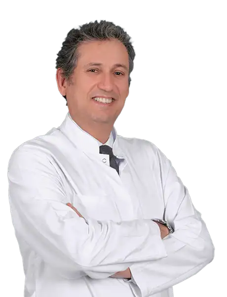
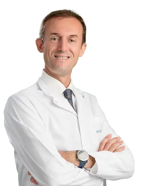
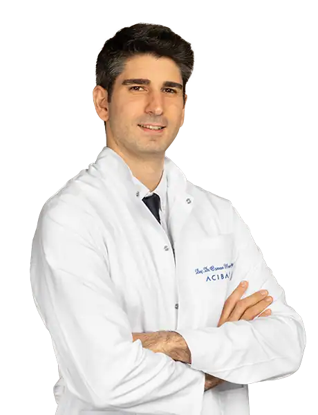
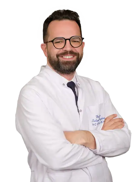


Available at these Hospitals


















Speak with our medical team
Share your case and our international patient team will guide you to the right specialist, free of charge.