

Urology
Kidney stones, an enlarged prostate, a raised PSA or blood in the urine investigated properly, plus prostate, bladder and kidney cancer surgery, continence, reconstruction and male fertility, with free remote review of the scans you already have.
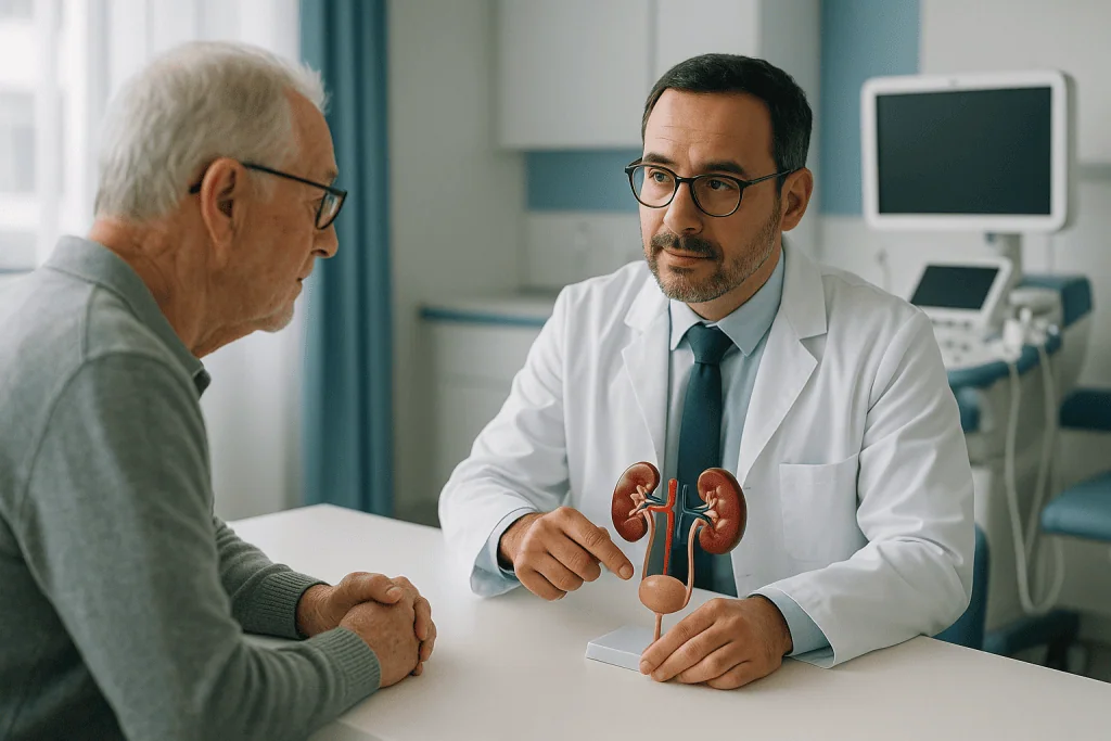
Stones, the prostate, and the findings that frighten people most
Most men arrive here with a stone on a CT, a PSA result they cannot interpret, or blood in the urine they were told to get checked. A great many of those findings need watching rather than operating, and knowing which is which is the whole job.
Stones and the kidney
From the stone that will pass on its own to the one that has blocked and infected a kidney, and the work-up that stops the next one forming.
Prostate and urologic cancer
Benign enlargement and the operations that relieve it, and cancers of the prostate, bladder and kidney planned with the wider team.
Bladder, continence and male health
Bladder symptoms investigated properly before they are treated, reconstruction, and the male fertility work-up that is too often skipped.
The stone that passes, the prostate that waits
Urology is unusual in how much of it is a decision not to operate. A small stone in the lower ureter will very often pass with time and the right analgesia. A prostate that has been called enlarged for a decade may never need surgery. A PSA of five is far more likely to be benign than malignant.
So the images are read before an operation is offered, the prostate gets an MRI before a needle, and the bladder is measured before it is medicated. What we will not do is treat a number.
What we will not do
- Operate on a stone the imaging says will pass on its own.
- Treat a raised PSA as a diagnosis before an MRI has been read.
- Repeat a urethral dilatation that has already failed twice.
- Let a man consent to prostate surgery without discussing ejaculation.
- Send you home with a stent and no removal date written down.
Who would actually operate
Urology subspecialises sharply: the surgeon who clears a complex stone is rarely the one who reconstructs a urethra or manages male infertility. Every profile is readable before you decide.
How this works when you are travelling for it
Send the images, not just the report
The CT or ultrasound images themselves, the stone analysis if you have one, and your PSA as a dated series rather than a single number.
Consultant review
What the existing scans already settle, and what genuinely needs repeating: a second reading changes the plan more often than people expect.
Tests on arrival
Low-dose CT, flow rate and residual volume, MRI before any prostate biopsy, urodynamics where the bladder rather than the prostate is the question.
Surgery and the night after
Most stone and endoscopic prostate operations are day-case or one night; robotic cancer surgery and reconstruction stay longer.
Follow-up before and after you fly
Stents removed and pathology explained before departure where possible, then review remotely, with flying advice specific to your operation.
Six things worth knowing first
Testicular pain is an emergency
Sudden severe testicular pain, often with nausea, can mean torsion, where the testis twists on its own blood supply. It is saved in hours, which is why torsion is operated on as an emergency.
A stone with a fever is different
Stone pain alone is managed with medication. Stone pain with fever and shivering means a blocked, infected kidney, and that is life-threatening.
A raised PSA is usually not cancer
Infection, an enlarged prostate, cycling and recent ejaculation all raise it. The number opens a conversation: it does not close one.
Blood in the urine is investigated
Even painless, even once, even on blood thinners. Anticoagulants do not explain visible blood away, and that belief costs diagnoses.
Stone size is a probability
The millimetre bands predict how likely passage is. Where the stone sits in the ureter decides as much as its diameter does.
Prostate surgery changes ejaculation
Retrograde ejaculation is the commonest consequence of the classic operations, and men are too often not warned before they consent.
Quick answer
Urology is the medical specialty that diagnoses and treats conditions of the urinary tract in women and men and the male reproductive system. At Acibadem in Turkey, urology care covers evaluation, imaging, laboratory testing, and treatment with medication, minimally invasive procedures, or surgery according to the condition.
What the urology unit covers, and how the work is organised
Almost nobody arrives at a urology department out of curiosity. People arrive holding something specific. A CT report that mentions a 6 mm stone in the left ureter and offers no further explanation. A blood test where the PSA figure has been flagged by a laboratory that does not interpret it. A dipstick from a routine check-up that found blood nobody could see. Or twenty years of getting up three times a night with a prostate that somebody once described as enlarged, and that nobody has measured since.
Urology is the surgical speciality responsible for the entire urinary tract, kidneys, ureters, bladder and urethra, in both men and women, together with the male reproductive organs. That is a wide territory, and it is why one department looks after a nineteen-year-old with his first attack of renal colic, a woman whose cystitis keeps returning, a man weighing up what to do about a prostate reading, and a patient whose kidney tumour was found while scanning for a slipped disc.
The work divides into six strands. Almost every referral belongs to one of them, and knowing which one yours falls into tells you most of what you need to know about what happens next.
- Stone diseasethe largest single reason people are referred urgently. This covers the diagnosis of a stone and the pain it causes, the question of whether it will pass on its own, the choice between shockwave treatment, telescopic laser surgery and keyhole kidney surgery, the temporary stents that sometimes go with them, and the metabolic work that follows afterwards to stop the next one forming. It runs from kidney stones through to stone prevention.
- Prostate: benign diseasean enlarged prostate is not a disease of the unlucky; it is what prostates do with age. The clinical question is never simply how big it is, but how much the bladder is suffering: weak stream, hesitancy, incomplete emptying, night-time waking, and at the far end, an inability to pass urine at all. Assessment, medication and the several different operations that reduce obstruction are covered in enlarged prostate and BPH surgery.
- Uro-oncology (kidney, bladder, prostate, testis)cancers of the urinary tract and male genital organs, from the first abnormal test through diagnosis, staging, surgery and the treatments that come before or after it. Because these decisions are rarely made by a surgeon alone, they run through a multidisciplinary meeting alongside medical oncology, radiation oncology, radiology and pathology. The detail sits in PSA testing, prostate biopsy, prostate cancer treatment, what follows prostate surgery, bladder cancer and kidney cancer. Most of these pathways begin with a single findingblood in the urinewhich is investigated the same way whatever eventually explains it. A lump in the testis belongs to the same pathway and is never a wait-and-see problem, see testicular cancer.
- Functional urology and continenceproblems of how the bladder behaves rather than what it looks like. Urgency and frequency that dominate the day, leakage after prostate surgery, bladders that empty poorly, and infections that keep coming back in a tract that is anatomically normal. See overactive bladder, urinary incontinence in men and recurrent UTIs.
- Reconstructive urologyrebuilding a urinary tract that has been narrowed or damaged, most commonly a scarred urethra after infection, instrumentation, catheterisation or injury. This is careful, planned surgery in which the choice of technique depends on where the narrowing is and how long it is, and in which repeat dilatation is usually the wrong long-term answer. See urethral stricture.
- Andrology and male fertilitythe male half of a fertility investigation, including semen analysis, varicocele, hormonal causes and surgical sperm retrieval; erectile and ejaculatory problems, which are frequently the first visible sign of vascular or metabolic disease elsewhere; and vasectomy, together with the reversal that some men later ask about. See male fertility, men’s sexual health and vasectomy and reversal.
Several tools cut across all six strands. Imaging decides more urological questions than examination does. Cystoscopya telescope passed into the bladder, is the only way to see the bladder lining directly, and it is the reason visible blood in the urine is investigated the way it is. The equipment used for stone and prostate work has changed considerably over the last decade, and what those changes actually mean for a patient is set out in technology rather than scattered through the clinical sections as advertising.
The sections that follow begin with the situations that cannot wait, the short list of urological problems where hours matter, and then move into stone disease, which is where most people’s questions start. Planning your care at the end explains what a first consultation involves and what to bring with you.
Red flags: the urological problems that cannot wait
Most urology is unhurried. A stone can be discussed next week, a prostate reading over a month, a slow stream over a year. A small number of situations are the exact opposite, and the cost of treating them like ordinary appointments is measured in lost organs. What follows is the short list.
- Sudden severe pain in one testicleoften waking you from sleep, frequently with nausea or vomiting, sometimes with the testis sitting higher or lying across instead of vertically. This may be testicular torsion: the cord that carries blood to the testis has twisted and the blood supply is cut off. The window in which the testis can be saved is measured in hours, not days, and it closes quietly. It is commonest in teenagers and young men, but it happens at any age, and it can follow trivial activity or nothing at all.
- A stone with fever or shaking chillsflank pain plus a temperature, rigors, feeling profoundly unwell, confusion, or a racing pulse. A kidney that is both blocked and infected is a closed, infected space. This is urosepsis, it is life-threatening, and antibiotics alone do not solve it: the obstruction has to be drained, usually with a stent or a nephrostomy tube, before anything else is done about the stone itself. Deterioration can be very fast, sometimes over a few hours in a person who seemed only moderately unwell.
- Complete inability to pass urineyou feel a desperate, mounting urge, the lower abdomen becomes distended and painful, and nothing comes. This is acute urinary retention. It is not a matter of trying harder or having a hot bath; the bladder needs draining with a catheter, and a bladder left over-distended can be damaged and can take the kidneys down with it. It most often affects men with an enlarged prostate, and is frequently triggered by constipation, alcohol, or a new medication such as a decongestant or an anticholinergic.
- Visible blood with clots, and difficulty or inability to urinateclot retention. Blood that has clotted inside the bladder blocks the outlet, and the bladder then bleeds more as it strains against the obstruction. It is more likely if you take an anticoagulant or antiplatelet medication. It needs a wide catheter and bladder washout, not a wait-and-see approach.
- An erection lasting more than four hourspriapism, particularly when it is painful and unrelated to arousal. Blood trapped in the erectile tissue loses its oxygen, and after several hours the tissue itself begins to be damaged, with permanent loss of erectile function a genuine risk. It can be triggered by sickle cell disease, some prescribed medications, injected erection treatments and recreational drugs.
- Major scrotal or flank traumaa direct blow, a sporting injury, a fall or a road accident followed by severe pain, rapid scrotal swelling or bruising, or blood in the urine. A ruptured testis repaired early can often be saved, and a significant kidney injury needs imaging to decide whether it can be managed without surgery.
Everything above shares a single feature: the tissue at risk is being damaged while the problem goes unaddressed.
Now the other dose, which matters just as much. A great deal in urology is urgent without being an emergency, and inflating it helps nobody. Painless visible blood in the urine is the clearest example. Passing red or cola-coloured urine with no pain at all is alarming to look at, and it must always be investigated, because it is the earliest sign of bladder and kidney tumours as well as of far more common and harmless causes. The investigation is imaging of the kidneys and a look inside the bladder. Crucially, the bleeding stopping does not cancel the investigation. Blood that appears once and clears completely is investigated exactly as thoroughly as blood that persists, and blood in the urine explains why.
Kidney stones: what they are, and why the pain behaves the way it does
A kidney stone is a solid crystal mass that forms inside the kidney’s drainage system. Urine is a concentrated solution carrying more dissolved mineral than it can comfortably hold, kept in solution by water and by natural inhibitors such as citrate. When the balance tips, too little fluid, too much of a particular salt, urine that is persistently too acidic or too alkaline, or an infection that changes its chemistry, crystals come out of solution, stick to one another, and grow. The process is slow and silent. By the time a stone announces itself it has usually been forming for months or years.
Stones are not all the same substance, and the type matters because it drives both treatment and prevention:
- Calcium oxalateby far the most common. Usually hard and dense, which makes some of them resistant to shockwave treatment. Linked to low fluid intake, high dietary oxalate in susceptible people, high urinary calcium and low urinary citrate.
- Calcium phosphateforms in urine that is persistently alkaline, and more often points to an underlying metabolic cause such as a renal tubular problem or an overactive parathyroid gland, which is why finding one usually prompts a wider work-up.
- Uric acidforms in urine that is persistently acidic, and is associated with gout, diabetes, obesity and high purine intake. These stones are invisible on a plain X-ray but show clearly on CT. They are also the one type that can sometimes be dissolved medically, by making the urine less acidic with treatment that is prescribed, timed and dosed by your own doctor and monitored with urine testing.
- Struvite, or infection stonesthese grow only in the presence of specific urine-splitting bacteria, which is why they are commoner in people with recurrent urinary infection or a catheter. They can enlarge quickly and fill the collecting system in a branched shape, which is where the word staghorn comes from. They are a reservoir of infection as well as an obstruction, and are almost always removed rather than watched.
- Cystinerare, and inherited. Cystinuria causes stones from childhood or early adulthood, they recur relentlessly through life, and they are notoriously hard, which makes shockwave treatment unrewarding. Lifelong specialist follow-up is the norm.
Here is the part that surprises people most. A stone sitting in the kidney generally does not hurt at all. Pain begins when it drops into the ureter, the narrow muscular tube to the bladder, and blocks it. Urine keeps being produced above the blockage, pressure rises inside the collecting system, and the ureter contracts hard against the obstruction. That is renal colic: the pain comes in waves because the ureter squeezes in waves, building to a peak that can be genuinely overwhelming and then easing before building again. There is no position that relieves it, which is why people with colic pace and cannot lie still: a useful distinction from the pain of peritonitis, where any movement is unbearable.
Kidney stone pain location follows the stone down. High in the ureter, the pain sits in the flank and loin, under the ribs at the back. As the stone descends it moves round to the lower abdomen and into the groin, and often into the testicle in men or the labia in women. When the stone reaches the last centimetres, where the ureter enters the bladder, the symptoms change character entirely: a constant urge to pass urine, going frequently and producing very little, and sometimes pain at the tip of the penis. People at that stage are often treated for a urinary infection when the stone is in fact almost out.
Among the most useful things to know about kidney stone symptoms is that the severity of the pain tells you nothing about the size of the stone. A 3 mm fragment wedged tightly in a narrow segment can produce the worst pain a person has ever experienced, while a large staghorn stone filling half a kidney can be completely painless and be discovered by accident. Pain reports obstruction, not diameter. This cuts both ways, and the second half of it is the dangerous half: a stone that stops hurting has not necessarily passed. Pain can settle while the blockage remains, and a silently obstructed kidney can lose function over weeks without producing a single symptom. That is why relief is confirmed with imaging and not assumed.
Nausea and vomiting accompany colic so often that they are part of the picture rather than a separate illness; the kidney and the gut share nerve supply, so the body interprets the signal as abdominal. Blood in the urine is common too, usually microscopic and detectable only on a dipstick, sometimes visible. Its absence proves nothing: plenty of obstructing stones produce no detectable blood at all.
A stone that sits quietly in a calyx of the kidney and obstructs nothing is a different clinical object from one in the ureter. It may be reasonable to leave it alone under periodic surveillance, and this is discussed in stone treatment options. What is never reasonable is to leave a stone that has caused an infection.
Which brings us to the line that matters. Colic with pain that prescribed analgesia is controlling, in someone who can drink and has no temperature, is managed as an outpatient problem. Colic plus fever or rigors is a different event altogether: a blocked and infected kidney, described in red flags.
The test that settles the question is a low-dose non-contrast CT of the kidneys, ureters and bladder. It finds essentially every stone regardless of composition, and it reports the three things treatment is planned around: the size, the exact position, and the density. Modern low-dose protocols use a fraction of the radiation of a standard abdominal CT, which matters because stone formers tend to be scanned repeatedly over a lifetime. In pregnancy, and as the first test in children, ultrasound is used instead and is the correct choice: it involves no radiation and reliably shows a swollen, obstructed kidney even when it cannot see the stone itself. Ultrasound and a plain X-ray are also frequently used for routine follow-up rather than repeating CT. The radiology department reports these studies formally, and the images, not the sentence summarising them, are the evidence.
Will it pass on its own? Size, time and the point where waiting stops
This is the question people actually want answered, usually at two in the morning with a scan report in one hand. The honest short answer is that most small stones pass without any intervention, most of them within a few weeks, and that size is the single best predictor, but only a predictor.
The size bands used in practice are these. Stones under about 4-5 mm usually pass spontaneously, and observation with adequate pain relief is the normal first plan. Stones between about 5 and 10 mm are genuinely unpredictable: many pass, many do not, and a decision to wait is made with a defined review point rather than left open-ended. Stones above about 10 mm are unlikely to pass on their own, and treatment is usually planned from the outset rather than after a period of hoping.
Now the framing that keeps those numbers honest. This is a kidney stone size chart in the sense that clinicians use one, as probabilities for planning, not as guarantees for individuals. Every experienced urologist has seen a 9 mm stone arrive in a strainer and a 4 mm stone refuse to move for two months. Three things blunt the numbers:
- Position matters at least as much as diameter. A stone already low in the ureter, close to the bladder, has a far shorter and easier journey than a stone of identical size sitting at the top where the renal pelvis narrows. Reports usually state the level; it is worth reading.
- The measurement itself is approximate. Stones are not spheres. A stone reported as 5 mm on one plane can measure 7 mm on another, and the widest dimension is the one that has to fit.
- The ureter is not a smooth pipe. It has three natural narrow points, where it leaves the kidney, where it crosses the pelvic blood vessels, and where it enters the bladder, and a stone can travel comfortably for most of its route and then stop at one of them.
On how long does it take to pass a kidney stone: most stones that are going to pass do so within days to a few weeks, and smaller, lower stones tend to be quicker. What is not reasonable is indefinite waiting. Prolonged obstruction damages the kidney, and the damage is silent, so observation is given a deadline, typically a review with imaging after a few weeks, and if the stone has not moved by then it is treated. The exact interval is set by your treating team based on the stone and on your kidney function, not by how you feel.
Medical expulsive therapy is medication given to help a stone pass, most often a drug from the alpha blocker class that relaxes the smooth muscle of the lower ureter. It is a doctor’s decision, not a self-help measure: it is most useful for larger stones in the lower third of the ureter, it is of little value for small or high stones, and it carries real side effects including dizziness, a drop in blood pressure on standing, and changes in ejaculation. It must be prescribed, timed and dosed by your own doctor, who will also check it against your other medications.
Pain relief follows the same rule. Anti-inflammatory medication of the NSAID class is generally the most effective for renal colic and works better than simple painkillers, but it is not automatically safe: it is restricted or avoided where kidney function is reduced, where there is a history of ulcers or bleeding, in pregnancy, and alongside certain anticoagulants. Take what has been prescribed for you, at the intervals you were given, rather than adding to it from a pharmacy shelf.
On hydration, be sceptical of the advice you will read online. Staying normally, steadily well hydrated is sensible and is the foundation of preventing the next stone. Forcing several litres in a short period to “flush the stone out” does not push it through a blocked ureter; it increases the pressure above the obstruction and can make the pain considerably worse. Drink to thirst and to pale urine, not against a stopwatch.
Strain your urine. This sounds trivial and it is one of the most valuable things you can do. Pass urine through a fine sieve or filter every time, and keep anything solid you catch, however small and unimpressive: a fragment the size of a grain of sand is enough. Sending it for analysis identifies the stone type, and the stone type is what turns generic advice into a specific prevention plan. Without it, prevention is guesswork. This analysis, together with blood and urine testing, is the starting point of stone prevention, and where kidney function or an underlying metabolic disorder is involved the assessment is shared with nephrology.
Finally, the hard rules. Watchful waiting is not a state you stay in regardless of what happens. It stops if any of the following occur:
- Fever or shaking chills. A blocked kidney that becomes infected is an emergency.
- Pain that your prescribed medication is not controlling, or pain that returns with increasing severity between doses, which often means the stone is not going to pass.
- Vomiting that prevents you keeping fluids or tablets down. You cannot take oral pain relief you cannot keep, and dehydration compounds everything.
- A single functioning kidney, a transplanted kidney, or stones obstructing both sides. Here an obstruction is not partial in any meaningful sense: it threatens everything you have. These situations are treated actively and early rather than observed.
- Pregnancy, reduced urine output, or existing chronic kidney disease. Each changes both the urgency and which treatments are available.
And the point worth repeating from the previous section: if the pain simply stops and you never see a stone, that is not proof of anything.
Stone treatment options: lithotripsy, ureteroscopy or keyhole surgery
When a stone will not pass, or should not be left, there are three principal ways of dealing with it. None is universally better. The choice is a matching exercise between the stone, the kidney it sits in, and the person it belongs to.
Extracorporeal shockwave lithotripsy (ESWL) is the least invasive. Focused shockwaves are generated outside the body, aimed at the stone under X-ray or ultrasound guidance, and used to crack it into fragments. There is no incision and no telescope; it is usually done with sedation and pain relief rather than a general anaesthetic, and people typically go home the same day. Its great limitation is the one that is most often glossed over: ESWL breaks a stone, it does not remove it. Every fragment still has to travel down the ureter exactly as the original stone would have. This is why “one session of lithotripsy” and “stone free” are not the same statement, why colic can follow the treatment rather than precede it, and why more than one session is frequently needed. Its specific complications are bruising and pain over the treatment site, blood in the urine, a bleed around the kidney that is uncommon but occasionally significant, infection when bacteria trapped inside a stone are released, and steinstrasseliterally “street of stones”, where a column of fragments lines up and blocks the ureter, sometimes requiring a stent or telescopic surgery to clear. It is not used in pregnancy, in uncorrected bleeding disorders, in untreated urinary infection, or where there is an obstruction below the stone that the fragments could never get past.
Ureteroscopy with laser passes a fine telescope up through the urethra and bladder into the ureter, and with a flexible instrument into the kidney itself. The stone is broken up with a laser and either dusted into particles or grasped and removed with a basket, so at the end of the operation the surgeon has seen the stone go. It works irrespective of how hard or dense the stone is, which is where ESWL fails, and it reaches places shockwaves handle poorly. It requires a general anaesthetic in most cases, and a temporary ureteral stent is often left afterwards, which is itself the commonest source of complaint in the following weeks. Its risks are ureteric injury or perforation, infection and sepsis, occasionally an inability to reach the stone at the first attempt in a narrow ureter, in which case a stent is placed and the operation repeated after the ureter has passively widened, and, rarely, ureteric narrowing developing later. The full account is in ureteroscopy.
Percutaneous nephrolithotomy (PCNL) is keyhole surgery on the kidney itself, and is the answer for large stones, generally above about 2 cm, and for staghorn stones. A tract is made through the skin of the flank directly into the kidney under imaging guidance, and the stone is fragmented and physically removed through it, which is why it clears a large volume of stone in a single operation when nothing else can. It is also the most invasive of the three. The complications are correspondingly real: bleeding, which is usually manageable but occasionally requires transfusion or a radiological procedure to block a bleeding vessel; injury to the lining of the chest when upper access is needed, causing air or fluid around the lung; infection and sepsis; leakage of urine; injury to bowel or other adjacent organs, which is rare; and residual fragments needing a second look. It involves a general anaesthetic and a hospital stay. Smaller-calibre “mini” variants reduce the tract size and some of the bleeding risk, at the cost of taking longer for very large stones.
The factors that actually decide between them are these, and they are worth asking about by name:
- Size and total stone burdena single 6 mm stone and three 6 mm stones in different calyces are different problems.
- Positiona stone in the lower pole of the kidney drains against gravity, so fragments there clear poorly after ESWL; a stone in the lower ureter is ideal territory for ureteroscopy.
- Density and compositionCT reports stone density in Hounsfield units, and hard stones such as cystine and calcium oxalate monohydrate resist shockwaves. This single number often decides against ESWL before anything else is considered.
- Anatomya horseshoe kidney, a narrow angle between the calyx and the renal pelvis, a calyceal diverticulum or a previous reconstruction all change what is feasible.
- Body habitusthe distance from skin to stone affects whether shockwaves can be focused effectively, and affects positioning for percutaneous access.
- Obstruction and infectionthis one is not a preference. A kidney that is blocked and infected is drained first with a stent or a nephrostomy, treated with antibiotics, and the stone is dealt with as a separate, later operation. Attempting definitive stone treatment on an infected obstructed system is how urosepsis is caused.
- Anticoagulants and antiplatelet medicationESWL and PCNL both carry bleeding risk and generally require these to be paused, which for some patients is itself hazardous. Ureteroscopy is usually the approach that can be performed with the least interruption to anticoagulation. That decision is made jointly with the doctor who manages the anticoagulant, never unilaterally, and never by the patient.
- A single kidney, pregnancy, or reduced renal functioneach narrows the options and raises the threshold for accepting any risk to the kidney.
On judging results, there is one measure that means anything: the stone-free rate, assessed on imaging after treatment. Not whether the operation felt straightforward, not whether you saw fragments in the strainer, not the number of sessions. Be cautious of any source that quotes a single figure for it, because the number depends entirely on the stone’s size, site and composition, on the patient’s anatomy, and on how the study defined “stone free”, some count any residual fragment, others allow fragments below a threshold on the assumption they will pass. Two centres quoting different numbers may simply be using different definitions. The useful questions to ask your surgeon are concrete ones: what proportion of stones like mine typically need a second procedure, what imaging will confirm the result, and when will it be done?
Two other routes deserve a mention. Small stones sitting quietly in the kidney and obstructing nothing may be watched with periodic imaging rather than treated, particularly in older patients or where treatment carries higher risk, though this is a decision to review, not to forget, since they can grow or drop into the ureter later. And uric acid stones can sometimes be dissolved medically by altering urine chemistry, under prescription and with monitoring, avoiding an operation altogether. Open surgery for stones is now rare.
Ureteroscopy: removing a stone through the natural passage
A ureteroscopy is an operation in which a fine telescope is passed through the urethra and bladder into the ureter to find a stone and remove it, with nothing cut on the outside of the body. It is keyhole surgery with no keyholes: the scope travels along the route urine already takes, through the urethra, into the bladder, and then upwards into the ureter towards the kidney. Nothing is cut on the outside of the body, and there is no scar afterwards. That absence of a wound is exactly why people underestimate it. It is a real operation, performed under anaesthetic, and it earns the same preparation and the same recovery as any other operation. Understanding what happens inside makes the days afterwards far less frightening.
How a ureteroscopy is done
There are two families of instrument, and the choice depends on where the stone sits. A semi-rigid ureteroscope is a straight, narrow scope used for stones in the lower and middle ureter: the part closest to the bladder, where most impacted stones cause colic. A flexible ureteroscope has a steerable tip that can be driven around the curve of the upper ureter and into the collecting system of the kidney itself, reaching individual calyces. When a flexible scope is used to treat a stone inside the kidney, the procedure is often called retrograde intrarenal surgery, or RIRS. A soft plastic ureteric access sheath is sometimes placed first, giving a protected channel for the scope to travel up and down and keeping pressure inside the kidney low.
Once the stone is in view, it is dealt with in one of two ways, and often both. A holmium laserthe long-standing workhorse, or a newer thulium fibre laser is passed down the working channel and used to break the stone up. The surgeon can either fragment it into pieces small enough to be picked out, or “dust” it into particles fine enough to wash out with the urine over the following days. Fragments are retrieved with a fine nitinol basket, which is opened beyond the stone and closed around it under direct vision. Baskets are never used blindly; grasping something that will not move damages the ureter.
Whatever comes out is sent for stone analysis. This matters more than most people are told. The chemical composition of your stone determines which blood and urine tests are worth doing, which dietary changes will actually help you rather than someone else, and whether preventive medication is likely to be useful. A stone thrown away is information lost. The whole logic of preventing the next stone starts here.
Anaesthetic, how long it takes, and going home
Ureteroscopy is almost always done under general anaesthetic, occasionally under spinal anaesthetic, and in many cases as a day case, in and home the same day. The operation itself is usually measured in tens of minutes rather than hours; a large or awkwardly placed stone, a tight ureter, or a kidney stone reached with a flexible scope all take longer, and the time you spend in the hospital is always far longer than the time you spend in theatre. An overnight stay is arranged when the operation was long, when there was bleeding, when you live far away, or when the team wants to see how you are passing urine. A urine sample is checked beforehand and an antibiotic is given at the start. If the urine is infected, the operation is postponed and the infection treated first; an infected, obstructed kidney is drained as an emergency, not operated on electively.
Ureteroscopy recovery, day by day
Recovery has a predictable shape, and knowing it in advance is what stops people phoning in alarm on the second night:
- The first 24 hours. Burning when you pass urine, urine that is pink or red and clears and returns again, and a need to go more often and more urgently. Take the painkillers you were prescribed on a schedule rather than waiting for pain to build.
- Days two to three. A dull ache in the flank or lower abdomen, and, if a stent was left, a sharp pull in the kidney at the moment of urinating. Both are expected, not a sign that something has gone wrong.
- The first week. Burning settles. Most people with desk-based work are back within a few days; heavy manual work and the gym wait longer, and your own team will give you the interval.
- One to two weeks. Small fragments may pass, occasionally with a short burst of colic. Strain your urine if you were asked to, so anything you catch can be analysed.
- Stent removal. If a stent was left, most symptoms disappear within a day of it coming out.
Drink normally; there is no benefit in forcing large volumes. What is not expected: a temperature with shivering, unrelenting pain that your medication does not touch, or being unable to pass urine at all.
A ureteral stent is not automatic. It is usually left when the ureter was swollen or tight, when an access sheath was used, when the stone was impacted, when access was difficult, when fragments remain, when you have a single functioning kidney, or when a second stage is already planned. It is often not left after a short, straightforward removal of a small stone from the lower ureter in an otherwise healthy ureter. Ask before your operation which is likely, and ask again afterwards whether one was placed: this is the single most useful question you can ask in the recovery bay.
Return to work is individual. Desk-based work is commonly resumed within a few days; physically demanding work, driving for a living or heavy lifting typically takes longer, and a stent in place tends to extend it because of urgency and discomfort rather than pain alone. Your surgeon will give you a figure based on what was actually done.
The risks, stated plainly
The honest complication list:
- Infection and sepsis. The most serious early risk. Bacteria released from a stone or pushed into the kidney under pressure can cause a rapid, severe illness, with fever, shivering, confusion or feeling profoundly unwell in the days after the operation.
- Ureteric injury. Minor grazing of the lining is common and heals. Perforation is uncommon and is usually managed by leaving a stent to let the ureter heal. Avulsion, the ureter being torn, is rare but serious and may require reconstructive surgery.
- Bleeding. Visible blood in the urine is expected. Bleeding heavy enough to form clots that block the bladder is uncommon, clots that stop the flow of urine and leave the bladder painfully full are clot retention.
- Stent symptoms, which are so consistent that they have their own section.
- Ureteric stricture. A late narrowing where the ureter healed, appearing months later and often causing no symptoms at all. This is the main reason a follow-up scan is arranged even when you feel completely well.
- A staged second procedure. If the stone burden is large, or if the ureter is too narrow for the scope to pass safely, the surgeon stops, leaves a stent for one to two weeks to let the ureter passively widen, and brings you back. This is a planned deferral, not a failed operation, and it is safer than forcing an instrument.
- Residual fragments, which is why the follow-up scan checks clearance rather than relying on how you feel.
- The general risks of anaesthetic, discussed with you by the anaesthetist.
Ureteroscopy or shockwave, which suits which stone
When is ureteroscopy preferred over shockwave therapy? Broadly, when the stone is in the lower ureter; when the stone is hard on CT density measurement or is a composition known to resist shockwaves; when body habitus puts the stone too far from the skin for shockwaves to focus well; when you take anticoagulant medication that cannot safely be stopped; when you have one working kidney; when shockwave treatment has already been tried without success; and when a single, definite result matters more than avoiding an anaesthetic, for example before a long trip, or in occupations where an episode of colic is unacceptable. Shockwave keeps its place: no instrument enters the body, but it may need repeating and it leaves fragments that still have to pass. The full comparison sits in stone treatment options.
Living with a ureteral stent
A ureteral stentalso called a JJ or double-J stent, is a soft, hollow plastic tube about the width of a thin drinking straw and roughly the length of your ureter. It has a curl at each end: one sits in the kidney, one sits in the bladder, and the curls are what stop it migrating. Urine drains both through it and around it. It is placed at the end of an operation, or sometimes as an emergency measure to relieve a blocked kidney before any stone is treated at all.
Stents are placed to keep the ureter open while it is swollen, to let fragments and debris drain past, to protect a place where the wall was grazed or perforated, to widen a tight ureter passively before a planned second procedure, and to guarantee drainage in a kidney that cannot afford to be blocked. A stent is a bridge, not a treatment.
Now the part people search for at two in the morning. Stent symptoms are common, and they are normal. They are not a sign that your operation failed. The usual set is: an ache or sharp pull in the kidney at the moment you pass urine, caused by urine travelling briefly back up the stent; a constant need to go, often with little to show for it; needing to get up at night; blood in the urine that comes and goes, typically worse after walking or exercise and then clearing again; and a dragging or pressure sensation low down, sometimes at the tip of the penis or in the vagina, because the lower curl rests against the bladder base. Discomfort when sitting, standing up or turning in bed is part of the same picture.
What reliably makes it worse: physical exertion, long car journeys, dehydration and concentrated urine, constipation and straining, holding on when you need to go, and bladder irritants such as strong coffee, fizzy drinks and alcohol. What tends to help: drinking steadily through the day rather than in bursts, emptying the bladder before it becomes very full, keeping the bowels comfortable, and pacing activity in the first week. Medication can genuinely reduce stent discomfort, there are medication classes used for exactly this, but they are prescribed, timed and dosed by your own doctor, who knows what else you take and what was done in theatre.
A stent usually stays for a few days to a few weeks. Occasionally one is kept for months, but only with an explicit plan to exchange it at set intervals. What must never happen is an open-ended stent. This is the one absolute rule of this section: a forgotten stent is a serious problem. Left far beyond its planned life, a stent becomes coated with mineral, encrusts, can block, can become fixed in place or fracture, and may then need several operations, sometimes from both ends, through the bladder and through the back, to remove. Kidney function can be lost in the process. That is why the removal appointment is booked before you leave hospital and written down. Keep the date somewhere you will find it. A simple scan or X-ray settles in minutes whether a stent is still in place.
Removal is much simpler than placement. In most cases it is done with a flexible cystoscope in the clinic, using local anaesthetic gel: the scope is passed into the bladder, the lower curl is grasped, and the stent is drawn out. The passing part takes seconds. Expect stinging when you pass urine, some blood and some urgency for a day or so afterwards, then rapid relief. Alternatively, a stent may be left with a string attached, taped to the thigh or to the penis, so it can be removed at home or by a nurse without any instrument at all. A string spares you a second procedure, but it needs care when washing and wiping, and it carries a slightly higher chance of the stent coming out earlier than intended. Neither approach is better in general; they suit different situations. Related detail on the scope itself is in cystoscopy.
When do stent symptoms stop being normal? A stent plus infection can become sepsis quickly, and a fever of 38°C or above, shivering or shaking chills, or feeling severely unwell are the signs of it. Clots that stop the flow of urine altogether are clot retention. Do not attempt to push a displaced stent back in or pull it out yourself.
Stopping the next stone: analysis, the 24 hour urine test and what actually changes
Most people who form a kidney stone will form another one at some point unless something changes. That is not a threat; it is the reason this section exists. Removing a stone treats today. Working out why your body made it is the only part of stone care that alters the years ahead, and it is the part most often skipped, because it happens after the pain has gone.
It begins with the stone itself. If you passed it, catch it: urinate through a strainer or a fine sieve and keep anything solid, however small, in a dry container. If it was removed surgically, fragments are sent for analysis routinely. Composition changes the plan completely. Calcium oxalate stones are the commonest and drive most dietary advice. Calcium phosphate stones raise questions about urine pH and about the parathyroid glands. Uric acid stones behave differently from all the others and are sometimes treatable without surgery at all. Struvite stones are infection stones: they grow in urine infected by particular bacteria, they can become large and branched, and the priority is complete clearance plus control of the infection rather than diet. Cystine stones come from an inherited transport defect, start young, recur relentlessly and need lifelong specialist management.
Alongside the stone, blood tests check kidney function, calcium, phosphate and urate. A persistently raised blood calcium prompts a parathyroid hormone test, because primary hyperparathyroidism is an uncommon but genuinely correctable cause of recurrent stones. Where kidney function is reduced or the stone picture is complicated, care is shared with nephrology.
The centrepiece of the metabolic work-up is the 24 hour urine test. You collect every drop of urine over a full day into supplied containers, kept cool, starting after your first morning void and ending with the first void of the next morning. It is inconvenient and it is worth it, because it measures what your kidneys are actually doing rather than what you think you eat. A complete collection reports: total volume; calcium; oxalate; citrate, the natural inhibitor that stops crystals sticking together; uric acid; sodium, which tracks your salt intake honestly; creatinine, which tells the laboratory whether you collected the whole day; and pH. It is usually done several weeks after the acute episode, on your normal diet, collecting during a week of unusually careful eating produces a beautiful result and a useless one. Two collections are sometimes requested, because a single day varies.
Fluid advice is best expressed as output, not as glasses. The general target for stone formers is a urine volume above roughly 2.5 litres a day, which for most people means drinking noticeably more than they currently do, spread across the whole day and into the evening rather than concentrated at breakfast. Hot climates, manual work and sport raise the requirement substantially. Pale urine through the day is a rough proxy between tests. If you have a heart or kidney condition, your doctor may set you a different target, and theirs overrides the general rule.
Then the counter-intuitive part, which is worth reading twice. Restricting dietary calcium makes calcium oxalate stones more likely, not less. Calcium eaten with a meal binds oxalate inside the gut, so the oxalate leaves in the stool instead of being absorbed and excreted into the urine. Cut the calcium out and more oxalate reaches your kidneys. The advice, therefore, is normal dietary calcium taken with mealsnot avoidance, and not high-dose supplements swallowed alone between meals, which behave differently and should be discussed rather than assumed.
The changes that carry the most weight after fluid are sodium and animal protein. High salt intake pushes calcium out into the urine, so reducing processed food, bread, cured meats and added salt lowers urinary calcium without touching your calcium intake at all. Large amounts of animal protein raise acid load and urinary uric acid and lower protective citrate. Oxalate-heavy foods, spinach, rhubarb, beetroot, nuts, chocolate, strong tea, are handled proportionately: for most people the answer is moderation and pairing them with a calcium-containing food in the same meal, not elimination. Citrus fruit and lemon in water add citrate naturally.
Uric acid stones follow a different logic entirely, and it is a logic about pH. They form when urine is persistently acidic, below about 5.5, regardless of how much urate is present. They are invisible on a plain X-ray and visible on CT, which is why they are sometimes discovered late. Because their formation is pH-driven, they can sometimes be dissolved by alkalinising the urine rather than operated on. That is a doctor-supervised programme with repeated pH monitoring, not a home remedy: push the pH too far and you encourage calcium phosphate stones instead. Low urine volume, obesity, insulin resistance and gout all sit in the background of this stone type.
Preventive medication exists for high urinary calcium, low citrate and uric acid overproduction, and it works. Every one of those options is prescribed, timed and dosed by your own doctor after the work-up results are in, with follow-up blood tests, because each has its own effects on potassium, blood pressure and urine chemistry. Nothing here should be self-started, and over-the-counter supplements are not neutral, high-dose vitamin C, for one, is converted to oxalate. Finally, expect a follow-up scan even when you feel entirely well: silent stones sitting in the kidney grow quietly, and finding one early keeps you in the territory of planned treatment rather than emergency colic. Imaging is arranged through radiology at intervals your team sets.
Enlarged prostate (BPH): what it is and what to do first
Prostate enlargementbenign prostatic hyperplasia, BPH, is the gradual growth of the inner part of the prostate from midlife onwards. Begin with the fact that most men come looking for: it is not cancer, it does not turn into cancer, and having it does not make cancer more likely. They are different processes in different zones of the same gland. A man can have both at the same time, entirely independently, which is precisely why urinary symptoms neither prove nor exclude cancer, and why a PSA result is read in the context of gland size and everything else rather than alone. That interpretation is set out in PSA testing.
The mechanics are simple. The prostate wraps around the urethra just below the bladder. As the inner zone grows it squeezes that channel, and the bladder muscle has to work harder to push urine through. Over time the bladder wall thickens and becomes irritable, and that is the reason storage symptoms can persist for a while even after the obstruction has been relieved. The bladder has changed too, not just the prostate.
Symptoms fall into two families, and knowing which family dominates yours shapes the treatment.
- Voiding symptomsa weak or slow stream, hesitancy before flow starts, straining, a stream that stops and starts, a feeling of not emptying, terminal dribbling, and leaking a little after you have finished and walked away.
- Storage symptomspassing urine often, sudden urgency that is hard to defer, getting up at night, and occasionally urgency leakage. These overlap with overactive bladder, which can exist alongside BPH or instead of it.
Nocturia deserves its own warning, because it disappoints more men than any other symptom. Getting up at night has causes well beyond the prostate: fluid, alcohol or caffeine late in the evening, diuretic tablets taken in the afternoon, heart failure, obstructive sleep apnoea, poorly controlled diabetes, leg swelling that redistributes when you lie flat, and the ordinary age-related shift in how much urine the body makes overnight. If nocturia is your main complaint, treating the prostate alone may not fix it. A simple frequency-volume chart, writing down times and volumes for three days, often identifies the real culprit before any treatment starts.
Another expectation worth resetting: prostate size correlates poorly with symptom severity. A large gland can cause almost no trouble, and a modest gland with a tight bladder neck can obstruct badly. The measurement on your scan report is used for planning an operation, not for deciding whether you need one. You are treated for what you experience and what your bladder is doing, not for a number in cubic centimetres.
Assessment is straightforward and mostly non-invasive: a validated symptom score to put a figure on how much this actually bothers you; the frequency-volume chart; a urine test to exclude infection and check for blood; a flow rate measurement, where you urinate into a machine that records the shape of the stream; an ultrasound post-void residual to see how much is left behind; blood tests for kidney function; PSA discussed and interpreted in context; and imaging of the gland where surgery is being considered. If the picture is contradictory, poor flow but a normal-looking prostate, or symptoms that do not fit, pressure-flow urodynamics can distinguish true obstruction from a bladder that is simply underactive, which matters enormously, because an operation does not help a bladder that is not obstructed.
Doing nothing, deliberately, is a legitimate treatment. Watchful waiting with review means: shifting fluid earlier in the day, reducing evening alcohol and caffeine, double voiding (waiting a moment and going again), allowing time rather than rushing, treating constipation, and reviewing medicines that worsen flow, some decongestants and some antidepressants among them, and the timing of diuretics. Review those with the doctor who prescribed them; do not stop prescribed medication on your own.
On “how to shrink the prostate”: no food, drink, exercise or herbal supplement has been shown in good trials to reliably reduce gland volume, and the popular plant extracts have generally not outperformed placebo when tested properly. What does exist is prescription medication. One class relaxes smooth muscle in the prostate and bladder neck and improves flow within days to weeks without changing gland size; its trade-offs include dizziness or a drop in blood pressure on standing, nasal congestion, and reduced or absent ejaculation, and it can complicate cataract surgery, so tell your eye surgeon you take it. A second class genuinely reduces gland volume, but takes six to twelve months, roughly halves the PSA reading so every future result must be interpreted knowing you take it, and can reduce libido and ejaculate volume. Medications for storage symptoms and for erectile function are sometimes added. Which combination suits you, and whether any of it is worth the trade-off, is your doctor’s decision made with you: this page describes classes, not prescriptions.
Two findings move a man from choice to necessity. The first is urinary retention: complete inability to pass urine, with a painfully full lower abdomen. The bladder needs draining with a catheter, and delay causes damage. The second is kidney impairment from chronic high-pressure retention, which is often painless and turns up on a blood test or as swollen kidneys on a scan. Recurrent urinary infections, bladder stones, repeated bleeding from the prostate and medication that has stopped working also push the conversation towards surgery.
BPH surgery compared: TURP, HoLEP, laser vaporisation, UroLift and Rezum
There is no best operation for an enlarged prostate. There is the operation that fits your gland, your medical situation and your priorities, and the honest comparison below is what that conversation should sound like. All of these procedures relieve obstruction by making a wider channel; they differ in how the tissue is dealt with, what the recovery costs you and how long the result lasts.
TURP
TURP surgerytransurethral resection of the prostate, remains the reference against which everything else is measured. A resectoscope is passed along the urethra and the obstructing tissue is removed in chips using an electrical loop; the chips are collected and sent to pathology, which occasionally reveals an unsuspected cancer. Modern bipolar systems use saline irrigation and have all but eliminated TUR syndrome, the dilutional problem that could occur with older monopolar resection when glycine irrigating fluid was absorbed, causing low blood sodium and confusion. A catheter usually stays for a day or two, with a short hospital stay. Durability is excellent and the evidence base is decades deep.
HoLEP and ThuLEP (enucleation)
HoLEP and its thulium equivalent, ThuLEP, take a different approach: the whole obstructing adenoma is enucleated off the surgical capsule, essentially peeled out the way you would remove the flesh of an orange from its rind, pushed into the bladder and then morcellated and removed. Because the technique is not limited by gland size, it suits large prostates that would otherwise mean a long resection or open surgery, and it removes more tissue than any other endoscopic option. Bleeding is typically low, which makes it attractive for men on anticoagulants when the team agrees the medication can be managed. Two honest caveats: it is a technically demanding operation with a long learning curve, and temporary urgency or stress leakage for some weeks afterwards is common and under-discussed. It usually settles, and pelvic floor training helps, see incontinence in men.
GreenLight (photoselective vaporisation)
Photoselective vaporisationthe green light laser prostate procedure, vaporises tissue rather than removing it. Bleeding is minimal, the catheter often comes out the next morning, and it is frequently chosen for men who cannot stop blood-thinning medication. The trade-off is that vaporised tissue cannot be sent to pathology, and it is best suited to small and moderate glands.
UroLift (prostatic urethral lift)
Two minimally invasive options preserve ejaculation, which is their whole point, and this is the first of them. The prostatic urethral lift (UroLift) places small permanent implants that hold the lobes of the prostate apart, mechanically opening the channel with no cutting and no heat. Recovery is quick and it can be done under light anaesthetic. It requires suitable anatomy, a large median lobe or a very big gland rules it out, and on average it improves symptoms less than resection and carries a higher chance of needing a further procedure later.
Rezum (water vapour therapy)
Water vapour therapy (Rezum) injects steam into the prostate tissue; the treated tissue is reabsorbed by the body over weeks to months. It can treat a median lobe, it preserves ejaculation, and it is done with minimal anaesthetic, but you keep a catheter for several days and the benefit arrives gradually rather than immediately, and long-term durability data are shorter than for TURP.
Bladder neck incision, simple prostatectomy and embolisation
Two more sit at the edges: a simple bladder neck incision for small glands with a tight neck, and simple prostatectomy, open, laparoscopic or robot-assisted, see robotic surgeryfor very large glands where enucleation is not the route chosen. Prostatic artery embolisation, delivered by interventional radiology, is used in selected men. Which of these is appropriate and available to you is a discussion with your surgeon.
The operations side by side
Read this as a map of trade-offs rather than a ranking. Nothing in it decides your operation on its own: your gland, your medication and what you care about do that.
| Operation | Gland size it suits | Ejaculation | Tissue to pathology | Notes |
|---|---|---|---|---|
| TURP | Small to moderate | Usually retrograde | Yes | The reference operation; decades of evidence, durable |
| HoLEP / ThuLEP | Any, including very large | Usually retrograde | Yes | Removes the most tissue; low bleeding; demanding technique |
| GreenLight | Small to moderate | Usually retrograde | No | Minimal bleeding; suits men who cannot stop blood thinners |
| UroLift | Small to moderate, no large median lobe | Preserved | No | No cutting or heat; quick recovery; more chance of further treatment later |
| Rezum | Small to moderate, median lobe treatable | Preserved | No | Benefit arrives over weeks; catheter for several days |
| Simple prostatectomy | Very large | Usually retrograde | Yes | Open, laparoscopic or robot-assisted; longer recovery |
Decide the comparison on these, in roughly this order:
- Gland size. Large glands favour enucleation; small and moderate glands open up the whole menu.
- Blood thinners. Anticoagulation or antiplatelet therapy that cannot be interrupted pushes towards the low-bleeding techniques.
- Ejaculation. If preserving it matters to you, say so out loud before the decision, not afterwards.
- Catheter time and hospital stay. These differ more between procedures than people expect.
- Durability and re-treatment. The gentler the intervention, generally the higher the chance of needing something further in the years ahead.
- Whether tissue is needed for pathology.
- Anaesthetic fitness and other medical conditions.
Now the consequence men are most often not warned about. Retrograde ejaculation is the commonest lasting effect of TURP, enucleation and vaporisation: most men who have these operations will ejaculate backwards into the bladder afterwards. Orgasm and sensation remain, the visible ejaculate largely does not, and it does not affect erections. It is not harmful, but it does end natural fertility, and it distresses men who were never told it was coming. If fatherhood is still a possibility for you, raise it before surgery rather than after; see male fertility. How this differs from the dry orgasm that follows cancer surgery is explained under what follows prostate surgery.
The rest of the risk list is real and should be given to you in writing: bleeding, which may require bladder irrigation, occasionally a transfusion or a return to theatre; clot retention; urinary infection and, uncommonly, sepsis; TUR syndrome, now largely historical with bipolar and laser techniques; urethral stricture or bladder neck contracture appearing months later, covered in urethral stricture; urgency and temporary leakage in the early weeks; lasting stress incontinence, which is uncommon but not zero; a small risk of change in erectile function; and the possibility of needing re-treatment years later. Expect blood in the urine on and off for several weeks, characteristically worse around the second week as the healing surface separates, drink steadily, avoid heavy lifting and straining, and do not be alarmed by it.
The PSA test, and what a normal PSA level actually means
The PSA test measures prostate-specific antigen, a protein made by prostate tissue and released into the blood. Read that name carefully, because almost every misunderstanding in men’s health starts here: PSA is prostate-specific, not cancer-specific. Healthy prostate cells make it. Enlarged prostate cells make more of it simply because there are more of them. Inflamed cells leak it. A number on a laboratory report tells you something is happening in the prostate. It does not tell you what.
Most raised PSA results are not cancer. That sentence is worth reading twice, because the gap between what men are told and what they hear at this point is enormous. The common benign causes are ordinary and often temporary:
- Benign prostatic hyperplasiathe age-related enlargement described in the section on the enlarged prostate. A bigger gland produces more PSA, permanently, with no cancer anywhere in it.
- Infection or prostatitisthis can push PSA up sharply, and it can stay up for weeks or months after the symptoms have gone. Testing during or just after a urinary infection produces a number that means very little.
- Catheterisation or any instrument passed through the urethra, including cystoscopy, and certainly a prostate biopsy, which raises PSA substantially for weeks.
- Recent ejaculationa modest, short-lived rise, usually settled within about 48 hours.
- Vigorous cyclingprolonged pressure on the perineum can nudge the result up. A long ride the day before a blood test is not a good idea.
- Digital rectal examinationthe effect is small, but if both are being done at one visit, the blood is taken first.
- Medication for an enlarged prostatethis is the one that works in the opposite direction, and it is the most consequential of the lot. The 5-alpha reductase inhibitors prescribed for benign enlargement roughly halve PSA after several months of treatment, so a result that looks reassuring may not be. Tell whoever interprets your PSA that you take one, and expect the reading to be compared only against other readings taken on the same medication.
- Acute urinary retention, which can raise PSA dramatically and briefly.
The practical advice that follows is simple. Avoid ejaculation and long cycle rides for about two days before the test. Do not have PSA measured while you have urinary symptoms suggesting infection, treat that first, then wait several weeks. Use the same laboratory where you can, because assays differ. And before anyone acts on a single abnormal result, repeat it. A striking number of raised PSA results are lower on a properly timed repeat.
PSA levels by age: the honest version. Age-referenced ranges do exist, and they are useful, because PSA rises naturally as the prostate grows. One commonly quoted set uses roughly 2.5 ng/mL for men in their forties, 3.5 in their fifties, 4.5 in their sixties and 6.5 in their seventies, while many laboratories still print a flat 4.0 ng/mL as the upper limit of normal for everyone. Different guidelines set different thresholds, and some deliberately avoid a threshold altogether and treat PSA as a continuous risk marker. There is no single dangerous number, and any source that gives you one is oversimplifying. A normal PSA level is best understood as a band that shifts with age and gland size, read alongside everything else about you, not as a pass mark.
Two facts follow from that, and they point in opposite directions. A normal PSA does not exclude prostate cancer; some cancers, including aggressive ones, produce very little PSA. And a raised PSA usually is not cancer. This is precisely why the number is a signal to investigate rather than a diagnosis, and why the next step is almost never surgery: it is a repeat test, a careful history, and in the modern pathway an MRI before any needle is considered.
The refinements that make one number more informative. Where a result sits in a grey zone, three adjustments help:
- PSA density divides the PSA by the prostate volume measured on MRI or ultrasound. It separates “high PSA because the gland is large” from “high PSA for this size of gland”. A figure around 0.15 is often used as a rough dividing line, and low density is one of the strongest arguments against rushing to biopsy.
- Free-to-total PSA ratio measures how much of the PSA circulates unbound. A lower free fraction leans towards cancer; a higher one leans towards benign enlargement. It shifts the odds; it does not settle them.
- PSA velocity is the rate of change over time. It is the most contested of the three, because it is only meaningful with several readings from the same assay over a decent interval, and short-term fluctuation is normal. A steady upward trend across years carries more weight than any single value.
Newer blood and urine tests that combine markers exist as well, and their role is to help decide whether a biopsy is needed rather than to replace PSA.
Whether to test at all is a decision, not a formality. This is the part that gets skipped. A PSA test can find a cancer early enough to be curable. It can also find a slow-growing cancer that would never have caused harm, and once found, that cancer generates biopsies, scans, anxiety and sometimes treatment with lasting effects on continence and erections. That is the over-diagnosis and over-treatment problem, and it is real. It is not an argument against testing; it is an argument for understanding what the test opens before you have it, rather than afterwards. Guidelines around the world differ on population screening for exactly this reason, and most of them converge on the same practical answer: an informed conversation between a man and his doctor, usually somewhere from the mid-forties to the fifties, revisited over time. Active surveillance, described in prostate cancer treatment, exists largely to defuse the over-treatment half of the problem.
Some things move that conversation earlier. A father or brother diagnosed with prostate cancer, particularly under 60, raises your risk. Several affected relatives, or a family history of breast, ovarian or pancreatic cancer, may point to an inherited BRCA1 or BRCA2 change, which is associated with more aggressive disease. Men of Black African or Caribbean ancestry have a higher lifetime risk and tend to be diagnosed younger. Any of these makes a discussion in your forties reasonable rather than premature.
PSA also has a second life as a monitoring tool, and there it is far more precise than it is as a screening test, following a treated cancer, a change in PSA is measured against your own previous values rather than against a population range. That use is covered in the section on life after prostate surgery. A PSA result carries meaning only when it is read alongside age, gland size, symptoms and family history. A screening number is a signal, not a diagnosis.
Prostate biopsy: MRI first, then targeted sampling
A prostate biopsy takes small cores of tissue from the prostate so that a pathologist can look at the cells. It is the only way to confirm or exclude cancer. What has changed profoundly in the last decade is not the needle: it is everything that happens before it.
Multiparametric MRI comes before biopsy, not after. The modern pathway is imaging-led. An mpMRI combines several sequences, anatomical images, diffusion-weighted imaging that detects densely packed cells, and dynamic contrast imaging of blood flow, to build a picture of where a suspicious area is, and whether one exists at all. It is done before biopsy for a practical reason as well as a clinical one: biopsy causes bleeding inside the gland that obscures the images for weeks. Reporting quality matters here more than the machine, which is why prostate MRI is read by radiologists who do a lot of it; the imaging side of this work sits with radiology.
The report will carry a PI-RADS score from 1 to 5. Read it as a probability scale, not a verdict. PI-RADS 1 and 2 mean clinically significant cancer is unlikely. PI-RADS 3 is genuinely equivocal: the score that means the MRI could not decide, and the score where PSA density does most of its work. PI-RADS 4 and 5 mean a lesion is likely or highly likely to be significant. A PI-RADS 4 is not a diagnosis of cancer, and men are given that number and left to read it as one far too often. A PI-RADS 2 is not a guarantee of a clean prostate either. A second read of a prostate MRI changes the plan more often than people expect.
A negative MRI can mean no biopsy at all. When the MRI shows no suspicious lesion and the PSA density is low, the chance of a significant cancer being missed is small enough that many men reasonably choose PSA monitoring instead of a biopsy. That is a shared decision, weighted by family history, ethnicity, examination findings and how the PSA has behaved over time, not an automatic discharge. Avoiding an unnecessary biopsy is a real benefit, not a corner cut.
Transperineal versus transrectal. Two routes reach the same gland. The transrectal route passes the needle through the wall of the rectum; the transperineal route passes it through the skin between the scrotum and the anus. The transperineal approach has become the preferred one in most units for a single dominant reason: it does not carry bowel bacteria into the prostate, and the risk of serious infection and sepsis is substantially lower. It also reaches the front and apex of the gland more easily, which is where transrectal sampling has always been weakest. It was historically the more uncomfortable option because it needed a general anaesthetic; it is now commonly done under local anaesthetic, with sedation or general anaesthesia available where a man prefers it or the anatomy demands it.
Targeted fusion biopsy versus systematic cores. In an MRI fusion biopsy, the MRI images are overlaid onto the live ultrasound picture so that the needle can be aimed directly at the lesion the radiologist marked. Targeted cores find significant cancer more efficiently and take fewer needles to do it. Systematic cores sample the gland in a set pattern regardless of what the MRI showed. In practice most men have both, because MRI does not see everything: targeted cores catch what the MRI identified, and systematic cores catch what it did not.
What the day involves. You will have a urine test beforehand to make sure there is no active infection. Antibiotic cover is given according to the local protocol and the route used, and blood-thinning medication may need adjusting: a decision made by the doctor who prescribed it, never by you. The procedure itself takes around twenty to thirty minutes. Local anaesthetic is injected around the prostate; you will feel pressure and some sharp moments. Most men go home a few hours later, and anyone who has had sedation or a general anaesthetic needs someone to take them home. Heavy exertion and cycling are avoided for a couple of days.
The complications are not trivial and should be described before you consent. Blood in the urine is expected and settles over days. Blood in the semen is also expected and lasts far longer, commonly several weeks, sometimes six to eight, and the colour can be rust-brown rather than red. It alarms men badly when nobody warned them. Rectal bleeding can occur after the transrectal route. Urinary retention, being unable to pass urine at all, happens in a small number of men, more often in those with large glands and existing obstruction, and the bladder has to be drained with a catheter. Infection is the serious risk, and it can progress to sepsis quickly, with a temperature, shaking chills and feeling profoundly unwell in the days after a biopsy.
A negative biopsy is not always the end. Needles sample a fraction of the gland, and some cancers are invisible on MRI. If the PSA keeps rising, or the pattern of the cores was worrying without being diagnostic, you may be offered continued PSA monitoring, a repeat MRI, or a repeat biopsy targeting a different part of the gland. That is not a failure of the first biopsy; it is how a sampling test behaves.
Reading the pathology report. If cancer is found, the pathologist grades how far the cells have departed from normal glandular architecture using the Gleason score. Patterns are graded 3 to 5, and the two most relevant patterns are added together, so 3+3=6, 3+4=7, 4+3=7, and so on. The order matters: 4+3 means the more aggressive pattern is the dominant one, and it behaves differently from 3+4. Because a score of 6 on a scale that appears to run to 10 sounds mid-range when it is in fact the lowest grade reported, pathologists now also give an ISUP grade group from 1 to 5: Gleason 6 is grade group 1, 3+4=7 is group 2, 4+3=7 is group 3, Gleason 8 is group 4, and Gleason 9–10 is group 5. Grade group 1 disease is the kind most often managed by active surveillance rather than immediate treatment. The report will also state how many cores contained cancer and what proportion of each core was involved, and those figures feed directly into the decision described next.
Prostate cancer treatment: a choice between options, not a ladder
Most men picture prostate cancer treatment as a staircase: watch it, then operate, then irradiate, then medicate, each step taken when the last one fails. That is the wrong mental model and it leads to bad decisions. These are parallel options with genuinely different trade-offs, and for a large group of men more than one of them is a defensible choice. The work of the first few weeks after diagnosis is not to hurry: it is to establish the risk group accurately and then decide what you are willing to trade.
Staging drives the conversation. Risk is assembled from the PSA, the ISUP grade group, the extent of disease in the biopsy cores and the clinical stage. MRI shows whether the tumour is confined within the capsule or extending beyond it, and whether the seminal vesicles are involved. Where the risk category warrants it, a PSMA PET scan looks for disease outside the prostate with far more sensitivity than the older bone scan and CT combination, and it changes the plan in a meaningful minority of men. Staging is done before treatment is chosen, not to delay it, but because the correct choice differs entirely between disease confined to the gland and disease that has spread.
Active surveillance is treatment, not neglect. For low-risk disease, typically grade group 1, limited core involvement, PSA not high, the evidence supports monitoring rather than immediate intervention, because the cancer is unlikely to threaten your life and the treatments carry lasting side effects. Surveillance is a structured programme, not being sent away: PSA at defined intervals, clinical review, repeat MRI, and a confirmatory or repeat biopsy at set points. If the grade rises, the volume grows or the MRI changes, treatment is offered then, and being on surveillance first does not close any door. The honest cost is psychological, living with a known, untreated cancer is difficult for some men, and that is a legitimate reason to choose treatment instead. Watchful waiting is a different thing again: for older men or those with serious other illness, where the aim is to treat symptoms if they ever appear rather than to monitor for progression.
Radical prostatectomy removes the whole prostate and the seminal vesicles, and joins the bladder to the urethra; pelvic lymph nodes are removed at the same time when the risk of nodal spread justifies it. It can be done open, laparoscopically or with robotic assistance. In a robotic prostatectomy the surgeon works from a console controlling wristed instruments under a magnified three-dimensional view: the system never moves on its own. In the narrow male pelvis that view and that instrument precision help, and the approach is generally associated with less blood loss and a shorter stay. What it does not do is convert a difficult cancer into an easy one, see the technology section for what the platform changes and what it does not. The complications belong in the same breath as the benefits: urinary incontinence, erectile dysfunction, permanent loss of ejaculate, infertility, narrowing where the bladder and urethra are joined, lymphocele, bleeding, infection, hernia and the ordinary risks of a general anaesthetic. What actually happens afterwards is set out in the next section, deliberately and in full.
Nerve-sparing, explained properly. The nerves responsible for erections run in bundles along either side of the prostate, close enough that removing the gland can damage or remove them. Sparing them is attempted when the cancer is not at or through the capsule on that side. Whether it is possible is judged from the MRI, which side and how much of the gland the biopsy cores involved, the grade, the examination, and what the surgeon finds during the operation. It can be full, partial or one-sided. Two things must be said plainly: cancer control comes first, and if sparing the nerve would leave disease behind, the nerve goes. And nerve-sparing is not a promise of function, spared nerves are frequently bruised by the dissection and recover slowly, if at all.
Radiotherapy is a genuine alternative, not a consolation prize. External beam radiotherapy is delivered in a course of daily treatments over several weeks, with modern schedules using fewer, larger fractions, and stereotactic techniques in selected men. Brachytherapy places the radiation source inside the gland, either as permanent seeds or as a temporary high-dose implant, sometimes as a boost alongside external treatment. For intermediate and high-risk disease, radiotherapy is often combined with hormone therapy, androgen deprivation, for a defined period of months to years, because the combination controls the cancer better than radiation alone. The side-effect profile differs from surgery rather than being milder: urinary and bowel irritation during and after treatment, fatigue, erectile function that tends to decline gradually over the following years rather than dropping immediately, a small long-term risk of a second cancer in the irradiated field, and, where hormone therapy is used, hot flushes, loss of libido, muscle and bone loss, weight change and mood effects. Planning, delivery and follow-up sit with radiation oncology, working alongside urology rather than downstream of it.
Focal therapyHIFU, cryotherapy and related techniques, treats the tumour and a margin around it rather than the whole gland, aiming to preserve continence and erectile function. It is a selective option, not a general one: it suits a single MRI-visible lesion, intermediate-risk disease and a gland shape the technique can reach. Long-term outcome data are less mature than for surgery and radiotherapy, follow-up with PSA and repeat MRI continues indefinitely, and further treatment of the residual gland may be needed. Anyone offering it as a soft alternative for everyone is overselling it.
When disease has spread, the approach becomes systemic: hormone therapy as the backbone, with additional agents added up front in defined settings, chemotherapy, targeted treatment where a genetic change is present, and radionuclide therapy in specific circumstances. Radiotherapy is still used for the primary tumour or for symptomatic sites. All of it is prescribed, timed and dosed by your own oncologist, and that care is shared with medical oncology. Advanced prostate cancer is managed over years, not cured, and it is managed well.
How the decision is actually made. Cases are reviewed by a multidisciplinary team, urology, radiation oncology, medical oncology, radiology and pathology in the same room, so that the options presented to you have already been argued over by people with different instruments in their hands. Then the choice becomes yours, weighted by your age, your other medical conditions, how your bladder works now, and what you are least willing to lose. Ask for the trade-off to be stated as a pair, because that is what it is: cancer control on one side, continence and erectile function on the other, with the balance point different for each treatment and each man. Ask for the figures that apply to you, with your stage, grade, age and plan attached to them, in your own consultation, in writing.
After prostate surgery: continence, erections, ejaculation and PSA
This is the section men say nobody gave them before their operation. It is written for the man who has already had the surgery and for the man deciding whether to, because the two need the same information.
Urinary control follows a pattern. A catheter stays in for roughly one to two weeks after a radical prostatectomy while the join between bladder and urethra heals; it is removed at a clinic visit, usually after a check that the join is watertight. Almost every man leaks after that catheter comes out. The prostate sits directly on one of the two mechanisms that hold urine in, and removing it leaves the external sphincter doing a job it previously shared. In the usual pattern, leakage is worst in the first days, improves steadily over the following weeks, and continues improving for six to twelve months, sometimes longer. Men typically move from several pads a day, to one, to a small security pad, to none. It is exhausting and demoralising in the early weeks, and it is also the normal course.
For a minority, it does not fully resolve. Incontinence after prostatectomy that persists beyond about a year is treated in its own right, with options that include a male sling or an artificial urinary sphincter, set out in the section on urinary incontinence in men. Persistent leakage is a treatable condition, not a permanent sentence and not something to be endured quietly out of embarrassment.
Pelvic floor training has to be taught, not guessed. Many men squeeze the wrong muscles entirely, buttocks, abdomen, thighs, and conclude the exercises do not work. A pelvic floor physiotherapist teaches the correct contraction, checks that you are doing it, and builds a programme of endurance and quick-contraction work, ideally started before the operation so the technique is already learned. Alongside it: treat constipation, because straining loads the same muscles; keep caffeine and alcohol moderate, as both irritate the bladder; and do not restrict fluids to reduce leakage, because concentrated urine irritates the bladder and makes urgency worse.
Erectile function recovers slowly, and for many men incompletely. Even when both nerve bundles are spared, they are stretched and bruised during the dissection, and nerve tissue recovers over months rather than weeks. Meaningful recovery is usually judged at twelve to twenty-four months, not at three. How much comes back depends on your erectile function before surgery, your age, whether nerve-sparing was possible and how complete it was, and other conditions such as diabetes or vascular disease. Where the nerves had to be removed for cancer control, spontaneous erections will not return, and other options are used instead.
Penile rehabilitation is the term for trying to maintain the health of the erectile tissue during that recovery window, typically regular PDE5 inhibitor medication, a vacuum erection device, and in some men intracavernosal injections. It is discussed rather than promised: the evidence that any protocol changes the eventual outcome is mixed, and it is offered as a reasonable approach rather than a guaranteed one. Every medication involved is prescribed, timed and dosed by your own doctor, and none of it should be self-started or bought online. Where erections do not recover, the options in men’s sexual health remain open, including implants for men in whom nothing else works. Other changes deserve mention because they surprise people: the penis can feel and appear shorter, particularly early on; sensation and orgasm intensity may change; some men leak a small amount of urine at orgasm; and curvature can develop.
Dry orgasm and retrograde ejaculation are not the same thing. Men confuse these constantly, and the difference is mechanical and absolute. After a radical prostatectomy, the prostate and seminal vesicles, the organs that make almost all of the fluid, are removed, and the tubes carrying sperm are divided. There is no semen to go anywhere. Orgasm is still possible and can still feel good, but it is permanently dry, and this is certain rather than a risk. After surgery for benign enlargement, described in BPH surgery, the prostate is still there and still producing fluid, but the bladder neck has been opened up, so at orgasm the semen takes the path of least resistance backwards into the bladder and leaves in the next urine, which looks cloudy. That is retrograde ejaculation: the sensation of orgasm is preserved, the volume is lost or greatly reduced, and it is common after most obstruction-relieving prostate procedures. Both outcomes end natural fertility. One follows cancer surgery and is inevitable; the other follows benign surgery and is a likely, though not universal, side effect. Neither is harmful to your health, and neither means the orgasm itself disappears.
Fertility has to be dealt with before treatment, not after. Prostatectomy ends natural conception permanently. Radiotherapy can damage sperm production, and hormone therapy suppresses it. If there is any possibility that you will want children, including a man in his fifties with a younger partner, which is the case most often overlooked, sperm banking is arranged before the first treatment of any kind. Afterwards, the options narrow sharply and some close completely. See male fertility for how storage and later use work.
PSA afterwards is a different test with a different meaning. Once the prostate has been removed, PSA should fall to undetectable levels, usually reported as below 0.1 ng/mL, within about six to eight weeks. From then on it is measured at intervals, commonly every three to six months at first, then less often, and it is compared with your own previous results rather than with any population range. A confirmed rise to around 0.2 ng/mL and climbing is termed biochemical recurrence. After radiotherapy the pattern is different: the prostate is still in place, so PSA falls slowly over a year or more to a low point, and recurrence is defined as a rise of about 2 ng/mL above that nadir. Radiotherapy can also produce a temporary PSA bounce a year or two afterwards that resolves on its own and does not mean the cancer is back.
A rising PSA triggers a process, not a panic. The test is repeated and confirmed, because laboratory variation and timing matter. How fast it is rising is calculated, since a slow doubling time behaves very differently from a fast one. Imaging follows, usually PSMA PET at low PSA levels where it is available. The case goes back to the multidisciplinary team, and the options, salvage radiotherapy after surgery, salvage local treatment after radiotherapy, hormone therapy, or continued observation, are chosen on what the imaging shows. Biochemical recurrence can precede any detectable disease by years, and many men treated at this stage do very well. Bring the actual numbers and dates to that appointment; a printed list of your PSA values over time is the single most useful thing you can carry into the room.
Cystoscopy: looking inside the bladder
A cystoscopy is a direct look inside the urethra and bladder with a thin telescope passed through the water pipe. Everything else in urology infers what is going on from scans, blood tests and symptoms. This is the one investigation that simply looks, which is why it sits at the end of so many diagnostic pathways and why a normal result carries real weight.
Flexible or rigid, two different procedures with the same name. A flexible cystoscopy uses a soft, bendable scope about the width of a drinking straw. It is done in an outpatient room, awake, with local anaesthetic gel instilled into the urethra a few minutes beforehand, and it takes minutes rather than an hour. You can usually watch the screen if you want to, and you go home straight afterwards and drive yourself. A rigid cystoscopy uses a wider, straight instrument and requires a spinal or general anaesthetic. It is chosen when something has to be done rather than seen: taking biopsies, resecting a bladder tumour, washing out clots, removing a stone or a stubborn stent, dilating or incising a stricture, or injecting the bladder wall. The rigid scope has a larger working channel and better irrigation, which is what makes real work possible through it.
Why it is done. The commonest reason by far is blood in the urine, visible or, in selected cases, microscopic, where cystoscopy is the part of the pathway that inspects the bladder lining directly, since small tumours and inflammatory patches are not reliably seen on any scan. See blood in the urine for the wider workup. Other reasons include surveillance after treatment for bladder cancer, which is a lifelong programme of repeat looks; recurrent urinary infections where an underlying cause is suspected; suspected urethral stricture; storage symptoms such as urgency, frequency or bladder pain that have not responded to treatment, as in overactive bladder; suspected bladder stones; assessment before certain prostate procedures; and removal of a ureteral stent.
What it actually feels like: the honest answer. The gel stings for a few seconds going in, and that is the sharpest moment for many people. As the scope passes, men feel a distinct pressure or brief sharp sensation at the point where it crosses the prostate and the sphincter, because the male urethra is long and curved. Once inside the bladder, sterile fluid is run in to open it up, and that produces a strong urge to pass urine, which is uncomfortable rather than painful. Most men describe the whole thing as unpleasant but very tolerable, and are surprised how quickly it is over; a minority genuinely find it painful, and that is not weakness or imagination. In women the urethra is short and straight, so flexible cystoscopy is usually quicker and easier, though the urge sensation is the same. Two practical things help more than any preparation: keep breathing out slowly as the scope passes rather than tensing, and tell the operator if it hurts: they can pause, add more anaesthetic, or stop.
Preparation is short. A urine sample is checked beforehand to make sure there is no active infection, because passing an instrument through infected urine can push bacteria into the bloodstream. If an infection is found, the test is postponed and treated first. There is no fasting for a flexible cystoscopy and you can eat and drink normally. Blood-thinning medication is usually continued for a flexible look, but tell the unit what you take and follow the instruction of the doctor who prescribed it, never stop it on your own. For a rigid cystoscopy under anaesthetic, fasting instructions apply and you will need someone to take you home. Empty your bladder just before you go in.
If something is seen. Findings are photographed and documented, and where the bladder lining looks abnormal, a small biopsy may be taken. During a flexible cystoscopy the scope is too narrow for anything but the smallest sampling, so the usual result of an abnormal finding is a plan: a second procedure under anaesthetic, most often a transurethral resection of the bladder tumour, which both removes what is there and provides the tissue for a diagnosis. If a stricture is found, its length and position determine what follows. Sometimes what is seen is inflammation, a stone, an enlarged prostate lobe or a trabeculated bladder from years of obstruction, findings that explain symptoms without being sinister. You are entitled to ask what was seen before you leave the room, and to have it written down.
Afterwards. Expect stinging when you pass urine for a day or two, some pink or blood-tinged urine, and a need to go more often than usual. Drink plenty of fluid for the first day or so, because dilute urine stings less and flushes the bladder. Simple painkillers of the kind your own doctor has already approved for you are enough for most people. If a biopsy was taken, avoid sex and heavy exertion for a few days, and expect the blood-staining to last a little longer.
Some symptoms are not part of the expected recovery:
- Fever, shivering or shaking chills, or feeling suddenly very unwell in the hours or days afterwards can mean infection entering the bloodstream. This is the one true emergency on the list.
- Being unable to pass urine at all, with a bladder that feels full and painful, is acute urinary retention. The bladder has to be drained with a catheter.
- Clots that stop the flow, so that urine cannot be passed, are clot retention and are handled the same way.
- Heavy bleeding while you are still passing urine normally, rather than the expected pink tinge, is an emergency if it comes with dizziness, breathlessness or feeling faint.
- Burning that is getting worse after two or three days rather than settling suggests an infection that needs a urine test and treatment.
A normal cystoscopy is genuinely reassuring. That deserves saying plainly, because patients are so often told that a test was “clear” without being told what clear means. In the haematuria pathway, a normal cystoscopy combined with normal upper-tract imaging makes a bladder or urethral tumour very unlikely, and it is the reason most men and women investigated for blood in the urine are told the cause was benign. It is a snapshot of that day, not a permanent guarantee, so if visible bleeding returns after a normal workup, it is investigated again rather than dismissed as already answered. And it works in the other direction too: cystoscopy is a targeted investigation for a specific reason, not a screening test to be done on people with no symptoms and no findings.
Blood in urine (haematuria)
Blood in urine has a medical namehaematuriaand it comes in two forms that feel entirely different to the person experiencing them. Visible haematuria (also called macroscopic or gross haematuria) is blood you can see: pink, red, rust, tea-coloured or cola-coloured urine, sometimes with clots. Microscopic haematuria is blood you cannot see. It is found when a urine sample is dipped or examined under a microscope, often during a routine health check or a test done for something else entirely. The urine looks normal. The report does not.
The single most important sentence in this section is this one: painless visible blood in the urine is investigated every time. Not if it comes back. Not if it happens twice. The first episode is the one that is investigated, in men and in women, at any adult age, even when it clears up the next day and never returns. Blood that appears once and disappears is behaving exactly the way early bladder tumours behave. That is not a reason to panic, most people who are investigated do not have cancer, but it is the reason the work-up is not optional.
There is a widespread and genuinely dangerous misconception attached to this. Many people taking an anticoagulant or antiplatelet medication are told, or assume, that the blood is simply “the blood thinner”. It is not. Anticoagulation does not create bleeding from a healthy urinary tract; it unmasks bleeding from something that was already there. Visible haematuria in someone taking these medicines is investigated in exactly the same way, to exactly the same standard, as in someone taking nothing at all. If a clinician attributes your visible haematuria to warfarin, a direct oral anticoagulant, aspirin or clopidogrel and offers no imaging and no camera test, that is the moment to ask for the standard work-up. Do not stop an anticoagulant yourself to test the theory: those medicines are prescribed, timed and dosed by your own doctor, and stopping them carries its own serious risks.
The standard work-up is short, and it is the same in most units worldwide. It has three parts, and each part looks at a different level of the urinary tract.
- Urine testing. A sample is cultured to exclude infection, because infection is a common and treatable cause and it changes the interpretation of everything else. Urine may also be sent for cytology, which looks for abnormal cells shed into the urine. Cytology is useful when positive and does not exclude anything when negative.
- Imaging of the upper tract. Usually a CT urograma scan timed so that contrast fills the kidneys, the collecting systems and the ureters, which lets radiology see stones, kidney masses and the rare tumours that arise in the ureter or renal pelvis. Where contrast or radiation is a problem, ultrasound and MRI are alternatives, with the trade-off that they see the ureters less well.
- Looking inside the bladder. A flexible cystoscopy, usually under local anaesthetic gel in a clinic room. No scan sees the bladder lining as well as a camera does, and flat lesions in particular can be invisible on CT.
Microscopic haematuria is handled with a little more nuance. A dipstick alone is not proof: the dipstick reacts to haemoglobin and myoglobin, so it can read positive when there are no red cells in the urine at all. A confirmed finding on microscopy is what triggers the work-up, and the extent of that work-up is then matched to your risk, age, smoking history, occupational exposure, previous episodes, urological symptoms. A single dipstick trace in a young non-smoker after a marathon is not the same clinical problem as persistent microscopic haematuria in a 65-year-old former smoker, and it should not be worked up as if it were.
Most explanations turn out to be benign, and it is worth knowing them so the waiting period is less frightening. Urinary tract infection is the commonest. Stones anywhere in the tract can bleed, usually with pain. An enlarged prostate bleeds from its surface vessels, sometimes briskly. Vigorous exercise, especially long-distance running, can produce transient haematuria. In women, menstruation contaminates the sample and the test is simply repeated at a different point in the cycle. Recent catheterisation, cystoscopy or sexual activity can all account for a trace.
Some things colour urine red without any blood being present at all. Beetroot is the classic; blackberries, rhubarb and food dyes do it too. Certain medicines discolour urine as a known and harmless effect, including some antibiotics and the urinary analgesic dyes. Muscle breakdown after extreme exertion releases myoglobin, which turns urine dark and makes the dipstick positive without red cells: that one is not harmless and needs assessment because of its effect on the kidneys. A dark stream that clears within a day after an unusually hard workout, with a negative microscopy, is a different story from bleeding.
There is one pattern that points away from urology entirely. Blood accompanied by significant protein in the urine, red cell casts on microscopy, raised blood pressure or a rising creatinine suggests bleeding from the filtering units of the kidney rather than from the drainage system. That belongs with kidney physicians, not with a cystoscope, and the referral goes in a different direction. Your urine result and your blood tests together decide which door you go through.
One situation is a genuine emergency. Passing clots and then being unable to pass urine at all means the bladder is blocked by clot. This is painful, the bladder distends, and it does not resolve on its own. The same applies to heavy continuous bleeding with dizziness, breathlessness or feeling faint. The urological problems that genuinely cannot wait are listed under red flags.
Small details help: the colour, whether the blood appeared at the beginning of the stream, throughout it or only at the end, whether there was pain, whether clots were passed and what shape they were, whether there was fever, and what medicines you take. And hold on to the reassurance honestly: the majority of haematuria work-ups find nothing sinister. That reassurance is only worth anything because the investigation was done.
Bladder cancer and TURBT
Bladder cancer is one of the more common urological cancers, and it is unusual among cancers in how much of its management happens through the urethra rather than through an incision. It is also unusual in how long it stays with you: even when the first treatment goes well, the follow-up programme is measured in years, and that fact belongs in the conversation from the beginning rather than as a surprise afterwards.
The dominant modifiable risk factor is smoking. Carcinogens from tobacco are filtered by the kidneys and sit in contact with the bladder lining, which is why the bladder is affected at all. Risk falls after stopping, though not immediately and not to baseline, and stopping still matters after a diagnosis because it reduces the chance of recurrence. Occupational exposure to aromatic amines is the other established cause, historically in the rubber, dye, paint, textile, leather, printing and chemical industries, and the lag between exposure and disease can be decades, so a job you left in your thirties is still relevant history at seventy. Chronic bladder irritation from long-term indwelling catheters, previous pelvic radiotherapy, some chemotherapy agents and, in endemic regions, schistosomiasis, make up most of the rest.
The classic presentation is painless visible haematuria. Some people instead have persistent urgency, frequency and burning without infection, which is the typical picture of carcinoma in situ: a flat, high-grade disease that hides from scans and is found by looking. That is why urinary symptoms that do not respond to treatment and keep culturing negative earn a camera test rather than another course of antibiotics.
Diagnosis is made by cystoscopy with imaging of the upper tract and, in many units, urine cytology. If something is seen, the next step is both the diagnosis and the first treatment.
TURBTtransurethral resection of bladder tumour, is done under general or spinal anaesthetic with a resectoscope passed through the urethra. The visible tumour is resected in layers, the base is sampled, and the specimen must contain detrusor muscle, because whether muscle is invaded is the question the entire pathway hangs on. Some units add blue-light or narrow-band imaging to make flat disease visible. A single instillation of intravesical chemotherapy in the hours after resection is given in selected low-risk cases to reduce early recurrence. You wake with a catheter, usually for a day or two, and pass blood-tinged urine for a while afterwards. The real risks are bleeding needing bladder washout or return to theatre, infection, bladder perforation (which can require a longer period of catheter drainage or, rarely, repair), temporary irritative symptoms, and later urethral stricture from the instrumentation itself.
A restaging or second-look TURBT a few weeks later is recommended in specific circumstances: high-grade T1 disease, an incomplete first resection, a large or multifocal tumour, or a specimen with no detrusor muscle in it. This is not a sign the first operation was botched. Understaging on the first resection is common enough that the second look changes management in a meaningful minority of cases, and it is better to find that out now than after a year of the wrong treatment.
Pathology then sorts the disease into two worlds, and the fork is absolute. Non-muscle-invasive disease (Ta, T1 and carcinoma in situ) stays in the bladder and is managed with resection plus drugs put into the bladder. Muscle-invasive disease (T2 and beyond) has grown into the bladder wall muscle and is managed with treatment aimed at the whole bladder and at the body, because the risk of spread is real.
For non-muscle-invasive disease, intravesical therapy means instilling a drug through a catheter and holding it in the bladder. Intermediate-risk disease is usually treated with a chemotherapy agent; high-risk disease and carcinoma in situ are usually treated with BCG, an immunotherapy given as a course of weekly instillations followed by maintenance over a period of years. BCG works by provoking an immune reaction in the bladder lining, and the side effects follow from that: burning, urgency, frequency and blood in the urine for a day or two after each instillation, flu-like symptoms, and rarely a systemic BCG infection needing anti-tuberculous treatment. Global BCG supply has been intermittent, and substitution or dose reduction is sometimes a supply decision rather than a clinical one: it is fair to ask which you are getting and why.
For muscle-invasive disease, the two established routes are surgery and bladder preservation, and both start with a conversation with medical oncology, because cisplatin-based chemotherapy before surgery is standard for those fit enough to receive it.
Radical cystectomy removes the bladder, the regional lymph nodes, and in men usually the prostate; in women it usually includes the uterus and part of the vaginal wall. Urine then has to leave the body another way, and the two common diversions are genuinely different daily lives. An ileal conduit uses a short segment of bowel to bring urine to a stoma on the abdominal wall, draining continuously into an appliance that you empty and change; it is simpler surgery, it is reliable, and it is visible. An orthotopic neobladder builds a reservoir from bowel and joins it to the urethra so you pass urine the usual way; it has no bag, and it has to be learned, timed voiding by the clock, emptying by relaxing the pelvic floor and using abdominal pressure, night-time leakage that is common at first and sometimes permanent, and the possibility of needing to self-catheterise to empty completely. Both are major operations with a long recovery: bowel leak, ileus, infection, hernia, ureteric stricture, metabolic and salt disturbances, vitamin B12 depletion over years with some diversions, and effects on sexual function and fertility that should be discussed before, not after.
Trimodal therapya maximal TURBT followed by radiotherapy with concurrent radiosensitising chemotherapy, preserves the bladder in carefully selected people: a single tumour, complete resection, no widespread carcinoma in situ, no hydronephrosis, and a bladder that already works reasonably well. It is not the soft option. It carries bowel and bladder radiation effects, it requires the same intensive surveillance, and salvage cystectomy is the plan if the bladder recurs, technically harder after radiotherapy than it would have been up front.
Then there is the part that is easy to underplay. Recurrence is common enough in non-muscle-invasive disease that lifelong cystoscopic surveillance is the norm, frequent at first, spaced out later, with upper tract imaging at intervals for higher-risk disease. Each recurrence may mean another resection. That ongoing burden is not a footnote; for some people it weighs on the choice between preserving a bladder that needs constant checking and removing it. Survival depends on stage, grade, completeness of resection and your own fitness, so ask your own team to put your pathology report in front of you and talk through what it means for you.
Kidney cancer, nephrectomy and partial nephrectomy
Most kidney cancer today is found by accident. Someone has an ultrasound for abdominal pain or a CT after a fall, and there is a mass in the kidney that has caused no symptoms at all. The old teaching triad, blood in the urine, flank pain and a palpable lump, now describes advanced disease and is uncommon as a presentation. That shift matters, because incidentally found masses are on average smaller, earlier and more treatable than the ones that announce themselves.
The first thing to establish is that not all renal masses are cancer. An angiomyolipoma contains macroscopic fat, is usually confidently identified on CT, and is generally managed by observation unless it grows large enough to risk bleeding. An oncocytoma is benign but, frustratingly, cannot be reliably distinguished on imaging from some subtypes of renal cell carcinoma. Cysts are graded by their complexity, and simple cysts need nothing at all. Infection, infarction and rarely lymphoma or a deposit from another cancer can all present as a mass. So the assessment starts with dedicated multiphase CT or MRI to characterise what is actually there, plus kidney function tests and a proper look at the other kidney, because that other kidney is about to become important.
A small renal massbroadly one under 4 cm, is not automatically operated on, and this is the part patients are most often not told. Many grow slowly or not at all, and a significant proportion are benign. Three options are legitimate depending on your age, fitness, other illnesses and kidney function:
- Active surveillance with interval imaging, moving to treatment if growth or worrying features appear. This is a reasonable primary strategy in older or medically frail people, where the risk of surgery may exceed the risk of the mass.
- Thermal ablationcryoablation, radiofrequency or microwave, usually placed percutaneously under imaging guidance. Less invasive, faster recovery, and an honestly higher rate of local recurrence than surgery, so it is chosen deliberately rather than as a lesser version of an operation.
- Surgical removal, which for most small masses means removing the tumour and not the kidney.
Renal mass biopsy has a limited but expanding role. It is increasingly used before ablation, before committing to surveillance in a younger person, when the imaging suggests the mass might be lymphoma or a metastasis, and when the result would genuinely change what happens next. Older fears about seeding the needle track are now considered very rare. The real limitations are that a small proportion of biopsies are non-diagnostic and that a benign result does not always settle the question, so biopsy is offered where the answer will be acted on, not as a routine step.
Partial nephrectomyremoving the tumour with a rim of normal tissue and reconstructing the kidney, is the standard treatment for small, localised tumours wherever it is technically feasible. The reason is not cosmetic. Removing a whole kidney reduces total filtering capacity permanently, and reduced kidney function over decades is associated with cardiovascular disease and with progression towards chronic kidney disease, particularly in people who also have diabetes, hypertension or a single functioning kidney. Preserving nephrons preserves options later, which is why nephrology input is worth having before as well as after the operation if your baseline function is already borderline. The specific risks of partial nephrectomy are bleeding (occasionally needing embolisation afterwards), urine leak from the collecting system, positive margins, the effect of the period of clamped blood supply on the remaining kidney, and conversion to complete removal if bleeding or anatomy dictates it.
Radical nephrectomyremoving the whole kidney with its surrounding fat, remains the right operation when the tumour is large, centrally placed, involves the hilum or has invaded beyond the kidney, or when partial surgery would leave too little functioning tissue to be worth it. It is done laparoscopically or with a robotic platform in most cases (see technology), open where the tumour size or extension demands it, and the recovery from a minimally invasive approach is usually measured in weeks rather than months. Risks include bleeding, injury to adjacent organs, hernia at port or incision sites, and the long-term consequence of living with one kidney, which most people tolerate well but which changes how future medications and contrast studies are handled. Tell any future clinician that you have a single kidney; it belongs on the same mental card as an allergy.
Advanced disease is a different specialty’s territory. Kidney cancer that has spread is treated with systemic therapy, immune checkpoint inhibitor combinations and targeted tyrosine kinase inhibitor classes, chosen by risk group and prescribed, timed and dosed by an oncologist, with surgery reserved for selected situations such as removing the primary tumour in some patients on systemic treatment, or removing a small number of isolated deposits. Those decisions are made jointly, and this page deliberately gives no response or survival figures, because they depend on subtype, grade, stage and treatment era, and a number without your histology attached is not information.
After treatment for localised disease, follow-up is imaging-based and risk-adapted, with attention to the function of whatever kidney tissue remains. Blood pressure control, avoiding unnecessary anti-inflammatory drugs, staying hydrated and keeping diabetes well managed are the unglamorous parts that protect the kidney you kept.
Testicular cancer: the lump you can feel, and why it is scanned within days
Testicular cancer is uncommon set against cancer as a whole, and it is nonetheless the commonest solid tumour in men roughly between fifteen and forty-five. That fact shapes everything here. It arrives at an age when men are not thinking about cancer, there is no screening programme for it, and no routine blood test picks it up, so the man himself is almost always the person who finds it. Self-examination is therefore the actual mechanism of early diagnosis rather than a health-magazine platitude. It takes a minute after a warm shower: roll each testis gently between thumb and fingers, feel the whole surface, and learn where the epididymis sits as a soft ridge behind, so you are neither alarmed by normal anatomy nor reassured by an abnormal lump.
A painless lump or swelling in the testis is the classic presentation, firm, arising from the body of the testis rather than beside it, and not tender. Other presentations are heaviness in the scrotum, a dull ache in the groin or lower abdomen, a change in the size or firmness of the whole testis, and occasionally breast tenderness from hormones the tumour produces. Pain is not the usual signal, and that is exactly why men wait: the absence of pain reads as reassurance when it is nothing of the kind. A painless testicular lump is never a wait-and-see finding.
Distinguish it from testicular torsion, the opposite picture and a true emergency: sudden agonising pain in one testicle, often with nausea, frequently waking you from sleep. The blood supply is cut off and the window is measured in hours.
Most scrotal lumps are not cancer, and ultrasound sorts them out quickly. An epididymal cyst sits behind the testis and separate from it. A hydrocele is fluid around the testis, so the scrotum enlarges smoothly and the testis becomes hard to feel, itself a reason to scan rather than assume. A varicocele is a collection of dilated veins, usually on the left, covered under male fertility. Epididymitis is painful, builds over days and usually follows infection. The point is not that a lump is probably harmless; it is that the distinction between a mass inside the testis and one beside it is made by imaging, and swelling that fails to settle on antibiotics is re-scanned rather than given a second course.
Risk factors, honestly. An undescended testicle is the strongest recognised risk, and it still counts if it was corrected surgically in childhood: the operation lowers the risk and makes the testis examinable, which is part of why it is done, but it does not abolish it, and the other testis carries a slightly raised risk too. A previous testicular cancer means the remaining testis warrants attention for life, and a father or brother affected raises risk modestly. Nothing you did caused this: injury does not cause testicular cancer, it merely draws attention to a lump already there.
The pathway is unusual. A scrotal ultrasound comes first, fast, no radiation, decisive about whether a mass is inside the testis. Blood is taken for tumour markers: AFP, beta-hCG and LDH. These are not a screening test and not a diagnosis; normal markers do not exclude cancer, and one common type does not raise AFP at all. What they do is help classify the tumour, contribute to staging and give a baseline against which response and follow-up are measured, which is why they are taken before treatment rather than after. A staging CT of chest, abdomen and pelvis then examines the lymph nodes at the back of the abdomen.
The crucial point is that diagnosis is not made by needle biopsy of the testis. The testis drains along the path it travelled in development, to nodes high at the back of the abdomen; scrotal skin drains to the groin instead. A needle or an incision through the scrotum breaches that boundary and risks seeding tumour into a compartment that was never involved, which changes the treatment that follows. So the testis is removed whole through a groin incisioninguinal orchidectomywith the cord secured high before the testis is mobilised, and that single operation both makes the diagnosis and treats the primary tumour. A prosthesis can be placed at the same time or later; decide beforehand rather than being asked on the morning.
Sperm banking before any treatment is the most important practical instruction in this section, because it is time-critical and frequently missed in the rush of a diagnosis. Semen quality is already reduced at diagnosis in a proportion of men, so storage is arranged before the operation where timing allows, and certainly before any chemotherapy or radiotherapy. If nobody has raised it, raise it yourself, on the day you are told. See male fertility for how storage and later use work.
What follows orchidectomy depends on the tumour type, the stage and what the markers do afterwards. For a substantial group the answer is surveillancean active choice, not a discharge: a structured programme of marker tests, imaging and clinic visits at defined intervals over years, on the reasoning that treatment can be given if and when it is needed rather than given to everyone. It only works if you attend. The alternatives are chemotherapy, prescribed, timed and dosed by medical oncology, and radiotherapy in selected cases of one tumour type, planned by radiation oncology. That decision is made in a multidisciplinary meeting, not by a surgeon alone. Retroperitoneal lymph node dissectionremoving the nodes at the back of the abdomen, most often for a residual mass after chemotherapy, is the specialist operation of the group, with nerve-sparing technique intended to preserve normal ejaculation, and it belongs with teams who perform it regularly.
The honest consequences. One remaining testis is usually enough for normal testosterone and for fertility, but usually is not always: testosterone is checked afterwards and some men need replacement, and fertility may already have been affected by the disease or by what treated it. Follow-up is a genuine burden rather than a formality, repeated visits, markers and scans over years. The late effects of chemotherapy are why it continues long after the cancer has gone: hearing changes and tinnitus, numbness in the fingers and toes, cold sensitivity, and raised cardiovascular and metabolic risk that deserves your family doctor’s attention for life. Testicular cancer responds very well to treatment, including in men whose disease has already spread, and the treatment required for early disease is smaller than the treatment required for late disease.
Overactive bladder, urodynamics and neuromodulation
Overactive bladder is not a disease of the bladder wall that shows up on a scan. It is a symptom syndrome, defined by urgencya sudden compelling need to pass urine that is difficult to defer, usually with frequency and waking at night to void, with or without urgency incontinence, and diagnosed only once infection and other identifiable causes have been excluded. That definition is doing real work. It means the diagnosis is made by taking a history properly and ruling things out, and it means anyone who diagnoses it in ninety seconds and reaches for a prescription has skipped the part that matters.
The most consequential distinction is between urgency incontinence and stress incontinence. Urgency incontinence is leakage that follows an overwhelming urge: you leak on the way to the toilet, at the front door, at the sound of running water. Stress incontinence is leakage on coughing, sneezing, lifting or standing up, with no urge at all. They have different mechanisms, different treatments, and treating one as if it were the other wastes months. Many people have both, and even then the balance decides what is treated first. Getting the type right is the whole game. Men have their own version of this problem after prostate treatment, covered under male incontinence.
Before anything is labelled overactive bladder, these are excluded: urinary tract infection and recurrent UTI; blood in the urine, because urgency plus haematuria can be carcinoma in situ and that must not be treated as a bladder-behaviour problem; bladder stones; bladder outlet obstruction from an enlarged prostate, which produces almost identical storage symptoms; neurological disease; poorly controlled diabetes; diuretics taken late in the day; and, for night-time symptoms specifically, nocturnal polyuria, sleep apnoea and leg oedema that reabsorbs when you lie down. Several of those are treated entirely differently and some are treated by someone else.
A bladder diary kept for three days is the single most useful test in this whole area, and it costs nothing. You record the time and volume of every void, what you drank and when, every episode of urgency and leakage, and pad changes. It reveals things that a consultation never does: that the total urine output is simply too high, that most of it is produced overnight, that voided volumes are large rather than small, that the trigger is four coffees before ten in the morning. It regularly changes the diagnosis, not just the treatment.
First-line treatment is behavioural, it is unglamorous, and it works for a substantial number of people who never need anything else.
- Fluid adjustmentmoderating total intake rather than restricting it to the point of concentrated urine, which irritates the bladder and makes things worse. Shifting intake earlier in the day for night-time symptoms.
- Caffeine, alcohol and fizzy drinks reduced or removed for a trial period long enough to judge, which means weeks rather than days.
- Bladder retrainingscheduled voiding with gradually lengthening intervals, plus urge suppression technique: stop moving, contract the pelvic floor firmly a few times, let the wave pass, then walk calmly. Running to the toilet makes urgency worse.
- Pelvic floor muscle training taught by a specialist physiotherapist, with a check that you are contracting the right muscles. Squeezing occasionally when you remember is not the treatment; a supervised programme over about three months is.
- Weight reduction where relevant, treating constipation, and reviewing medicines that increase urine output.
If symptoms persist, medication is added rather than substituted for the behavioural work. Two classes are used: the antimuscarinic (anticholinergic) class and the beta-3 agonist class. Both are prescribed, timed and dosed by your own doctor, both take several weeks to judge, and both reduce symptoms rather than abolish them. Antimuscarinics cause dry mouth, constipation, blurred vision and, in men with outlet obstruction, a risk of retention. The important caveat is anticholinergic burden in older adults: these drugs add to the cumulative anticholinergic load of everything else on the prescription list, and that cumulative load is associated with confusion and cognitive concerns in older people, which is why the beta-3 class is often preferred over the age of about 65. Beta-3 agonists require blood pressure monitoring. Neither class is a lifelong commitment by default, periodic review of whether the drug is still helping is part of proper care.
Urodynamics is the test people are most often given unnecessarily and most often denied when they need it. It measures what the bladder actually does: fine catheters record bladder and abdominal pressure while the bladder is filled, so the study can show involuntary detrusor contractions during filling, how compliant the bladder is, at what pressure leakage occurs, and, during the voiding phase, whether a poor flow is caused by obstruction or by a weak bladder muscle. That last distinction cannot be made from symptoms alone and it changes the operation you are offered. Urodynamics is genuinely indicated before irreversible surgery, when conservative and drug treatment have failed, when the picture is mixed or contradictory, in neurological bladder disease, and after previous incontinence or prostate surgery. It is not indicated as a routine first step before behavioural treatment. It involves catheters, it is undignified rather than painful, and it carries a small risk of infection.
When conservative treatment and medication are not enough, there is a genuine escalation ladder, and each rung has an honest downside.
- Intravesical botulinum toxin, injected into the bladder wall through a cystoscope, usually under local anaesthetic. The effect wears off, so treatment is repeated every several months, and the effect can diminish over successive cycles. The warning that must be given before you consent: it can cause incomplete emptying or frank urinary retention, and if that happens you will need to perform clean intermittent self-catheterisation until the effect wears off. You should not have this treatment unless you are willing and able to learn that. Urinary infection after treatment is common.
- Percutaneous tibial nerve stimulationa fine needle electrode near the ankle stimulating a nerve that shares spinal segments with the bladder, typically as a course of weekly sessions followed by maintenance. Low risk, low burden, but it requires repeated attendance indefinitely to hold the benefit.
- Sacral neuromodulation, which has a feature the others do not: a test phase. A temporary lead is placed near a sacral nerve root and connected to an external stimulator for an evaluation period, and the permanent implanted device is only offered if you record a meaningful improvement during that trial. That structure protects you from an implant that was never going to work. The permanent system has a battery that eventually needs replacing, conditions attached to future MRI scanning, and the possibility of lead migration, site pain, loss of effect and revision surgery.
One honest framing to end on: overactive bladder is managed, not cured. The realistic goal is fewer urgent episodes, fewer accidents, more predictable days and uninterrupted nights, enough control that the bladder stops dictating where you sit in a room. Most people reach that. Very few reach it with a single tablet and no changes to anything else.
Urinary incontinence in men and the artificial urinary sphincter
Incontinence in men is a different problem from incontinence in women, and it usually has a traceable cause. In the great majority of cases it follows treatment of the prostate. Leakage after radical prostatectomy happens because the operation removes one of the two continence mechanisms and leaves the external sphincter doing the work alone. Leakage after BPH surgery is less common but real. Radiotherapy contributes its own pattern, often with urgency, and its effects can appear years later.
Before any of that is assumed, one cause must be excluded because missing it damages kidneys: overflow incontinence from chronic retention. Here the bladder never empties, sits permanently full, and leaks over the top, continuous dribbling, wetting at night, sometimes with little sensation of needing to go. It is the version most often mislabelled as a “weak bladder” and treated with a bladder-relaxing drug, which makes it worse. A post-void residual measurement takes two minutes with an ultrasound scanner and settles the question. If retention is present, relieving it comes first and everything else waits.
Assessment otherwise is straightforward. A history that establishes the pattern, leakage on coughing, lifting and standing points to sphincter weakness; leakage preceded by an overwhelming urge points to overactive bladder; many men have both. A bladder diary. Pad testing, where pads are weighed over 24 hours, because “how many pads a day” is a poor measure, pad counts vary with habit and anxiety, whereas grams of urine do not, and the number matters when choosing between operations. Examination, flow rate and residual volume. Cystoscopy to check the anastomosis, exclude a urethral stricture or bladder neck contracture, and assess the sphincter. Urodynamics where surgery is being considered or the picture is mixed.
Conservative management is not a holding pattern. Pelvic floor rehabilitation with a physiotherapist who works with men, started before surgery where possible and resumed once the catheter is out, improves the speed of recovery. Fluid and caffeine adjustment, treating constipation, weight reduction and well-chosen containment products all help you live normally while recovery happens. Penile clamps exist and can be useful for specific activities, but they compress the urethra and must be used with care and released regularly; they are not a solution to wear all day.
Timing is the part men most want to argue with. Most spontaneous recovery after prostatectomy happens in the first three to six months and continues more slowly after that, which is why surgery for incontinence is generally not considered until around twelve months have passed and the leakage has been stable for a period. Operating early risks operating on a man who was going to become dry anyway. Waiting is not being fobbed off; it is the only way to know what you are treating. Previous pelvic radiotherapy shifts the whole calculation and is discussed specifically, because irradiated tissue behaves differently with any implanted device.
For persistent mild-to-moderate leakage, a male sling is the usual first surgical option. A mesh sling placed through a perineal incision repositions and supports the urethra. Its attraction is that there is nothing to operate: you pass urine normally, with no pump and no mechanics. Its limitations are equally clear: it is not adjustable after implantation, results are less reliable with severe leakage and after radiotherapy, and the risks include persistent or recurrent leakage, perineal pain that can last months, temporary or occasionally prolonged difficulty emptying, infection and erosion requiring removal.
For severe leakage, the reference standard is the artificial urinary sphincter. It has three parts implanted in one operation: an inflatable cuff around the urethra that keeps it closed, a pressure-regulating balloon in the abdomen, and a control pump placed in the scrotum. To pass urine you squeeze the pump, which moves fluid out of the cuff and opens the urethra; the cuff refills automatically over the next couple of minutes and closes again. It is worth being blunt about what that means. It is a mechanical device inside your body that you operate by hand, several times a day, for the rest of your life. It requires enough manual dexterity to find and squeeze a scrotal pump and enough cognitive capacity to remember to do it before voiding, which is why it is not the right choice for everyone, and why dexterity and memory are assessed honestly before implantation rather than discovered afterwards.
The device is left deactivated for several weeks after implantation to allow healing, so you continue to leak during that period and are then taught to use it at an activation visit. Its genuine risks are mechanical failure of any component, gradual thinning of the urethral tissue under the cuff causing recurrent leakage, erosion of the cuff into the urethra, and infection, any of which can mean revision surgery or removal of the whole device and a period without it. Devices do not last indefinitely; over a long enough horizon, revision is a realistic expectation rather than a rare misfortune. One safety point matters more than all the technical detail: never allow a urinary catheter to be passed while the cuff is closed. Tell every clinician you meet that you have an artificial sphincter, carry the device card, and consider a medical alert bracelet, because a catheter forced through a closed cuff can cause serious urethral injury.
The stated goal of all of this is social continence, dry, or dry enough that a single small pad covers a normal day and you stop planning your life around toilets. That is an honest target. A promise of never leaking again is not one anybody can make.
Urethral stricture
A urethral stricture is a narrowing of the urethra caused by scar tissue in the wall of the tube and the spongy tissue around it. Because scar does not stretch, the narrowing behaves like a fixed obstruction: the stream slows, the bladder works harder, and over years the bladder itself can change. Strictures are overwhelmingly a male problem: the female urethra is short and true strictures there are rare.
The common causes fall into a few groups. Instrumentation and catheters are now the largest single group: transurethral surgery, cystoscopy, a traumatic or repeated catheterisation attempt, or a catheter left in for a long period, particularly a large one. Infection, historically gonococcal urethritis, causes long and often multi-segment strictures. Trauma covers straddle injuries, where the bulbar urethra is crushed against the pubic bone, and pelvic fracture injuries, which are a distinct and more complex problem. Lichen sclerosusalso called balanitis xerotica obliterans, is a chronic inflammatory skin condition that produces a pale, tight, scarred glans and foreskin and narrows the meatus and penile urethra. Previous hypospadias surgery in childhood can present decades later. And a substantial proportion remain idiopathic, with no cause ever identified.
The symptoms are those of obstruction, and they build slowly enough that men adapt without noticing: a slow stream, a stream that sprays or splits or that you have to aim, straining to start and to keep going, an intermittent flow, a feeling of incomplete emptying, and dribbling after you finish. Complications follow the obstructionrecurrent urinary infections, prostatitis-like pelvic pain, bladder stones, and eventually acute retention. The overlap with an enlarged prostate is almost complete, which is why some men are treated for their prostate for a year before anyone considers the urethra. Two clues push towards stricture: a spraying or split stream, and obstructive symptoms in a man too young for significant prostate enlargement. In any man under about fifty with a poor flow, stricture belongs near the top of the list.
Diagnosis is not difficult when it is looked for. Uroflowmetry typically shows a flat, plateau-shaped curve rather than the normal bell shape, and post-void residual is measured at the same time. Flexible cystoscopy shows the narrowing directly, though a tight stricture may not allow the scope past it. A retrograde urethrogram, often combined with a voiding study, is the test that maps the stricture, where it is, how long it is, and whether there is more than one, and that map is what determines which operation is appropriate. Ultrasound urethrography adds information about the depth of scarring in some units, and MRI is used for pelvic fracture injuries.
Now the part that changes decisions. Dilatation and direct vision internal urethrotomycutting the stricture endoscopically, are quick, usually day-case, and low-risk in the short term. They also have a high recurrence rate. They are most likely to give a durable result in a single short bulbar stricture treated for the first time; for longer, denser, penile or previously treated strictures, recurrence is the expected outcome rather than the unlucky one. A second urethrotomy after a recurrence rarely produces lasting benefit, and a third almost never does. Repeated dilatation is not a treatment strategy. It can be a legitimate, deliberately chosen palliation, for a man unfit for reconstructive surgery, or one who prefers periodic intermittent self-dilatation to an operation, but it should be chosen with that framing, not drifted into one procedure at a time.
The reason this matters beyond convenience is that repeated endoscopic treatment is not neutral. Each cut and each forced dilatation adds scar. Strictures that were short become longer, spongiofibrosis becomes deeper, and the tissue a reconstructive surgeon needs to work with is consumed. A stricture that could have been cured by a short excision and re-join may, after several years of repeat urethrotomies, require a graft. The minor procedures make the definitive one harder.
That definitive option is urethroplasty: open surgical reconstruction, done once, with the intention of curing the stricture rather than managing it. For a short bulbar stricture, the scarred segment is excised and the healthy ends joined. For longer strictures, or where excision is not possible, the urethra is widened using a graft, most often a buccal mucosal graft taken from the inside of the cheek, which suits the job because it is thin, well-vascularised and used to a wet environment. The donor site heals without a visible scar but is sore for a week or two and can leave temporary tightness or numbness. Complex disease, extensive lichen sclerosus and some hypospadias failures are repaired in two stages, months apart. A catheter stays in for around two to three weeks and a contrast study is usually done before it is removed. Real risks include recurrence, wound infection, bleeding and haematoma, fistula, post-void dribbling from the reconstructed segment, curvature, and effects on ejaculation; effects on erections are uncommon but are discussed before consent.
Urethroplasty is the clearest instance of the point made in technology: ask how often the surgeon performs reconstruction and which techniques they use. If the answer is that they mainly perform urethrotomy and refer reconstruction elsewhere, that is useful information, not an insult.
Two additional points. If lichen sclerosus is the cause, it is a skin disease as well as a urological one: it needs dermatological co-management, genital skin should not be used as graft material, and long-term follow-up is advised because of a small risk of malignant change in affected skin. And complete inability to pass urine is acute retention. Say clearly that you have a known stricture, because repeated forced attempts to pass a urethral catheter can cause further injury, and a suprapubic catheter placed through the abdominal wall may be the safer route.
Recurrent urinary tract infections and interstitial cystitis
A urinary tract infection becomes “recurrent” when it meets a definition, not when it merely feels relentless. The usual threshold is two or more culture-proven infections in six months, or three or more in twelve months. The wording matters because it changes the plan: a single unpleasant infection is treated and forgotten, while a recurrent pattern is investigated for a reason behind it: a stone acting as a reservoir for bacteria, incomplete bladder emptying, a narrowing, a foreign body such as a long-forgotten stent, diabetes, or changes in the vaginal and urethral tissues after menopause.
The single most useful change most people can make is a urine culture before the antibiotic, not after it. Empirical treatment, a prescription based on symptoms alone, is reasonable for a first, straightforward infection. Repeated empirical treatment is how the cycle becomes permanent. Without a culture nobody knows which organism is involved, whether it is the same organism returning or a new one each time, or which antibiotics it is already resistant to. People arrive after two or three years of rotating prescriptions with no culture result in the file at all, and the first honest step is to stop, obtain a properly collected midstream sample while symptoms are present, and treat what actually grows. That is also why you should not start a leftover course from a previous episode: it removes the evidence and leaves the diagnosis unproven.
A urinary tract infection means something different in men. The male urethra is longer and infection is uncommon enough that a single confirmed episode is generally investigated rather than simply treated, with imaging of the kidneys and bladder, a measurement of how completely the bladder empties, and consideration of the prostate. Common findings are an enlarged prostate causing residual urine, a stone, or a urethral stricture. Prostatitis, which can be acute and dramatic or chronic and grinding, sits inside this same work-up. In women the investigation threshold is higher, because uncomplicated infection is common and most women with recurrence have a normal urinary tract; imaging and cystoscopy are reserved for those with stones, blood in the urine that persists after treatment, infections with unusual organisms, or a pattern that does not fit.
What genuinely reduces recurrence, described honestly. Drinking more fluid so that urine stays dilute has reasonable evidence behind it and costs nothing. Treating constipation and avoiding spermicide-based contraception help some women. After menopause, topical vaginal oestrogen restores the tissue and the local flora, and in the right patient it is one of the more effective measures available: it is a prescription, timed and dosed by your own doctor, and it is not the same as systemic hormone therapy. Non-antibiotic urinary agents such as methenamine hippurate are used in some countries as an alternative to long-term prophylaxis. Low-dose continuous or post-intercourse antibiotic prophylaxis works while it is being taken, and its cost is resistance and side effects, so it is a deliberate, time-limited decision made with a doctor and reviewed, not an open-ended repeat prescription. The evidence for cranberry products, D-mannose and probiotics is weaker and inconsistent; they are unlikely to harm you, and they are not a substitute for finding out why the infections keep happening. Vaccine-style immunostimulants exist in some markets with mixed evidence. Nothing on this list should be self-started on the strength of a web page.
Bacteria in the urine without symptoms is usually left alone. Asymptomatic bacteriuria, a positive culture in someone who feels well, is common in older adults, in people with long-term catheters, and after some operations. Outside pregnancy and a small number of urological procedures that break the lining of the urinary tract, treating it does not prevent later symptomatic infection and does cause harm: resistant organisms, Clostridioides difficile colitis, allergic reactions, and the erosion of the antibiotics you may badly need later. Cloudy or strong-smelling urine on its own is not a reason for antibiotics. This is a patient-safety argument, not an economic one: the person who is harmed by unnecessary antibiotics is the person who took them.
The pivot that changes lives: a painful bladder with sterile urine is not an infection. If cultures are repeatedly negative while the symptoms continue, the diagnosis is not a stubborn UTI. Interstitial cystitis, also called bladder pain syndrome, is pain or pressure that the patient relates to the bladder, with urinary frequency and urgency, lasting longer than six weeks, without infection or another explanation. The classic pattern is pain that builds as the bladder fills and eases briefly after voiding. People commonly reach this diagnosis after years of antibiotics that never helped, and being told that the tests are “normal” is not the same as being told there is nothing wrong.
The pathway is different from an infection pathway. It starts with a symptom and voiding diary, examination including the pelvic floor, urine testing to exclude infection and to check for blood, and exclusion of the other causes of these symptoms, stones, bladder cancer, overactive bladder, gynaecological pain, and in men chronic pelvic pain syndrome. Cystoscopy is not needed in every case; it is used where blood in the urine, a suspicious history or an atypical picture demands it, and it can identify the specific subtype with Hunner lesions, which is treated differently. Management is layered: education and dietary trigger identification, stress and pain-management support, pelvic floor physiotherapy from a therapist trained in this area, bladder instillations, and oral agents, all prescribed, timed and dosed by your own doctor. Pelvic floor exercises of the strengthening kind can make this condition worse, which is precisely why it needs a specialist physiotherapist rather than a generic leaflet. This is a condition that is managed, often well, rather than cured, and the aim is a life that is not organised around a bladder.
When an infection is an emergency. A urinary infection with fever, shaking chills or rigors, pain in the flank, confusion, a racing pulse or feeling profoundly unwell may be a kidney infection or urosepsis, and it can deteriorate within hours, particularly if a stone is obstructing the kidney, which turns it into a blocked, infected system that needs urgent drainage. The full list of urological warning signs is set out under emergency red flags.
Undescended testicle (cryptorchidism): when it needs treating
A testis does not begin life in the scrotum. It forms high at the back of the abdomen and travels down through the inguinal canal in the closing weeks of pregnancy, which is why undescended testiclecryptorchidismis much commoner in babies born preterm, and why it is one of the findings most often picked up at the newborn examination. The term describes a testis that has never reached the bottom of the scrotum and cannot be brought there: it may sit in the groin where it can be felt, or lie somewhere along the path and not be felt at all. A separate group of boys have an ascending testis, which genuinely was in the scrotum in infancy and rises out of it later in childhood.
The distinction that matters most to a worried parent is retractile versus undescended, because a retractile testis is normal and needs no operation. Small boys have a brisk cremasteric reflex: cold hands, a cold room, anxiety, even tickling pulls the testis up out of the scrotum, so a father who finds it at bath time one week and an empty scrotum the next is usually watching a reflex rather than a problem. The difference is settled by examination, not by imaging: a warm room, warm hands, an unhurried child, sometimes sitting cross-legged, and the examiner sweeping gently down along the groin. A retractile testis can be brought to the bottom of the scrotum and stays there for a moment once the hand is released; an undescended one either will not come down or springs straight back. Be firm about this: an ultrasound scan does not decide it. A scan reporting a testis in the groin changes nothing, and a scan that finds none does not prove absence. Retractile testes are reviewed rather than operated on, because a small number later ascend.
The window for spontaneous descent is short. A testis that is going to come down usually does so in the first months of life, and after around six months, counted from the due date rather than the birth date in a preterm baby, spontaneous descent becomes unlikely. Waiting past that is not neutral: a testis outside the scrotum sits warmer than it was built for, and the changes in the sperm-forming cells begin in early life rather than at puberty. That is the reasoning behind guidelines placing orchidopexythe operation that brings the testis down and fixes it in the scrotum, in a defined band in the first year and a half, commonly quoted as between six and eighteen months of age. Treat that as background to the conversation rather than a rule to apply yourself; the timing for your child is set by his surgeon, and prematurity, other medical problems and anaesthetic considerations move it. It is usually a day case through a small groin incision, with any associated hernia sac dealt with at the same time. The consequences belong in the same breath: the testis can ride up again and need a second operation, and uncommonly it fails to thrive afterwards and shrinks.
In a minority of boys nothing can be felt on that side: the impalpable testisand this is where laparoscopy earns its place. A telescope in the abdomen answers the question directly, and what it finds decides what happens under the same anaesthetic. The testis may be inside the abdomen and is then brought down, sometimes in two stages when the vessels are too short to reach the scrotum in one. Or the vessels may be seen ending blindly, meaning the testis is absent or vestigialthought to follow a twist in fetal life, and any remnant is removed. Parents should be told that possibility before the anaesthetic, not afterwards. One situation differs in kind: a newborn in whom both testes are impalpable is not a routine referral, and needs prompt assessment of hormones and of sex development shared with paediatrics before any surgical plan.
Why treat it at all? Two long-term associations, both stated carefully. The first is reduced fertility, more marked where both sides were affected, and the reason semen quality is worth checking in adulthood rather than assumed, see male fertility. The second is testicular cancer. The association is real, and here is the part usually glossed over: bringing the testis down reduces the risk but does not abolish it. What the operation reliably does is put the testis somewhere it can be felt, for life. That is exactly why self-examination from puberty matters as much for a man who had a childhood orchidopexy as for anyone else, and testicular cancer sets out what to feel for. A testis left in the groin is also more exposed to injury.
The adult who finds out late. Some men reach adulthood having been told in passing that a testis never came down, or discover that one cannot be felt. The calculation at that age is different and should be explained plainly. Bringing a long-undescended testis into the scrotum in adulthood does not recover sperm production lost in childhood, so fertility is no longer the argument for it. What remains is the cancer question, and the difficulty is an organ nobody can examine, which is why a high or intra-abdominal testis in an adult is often removed rather than repositioned, while one that can be brought somewhere examinable may be fixed there instead. Before deciding, the other testis is examined, testosterone is measured and a semen analysis is worth having; if removal is on the table, sperm storage is discussed first, not later.
One thing must never be confused with a descent problem. Sudden severe pain in a testicle is torsion, not a question of position, and pain in the groin of a boy whose scrotum is empty on that side is treated as torsion until proven otherwise, because an undescended testis can twist too.
Male fertility: semen analysis, azoospermia and varicocele
When a couple has difficulty conceiving, a male factor is involved in roughly half of cases, either alone or alongside a female factor. Despite that, the male work-up is frequently the last thing arranged, after months of ovulation tracking and sometimes after a cycle of treatment has already been planned. This is difficult to justify, because the male assessment is quick, inexpensive and non-invasive: a history, a physical examination and a semen analysis. Doing it at the start can change the whole direction of a couple’s care, and occasionally it finds a treatable medical condition that has nothing to do with fertility at all.
What a semen analysis actually measures. The laboratory reports volume, sperm concentration, total sperm number, motility, particularly progressive motility, morphology, vitality, pH and the presence of white cells. Preparation affects every one of those numbers, which is why the instructions are not bureaucracy: an abstinence interval of two to seven days, the whole sample collected including the first fraction, delivered to the laboratory promptly and kept near body temperature on the way. A sample produced after ten days of abstinence, or half spilled, or left in a cold car, produces a result that describes the journey rather than the man. Reference values are population thresholds, not a pass mark for fatherhood; men with figures below them do father children, and men with normal figures can still have difficulty conceiving.
One abnormal result is repeated, not acted on. Sperm production runs on a cycle of roughly three months, and output varies with fever, illness, alcohol, sleep and stress in the weeks beforehand. A single poor sample after a viral illness may say nothing about your baseline. Standard practice is a repeat test, usually after an interval of some weeks, before any conclusion is drawn, and any man told he is infertile on the basis of one sample has been told something the test cannot support. Where results are persistently abnormal, additional tests such as sperm DNA fragmentation may be discussed in specific situations, and their role is still debated; you should be told when a test is being used because it is informative and when it is being used because it is available.
Examination and hormones. The physical examination is not a formality. Testicular volume reflects the tissue that makes sperm; absence of the vas deferens on one or both sides changes the diagnosis immediately; and a varicocele is found by examining a standing patient. Hormonal assessment, typically FSH, LH, total testosterone and, where indicated, prolactin and oestradiol, is arranged when the sperm count is low, when the testes are small, or when there are symptoms of low testosterone. A high FSH with small testes points towards a production problem in the testis; a normal FSH with normal-sized testes and no sperm points towards a blockage.
When genetic testing is indicated. Severe oligozoospermia and azoospermia warrant a karyotype and testing for Y-chromosome microdeletions, because chromosomal conditions such as Klinefelter syndrome and deletions in specific regions of the Y chromosome are found in this group and change both the prognosis and what can be inherited. Where the vas deferens is absent, testing for CFTR mutations, the cystic fibrosis gene, is standard, and the female partner is tested too, because the combination determines the risk to a child. Genetic counselling belongs with these tests rather than after them, and it is one of the reasons this work is done jointly with reproductive medicine rather than in isolation.
Varicocele: common, often harmless, repaired only on indication. A varicocele is a collection of dilated veins in the scrotum, usually on the left, present in a substantial minority of all men, most of whom are fertile and have no symptoms. It is graded by whether it is visible, palpable at rest, or palpable only when straining. Repair is considered for a palpable varicocele in a man with abnormal semen parameters whose partner is fertile or treatable, for a varicocele causing genuine persistent aching, and for an adolescent whose affected testis is failing to grow. It is not repaired because an ultrasound found one in a man with normal semen parameters, and a varicocele detected only on ultrasound in an otherwise normal examination is generally not treated at all. Repair is usually microsurgical or laparoscopic; the recognised complications are hydrocele formation, recurrence, and injury to the testicular artery, and the honest expectation is a possible improvement in semen parameters over several months rather than a guaranteed pregnancy.
Azoospermia: no sperm in the ejaculate. The first step is to confirm it on a second, properly centrifuged sample, and to check a post-ejaculation urine sample for retrograde ejaculation, where semen travels backwards into the bladder: a completely different problem with a different solution. True azoospermia is then divided in two. Obstructive azoospermia means production is normal but the pathway is blocked: after vasectomy, after infection, after hernia or scrotal surgery, from ejaculatory duct obstruction, or from congenital absence of the vas. Non-obstructive azoospermia means the testis is producing little or nothing, from genetic causes, previous chemotherapy or radiotherapy, undescended testes, mumps orchitis, or without an identifiable cause.
Surgical sperm retrieval, what it can and cannot offer. Sperm can sometimes be recovered directly from the epididymis or the testis and used with ICSI. In obstructive azoospermia retrieval is technically straightforward, and reconstruction of the blockage may also be an option. In non-obstructive azoospermia the operation is micro-TESE, in which the testis is opened under an operating microscope and the tubules most likely to contain sperm are identified and sampled. The chance of finding sperm depends on the underlying cause, the testicular histology, the hormonal profile and the surgeon’s technique, so ask for the estimate that applies to your own case. What must be said plainly is that retrieval can fail, and that the possibility of finding no sperm at all has to be discussed, including what the couple will do that day, before the operation, not after it. The procedure is coordinated with the embryology laboratory, carries risks of bleeding, haematoma, pain, infection and a fall in testosterone afterwards, and warrants long-term follow-up of testosterone levels.
Lifestyle, stated proportionately. Heat matters modestly and reversibly: frequent hot tubs, saunas and laptops resting on the lap are worth avoiding while trying. Smoking, heavy alcohol use, obesity and poorly controlled chronic illness all affect semen quality. The point that surprises most men is that anabolic steroids and testosterone replacement suppress sperm production, sometimes to zero, testosterone taken from outside switches off the signal from the brain that drives the testis. Recovery after stopping may take many months and is not guaranteed. If you are taking testosterone and want children, say so before anything else is planned; there are other ways to manage low testosterone in a man who wants to conceive, and they are prescribed, timed and dosed by your own doctor. Some prescribed medicines also affect sperm, so bring your full medication list to the first appointment.
Men’s sexual health: erectile dysfunction and Peyronie’s disease
The most useful thing in this section for a man seeking erectile dysfunction treatment is this: erectile dysfunction is frequently the first presenting sign of cardiovascular disease and diabetes. The arteries that supply the penis are narrower than the coronary arteries, so the same process of vascular disease shows itself there first, often years before chest symptoms. That is why a competent assessment is a cardiometabolic assessment and not a prescription request. Men who obtain tablets online skip the one part of this that could genuinely extend their life. If you take nothing else from this section, take that.
The assessment. A proper first consultation covers how the problem began, gradual onset over months points towards a physical cause, sudden onset with preserved early-morning erections points towards a psychological or situational one, whether erections still occur on waking, whether the problem happens in all situations, and what has changed in your health, medication and life. It includes blood pressure, waist measurement, glucose or HbA1c, a lipid profile, a morning total testosterone repeated if low, and thyroid or prolactin testing where indicated. A medication review is essential, because several widely prescribed drug classes contribute: some antihypertensives, many antidepressants, antipsychotics, opioids, and the 5-alpha reductase inhibitors used for an enlarged prostate. Never stop a prescribed medication on your own to test this: the conversation belongs with the doctor who prescribed it.
The causes, properly grouped. Vascular disease is the commonest. Neurological causes include diabetic neuropathy, multiple sclerosis, spinal cord injury and pelvic nerve damage. Hormonal causes include genuine hypogonadism and raised prolactin. Drug-induced causes are as above, with alcohol, tobacco and recreational drugs alongside them. Psychological causes, performance anxiety, depression, relationship difficulty, are real causes, not a diagnosis of exclusion, and they frequently coexist with a physical cause rather than replacing it. Post-surgical and post-radiotherapy erectile dysfunction after treatment for prostate cancer is a distinct situation with its own rehabilitation pathway, covered under life after prostate surgery. Peyronie’s disease can cause it mechanically. Selected men are investigated further with a penile duplex ultrasound performed after a vasoactive injection, which distinguishes arterial inflow problems from venous leak; this is done when the answer will change the plan, typically in younger men, after trauma, or before surgery.
The treatment ladder, every step a doctor’s decision. The foundation is treating what is underneath: blood pressure, glucose, lipids, weight, sleep apnoea, smoking, alcohol, and physical activity. These are not a consolation prize; vascular function responds to them. Psychosexual therapy, alone or alongside medical treatment, has a real place, especially where anxiety has built on top of a physical problem.
Oral medication of the PDE5-inhibitor class is the usual first medical step, prescribed, timed and dosed by your own doctor. It is absolutely contraindicated with nitrates in any form, including a nitrate spray for angina and amyl nitrite “poppers”, because the combination can cause a catastrophic fall in blood pressure. It needs care with alpha-blockers, in significant cardiac disease and with certain other medicines, and it does not work if there is no sexual stimulation, which is the commonest reason men wrongly conclude it has failed. Counterfeit tablets bought online are a genuine hazard, with unknown contents and doses.
Where tablets are unsuitable or insufficient, a vacuum erection device is a non-drug option that works mechanically and suits some couples well, with bruising, a cool penis and a tethered sensation as its trade-offs. Intracavernosal injection therapy, a small injection into the side of the penis before intercourse, is effective for many men who do not respond to tablets, and it is taught in clinic with the first dose given under supervision, because the dose has to be titrated and because prolonged erection is a recognised risk. An intraurethral preparation exists in some countries. Low-intensity shockwave therapy is offered widely and remains investigational; you are entitled to be told that the evidence is not settled before you pay for a course of it.
A penile implant is the option when the others have failed or do not suit. A penile prosthesis is a device placed surgically inside the erectile bodies, either a three-piece inflatable system with a pump in the scrotum or a malleable pair of rods. It is worth understanding what it involves. The operation destroys the native erectile tissue, so it is not reversible, after an implant, the other treatments no longer work. Infection is the most serious complication and is more likely in men with diabetes or a previous implant; mechanical failure occurs over the lifetime of any device and may require revision surgery; the penis may feel or appear shorter than the man remembers; and sensation and orgasm are usually unchanged, since the implant addresses rigidity only. Chosen with full information, it is a durable solution for men who had run out of options. Chosen without it, it is a shock.
Testosterone. Replacement is appropriate only for men with symptoms and biochemically confirmed low testosterone on repeated morning samples, and it is prescribed, timed, dosed and monitored by your own doctor with blood count, PSA and symptom review. It is not a treatment for erectile dysfunction with normal testosterone, and it suppresses sperm production, see male fertility if children are still a possibility.
Peyronie’s disease is the formation of fibrous plaque in the tunica albuginea, the tough layer surrounding the erectile bodies, causing curvature, narrowing, an hourglass deformity, shortening, pain, and sometimes erectile dysfunction. It is thought to follow repeated minor injury during intercourse in susceptible men, and it is not caused by anything shameful. The condition has two phases, and the phase determines the treatment. In the acute phase, which may last many months, the curvature is still changing and erections are often painful. In the stable phase there is no pain and the curvature has not altered for at least three months. Assessment includes measurement of the curvature on a photograph of the erect penis or after an injection-induced erection in clinic, and ultrasound to map the plaque and any calcification.
Surgery waits for stability, for a straightforward reason: operating on a curve that is still changing risks correcting the wrong deformity. During the acute phase, treatment is aimed at pain and at limiting progression; oral agents have weak evidence, intralesional injection therapy is used where available and licensed, and mechanical traction is used in some programmes. In the stable phase the surgical options are plication, which shortens the longer side and is simpler but costs length; grafting, which opens the plaque and patches the defect, used for severe curvature or hourglass deformity and carrying a higher risk of erectile dysfunction and altered sensation; and, where significant erectile dysfunction coexists, an implant with straightening at the same time. The honest statement, which should be made before consent and not after: these operations address curvature and function so that penetrative sex becomes possible again. They do not restore the penis to the way it was.
One true emergency. A painful erection that persists beyond four hours without sexual stimulation is priapism. Blood trapped in the erectile tissue loses its oxygen supply, and permanent damage follows delay. It is a particular risk with injection therapy and in sickle cell disease.
Vasectomy and vasectomy reversal
A vasectomy is a small operation that divides and seals the vas deferens on each side, so that sperm can no longer reach the ejaculate. It is done as a day case under local anaesthetic, usually in well under an hour, and most men walk out afterwards. The no-scalpel technique uses a puncture rather than a cut, with a single small opening in the front of the scrotum, and needs no stitch or only one; it is associated with less bruising and less discomfort than the older incision technique. You will be advised to rest for a day or two, to wear supportive underwear, to avoid heavy lifting and sport for about a week, and to expect some bruising and aching.
What it does not change. The testes continue to make testosterone and release it into the bloodstream exactly as before, so there is no effect on hormone levels, libido, erections or orgasm. Sperm continue to be produced and are simply reabsorbed, as they already are between ejaculations. The volume of the ejaculate does not meaningfully change, because sperm make up only a very small fraction of it: the rest comes from the seminal vesicles and prostate, which are untouched. It offers no protection against sexually transmitted infection, and it does not affect the prostate.
It is intended to be permanent, and it must be chosen on that basis. Reversal exists, but a decision made in the belief that it can simply be undone is a decision made on the wrong information. Take the time you need. Men who later regret it are, as a group, those who were younger at the time, those whose circumstances changed, and those who felt pushed into it by someone else. If there is any real doubt, sperm banking beforehand is a reasonable and inexpensive precaution to discuss.
The step that gets skipped: the post-procedure semen analysis. A vasectomy does not work immediately. Sperm remain downstream of the cut for some time, and you must continue your existing contraception until a semen sample confirms clearance, typically checked around three months afterwards and after a number of ejaculations, according to your surgeon’s protocol. A meaningful proportion of men never return for this test, and unplanned pregnancies after vasectomy are far more often the result of an unconfirmed clearance than of the operation failing. If the sample still shows sperm, the test is repeated rather than assumed to mean failure. Rarely, the two ends rejoin later, late recanalisation, which is why an unexpected pregnancy after a cleared vasectomy is investigated rather than dismissed.
Complications, stated plainly. Bruising and swelling are common and settle. A haematoma, a collection of blood in the scrotum, occurs in a small number of men and occasionally needs drainage. Infection is uncommon and treatable. A sperm granuloma, a small tender lump at the cut end, may form and usually settles on its own. The complication that must be disclosed before consent, and often is not, is chronic scrotal pain after vasectomypost-vasectomy pain syndrome. It is uncommon, it is genuine, it can persist for months or longer, and it can be difficult to treat. Any man consenting to an elective operation is entitled to know about it in advance.
Reversal. A vasectomy reversal is microsurgery performed under an operating microscope, and it takes considerably longer than the original procedure. There are two operations, and which one is done is decided in theatre rather than in clinic. A vasovasostomy rejoins the two ends of the vas, and is possible when fluid from the testicular end contains sperm or has the right appearance. A vasoepididymostomy connects the vas directly to the epididymis, bypassing a secondary blockage that has developed there under back-pressure over the years; it is technically harder and needs a surgeon who performs it regularly. Because the choice is made on what is found, you should be consented for both.
The interval since the vasectomy is the dominant factor in what a reversal can achieve: the longer the interval, the more likely a secondary epididymal obstruction and the more likely the more complex operation. Two other things should be understood clearly. First, patency and pregnancy are different outcomes: patency means sperm reappear in the ejaculate, which can be confirmed by a semen analysis afterwards; pregnancy depends on that plus the couple’s overall fertility, and above all on the female partner’s age and ovarian reserve. A technically successful reversal is not a pregnancy. Second, sperm can also stop reappearing after initially returning, so semen analysis is repeated over time. Patency and pregnancy rates vary with the interval, the operation performed, the surgeon and the couple, so ask your surgeon what to expect for your own interval and situation.
The alternative route is surgical sperm retrieval from the epididymis or testis combined with IVF and ICSI, discussed under male fertility and planned jointly with reproductive medicine. Neither route is automatically better. Reversal offers the possibility of conceiving naturally, at home, and more than once, at the cost of a larger operation for the man. Retrieval with ICSI moves the treatment burden to the female partner and to the laboratory, and it is often the more sensible choice when the partner is older, when the interval since vasectomy is long, or when there is a female factor that will require IVF anyway. That comparison, with your ages, your history and your intentions in front of you, is the consultation worth having before either operation is booked.
Technology: what the equipment actually changes for you
An equipment list is easy to publish and tells you very little. What follows is the short version of what each piece of technology changes for the person on the table, and where it changes nothing at all.
Low-dose CT, because a stone former will be scanned again. Non-contrast CT is the most accurate test for urinary stones, and someone who forms stones is likely to be imaged repeatedly across decades, during episodes, before treatment, after treatment, and at follow-up. Radiation dose therefore matters more in this group than in almost any other. Modern low-dose and ultra-low-dose stone protocols, with iterative reconstruction, answer the question, is there a stone, where is it, how big, is the kidney obstructed, at a fraction of the dose of a standard abdominal CT, with the trade-off that image quality is lower for everything else. Ultrasound comes first in children, in pregnancy and in follow-up where it is adequate, and plain X-ray still has a role for tracking a stone already known to be visible on it. The useful question at any centre is not whether they own a CT scanner but whether they use a low-dose stone protocol and whether they can review the outside scan you already have rather than repeating it. Imaging strategy is set jointly with radiology.
Multiparametric MRI and fusion biopsy. What the scanner changes is whether a needle is used at all, and where it goes when it is: the reasoning, the PI-RADS scale and the limits of a negative scan are set out under prostate biopsy.
PSMA PET where it is indicated. What it changes is the map the treatment is planned on, sometimes revealing disease outside the field that was about to be treated: its place in staging and in a rising PSA is covered under prostate cancer treatment.
Holmium and thulium fibre lasers. The laser determines how a stone is broken. Dusting turns the stone into fine particles that pass on their own; fragmentation produces pieces that are removed with a basket. Thulium fibre lasers offer finer dusting and different settings, and both are used according to stone composition, size and position. The same laser technology underlies enucleation of the prostate, which allows very large glands to be treated endoscopically rather than through an open incision, described under surgery for an enlarged prostate. What this changes for you is fewer residual fragments, less need for a second procedure, and in the prostate a size limit that is no longer a barrier.
Flexible digital ureteroscopes and single-use scopes. A modern flexible ureteroscope with a chip at the tip gives a brighter, sharper image and deflects far enough to reach the lower pole of the kidney, which is the hardest place to clear. Single-use scopes remove the variability that comes from a reusable instrument late in its life and remove reprocessing as a source of infection. Ureteral access sheaths and, increasingly, pressure monitoring during the operation matter because the pressure inside the kidney during a long procedure is linked to post-operative infection. See ureteroscopy for what the operation involves.
The robotic platform, and what it does not change. In robotic surgery the surgeon operates every instrument from a console with a magnified three-dimensional view and wristed instruments that articulate inside the body; the system does nothing by itself. In urology it is used most for radical prostatectomy, partial nephrectomy and reconstruction, where fine suturing deep in the pelvis or close to the renal vessels is the whole task. The genuine benefits are smaller incisions, less blood loss and a faster return to normal activity. What it does not change: the cancer is the same cancer, and the pathology report, the stage and the grade drive the outcome. Continence and erectile recovery after prostatectomy depend on the position of the tumour, whether the nerves can safely be preserved, your age and your function beforehand, not on the brand of the platform. A patient told that the robot guarantees a better cancer result is being sold something.
Urodynamics. What the equipment changes is the ability to separate a bladder that cannot contract from an outlet that will not open, two problems with identical symptoms and opposite treatments. When it is genuinely indicated, and when being offered it routinely should prompt a question, is covered under overactive bladder.
The honest closing point. Equipment is necessary and it is not sufficient. The same laser in two hands produces different stone-free rates; the same robotic console produces different continence outcomes. The variable that consistently predicts results in urological surgery is how often the surgeon and the team perform that specific operation. So the better question at any hospital is not “what machines do you have” but “how many of these operations does this surgeon do in a year, who exactly will perform mine, and what is the plan if it does not work first time”. A centre that answers those three questions clearly is telling you more than any specification list.
Planning your care: second opinions, records and travelling for treatment
Most people who contact a urology department from abroad have already been told something. They have a scan report, a PSA figure, a biopsy result or a recommendation for an operation, and what they actually want to know is whether that recommendation is the right one.
What the material consists of. The images matter, not only the reports. A CT or MRI report is one radiologist’s summary of what they saw on one day; the images are the evidence, and a second look at them changes the plan more often than people expect. That means the actual DICOM files on a disc, a USB drive or through a hospital image-sharing link, not photographs of a printout and not a scanned report alone. Then, as they apply: the stone analysis if a stone has ever been retrieved or passed and sent to the laboratory, because composition determines prevention and is the single most useful piece of paper a stone former owns; a PSA history as a series of dated values rather than one number, because the trend and the interval between tests carry more information than the latest figure; operative notes from any previous urological, abdominal or pelvic surgery, which tell a surgeon what anatomy to expect; the pathology report in full, with the slides or blocks where a second pathological opinion is needed; recent blood tests including kidney function; a urine culture result if infection is part of the picture; a bladder diary if the problem is urinary symptoms; and a complete, current medication list, including blood thinners, testosterone and any supplement taken.
What a remote review can settle, and what it cannot. It can tell you whether the imaging supports the diagnosis you were given, whether the options presented to you were complete, whether a step has been missed: a urine culture before treating recurrent infection, an MRI before a biopsy, a stone analysis never sent, and whether a proposed operation is a reasonable choice for your situation or one of several. It can often tell you that nothing needs doing at all, which is a legitimate and common outcome. It cannot replace an examination, a flexible cystoscopy, a urodynamic study or a tissue diagnosis that has not yet been made, and it cannot promise that the plan will hold once you are seen in person. Any service that issues a firm surgical plan from a report alone, without images and without an examination, is overreaching.
How the pathway runs if you travel. The first appointment on arrival is an assessment, not a formality: history, examination, and whatever tests are needed to make the plan safe. Repeat imaging is common and is not a commercial exercise, outside scans may be months old, may have used a protocol that does not answer the surgical question, or may not be readable in the format supplied, and a stone can move or pass in the interval. Expect the possibility that the plan changes, and expect to be told why in writing if it does. Some procedures are day cases with a few hours of observation; others require an inpatient stay, and reconstructive and cancer operations require a longer one. You may leave with a catheter, a drain or a ureteral stent, and if a stent is placed you need to know before you fly home when and where it will come out, because a forgotten stent is a serious problem. Ask for the operation note, the pathology report, the imaging on a disc and a written follow-up plan in a language your own doctor reads, and ask who to contact if something goes wrong after you have travelled.
Flying. The interval before flying is set by the surgeon who performed the operation, not by a general rule, and it differs sharply between a short endoscopic day-case procedure and open or major laparoscopic surgery. What is common to all of them is the reason for caution: the risk of bleeding in the first days, the risk of venous thrombosis on a long flight after pelvic or abdominal surgery, and the difficulty of getting help at altitude. Ask for the answer in days, in writing, at discharge, and build the return ticket around it rather than the other way round.
Why this page publishes no prices, waiting times or outcome figures. A urological plan is not a fixed item. The cost of care depends on which operation is actually performed, whether a stent, a laser fibre, a basket or an implant is used, how long you stay, whether a second procedure is needed to clear a stone completely, and what your insurer covers. A number printed on a web page would be a guess, and a guess about your money is worse than no number at all. Scheduling works the same way: clinical urgency orders the list, so a suspected cancer and an elective vasectomy are not in the same queue, and publishing an average would misrepresent both. You will be given the figures and the dates that apply to your own plan once the plan exists, in writing, before you commit to anything.
Frequently Asked Questions
How long does it take to pass a kidney stone?
It depends far more on size and position than on anything you can do. A small stone already sitting low in the ureter, close to the bladder, often passes within days. A stone still high up near the kidney can take several weeks, and your urologist will set a review point with imaging: the interval belongs to your treating team, based on the stone and on your kidney function, rather than to a general rule, after which continued waiting is not sensible, because a ureter that stays blocked can lose function quietly. The bands and the reasoning are set out under will my stone pass. You are normally given a strainer to catch the stone, pain relief, and sometimes a medicine that relaxes the ureter, prescribed, timed and dosed by your own doctor. More in passing a stone.
What size kidney stone can pass on its own?
The honest answer is a range, not a threshold, and it is the same range set out under will my stone pass: stones under about 4-5 mm usually pass spontaneously, stones between about 5 and 10 mm are genuinely unpredictable, many pass, many do not, and stones above about 10 mm are unlikely to clear the ureter on their own. Position matters as much as millimetres: the same stone is far more likely to pass from the lower ureter than from the kidney end. Your CT report gives the size, and it should also say where the stone sits and whether the kidney above it is swollen, both of those change the plan as much as the measurement does.
Where is kidney stone pain actually felt?
Classic stone painrenal colicstarts in the flank, between the lower ribs and the hip at the back, and comes in waves that build and ease rather than sitting flat. As the stone moves down the ureter the pain tends to travel with it, around the side and into the groin, in men into the testicle and in women into the labia. Once a stone reaches the junction with the bladder, the pain can change character completely: urgency, burning, and a constant feeling of needing to pass urine. People often mistake that final stage for a simple urine infection.
What dissolves kidney stones fast?
Only one type dissolves, and nothing does it fast. Uric acid stones can shrink and sometimes disappear with alkalinising treatment that raises the pH of the urine, prescribed, timed and dosed by your own doctor, over weeks to months. Calcium oxalate, calcium phosphate, struvite and cystine stones do not dissolveno drink, supplement, herbal remedy or diet removes them, and any source claiming otherwise is selling something. Uric acid stones look different on CT, so the scan often suggests the type before you have ever passed one. Everything else clears either through the ureter with time, or through a procedure.
Can you have a kidney stone with no pain at all?
Yes, and it is common. Stones sitting quietly inside the kidney often cause nothing and are found by accident on a scan done for another reason. That is usually not an emergency. The situation that matters is a stone blocking the ureter without hurting much, which happens more often in people with diabetes, in older adults, and in a single or transplanted kidney, where the warning signals are blunted. Silent obstruction can damage a kidney over weeks. That is why a known stone is followed with imaging rather than forgotten about, even when you feel completely well.
Why does the ureteral stent hurt so much?
Because it is a foreign object touching two sensitive places at once. The upper coil sits in the kidney and the lower coil sits inside the bladder, where it rubs the bladder lining every time you pass urine, so most people get urgency, frequency, blood in the urine and a flank ache when the bladder contracts. This is expected, not a sign the stent is misplaced, and it stops when the stent comes out. Simple pain relief, and sometimes a bladder-relaxing medicine prescribed, timed and dosed by your own doctor, make it tolerable. Fever, uncontrollable pain or inability to pass urine are different, and are not part of the expected picture. More in ureteral stents.
When can I fly after kidney stone surgery?
Many people are cleared for a short flight within a few days of ureteroscopy, but that decision belongs to the surgeon who operated, not to a general rule. What matters is whether you still have a stent, whether there was infection before or during the operation, and how far you would be from care if you developed a fever in the air or on arrival. Flying with a stent is not forbidden, though the symptoms it causes are miserable in a cabin. Do not leave the country with a stent unless the date and place for removing it are written down and already arranged.
Will my kidney stones come back?
Recurrence is the norm rather than the exception, which is why prevention is treated as part of the treatment and not as an afterthought. The single most useful measure is unglamorous: enough fluid, spread through the day, to keep your urine pale. Beyond that, a stone analysis on the fragment you pass or the pieces retrieved, together with blood tests and a 24-hour urine collection, tells you which chemistry you are actually dealing with, and the advice for a calcium oxalate former differs from that for a uric acid or cystine former. Generic diet lists handed out without that testing are guesswork. See stone prevention and nephrology.
What is a normal PSA for my age?
There is no single normal number, and any source that gives you one is oversimplifying. PSA rises with age and with prostate volume, so laboratories often report age-referenced ranges, and the widely quoted figure of 4 ng/mL is a historical cut-off rather than a line between health and disease, cancers occur below it, and benign enlargement pushes many men above it. What your urologist reads is the pattern: your value against your age and gland size, the trend across repeated tests done at the same laboratory, the free-to-total ratio, and the examination. One number in isolation answers very little. See PSA testing.
Does a high PSA mean I have cancer?
No. A raised PSA means the prostate is releasing more of a particular protein into the blood, and the commonest reasons are benign enlargement, inflammation or infection of the prostate, a recent catheter or urine infection, and sometimes cycling or ejaculation shortly before the sample. Cancer is one possible cause among several. That is why a single high reading is repeated, usually after any infection has been treated, before anything invasive is discussed, and why the next step when it stays raised is imaging: an MRI before biopsyrather than an automatic biopsy.
Can an enlarged prostate turn into prostate cancer?
No: they are different diseases in different parts of the same organ. Benign prostatic enlargement arises in the transition zone wrapped around the urethra, which is exactly why it obstructs flow, while most prostate cancers begin in the peripheral zone at the outside, where they cause no symptoms early on. Having one neither causes nor protects you from the other. The two get confused because both are common with age, both can raise PSA, and both are assessed by the same examination, so a man being treated for an enlarged prostate still needs his PSA discussion handled on its own terms.
Does prostate size decide which operation I get?
Size is the biggest single factor, but not the only one. Broadly, small and moderate glands can be treated from the inside through the urethra; very large glands need an approach that removes the whole adenoma, such as enucleation or open or robotic simple prostatectomy; and a prominent middle lobe rules some minimally invasive options out. Your bladder matters too, how well it contracts, whether you are retaining urine, whether there are bladder stones. So does what you are prepared to accept, particularly around ejaculation, and whether you take blood thinners. Measurement and flow testing come before the choice, not after. See BPH surgery.
Which prostate operation preserves ejaculation?
The techniques designed with this in mind are the mechanical implant that holds the prostate lobes apart and water vapour therapy, both of which relieve obstruction without cutting away tissue at the bladder neck. They suit selected anatomy, modest gland size, no large middle lobe, and they generally relieve obstruction less completely than resection or enucleation, with a greater chance of needing a further procedure later. Standard TURP and enucleation give the strongest improvement in flow and commonly cause retrograde ejaculation. This is a genuine trade-off between flow and ejaculation, and the decision is yours to make rather than your surgeon’s.
What is retrograde ejaculation, and is it harmful?
Most men who have the classic prostate operations ejaculate backwards into the bladder afterwards: the sensation of orgasm remains, but little or no fluid appears and the next urine you pass looks cloudy. It is not physically harmful and it does not damage the bladder or the kidneys, though it is permanent, and it is a different thing from the dry orgasm that follows cancer surgery: the distinction is drawn under what follows prostate surgery. It does affect fertility, which is why it matters if you may want children, sperm can often be retrieved from urine for treatment, and that conversation belongs with reproductive medicine before the operation, not after it. Many men find the change unwelcome even when they were warned.
How long does the catheter stay in after TURP?
Usually a day or two. The catheter is there less to drain urine than to allow irrigationfluid running through the bladder to wash out blood and stop clots forming while the raw surface settles. It comes out when the fluid draining is clear enough, so a bloodier start means a longer catheter rather than a complication in itself. Some men cannot pass urine when the catheter first comes out and need it replaced for a few days; in hospital that is common and is not a sign the operation failed. A bladder that becomes full and painful after going home is acute urinary retention. Expect blood in the urine on and off for a few weeks afterwards, often looking far worse than it actually is.
Is active surveillance safe for prostate cancer?
For carefully selected men it is a recognised standard of care, not a way of doing nothing. It applies to low-risk diseasesmall volume, low grade, PSA in a low range, where immediate treatment would bring urinary and erectile side effects for a cancer that may never threaten you. Surveillance is only as safe as its schedule: repeat PSA, examination, MRI and repeat biopsy at defined intervals, with an agreed trigger to switch to treatment if the grade rises. Men who quietly drop out of follow-up lose the safety of the strategy.
What does blood in the urine mean?
It means bleeding somewhere along the urinary tract, and the whole point of investigating is to find out where. Common and benign causes include infection, stones and an enlarged prostate. The reason it is always taken seriously is that bladder and kidney cancers often announce themselves this way and in no other way. Visible bloodfrank haematuriais investigated every time, even if it happened once and then stopped. Blood found only on a dipstick is followed up too, though less urgently. The standard work-up pairs imaging of the kidneys with a look inside the bladder. See blood in the urine.
I take blood thinners, does that explain the blood in my urine?
No. This is one of the commonest and most costly assumptions in urology. Anticoagulant and antiplatelet drugs can make bleeding heavier and more visible, but they do not create the bleeding point: they expose it. People on these drugs who develop visible blood in the urine are found to have the same underlying causes, tumours included, as everyone else. So the investigation does not change: imaging of the upper tract plus cystoscopy. Do not stop a blood thinner because of this, and do not accept the tablets as the explanation. Any change to that medication is a decision for your cardiologist and urologist together.
Does a cystoscopy hurt?
A flexible cystoscopy is uncomfortable rather than painful for most people. Anaesthetic gel goes into the urethra first and is given a few minutes to work; the scope is thin and bends, and the look around the bladder takes only minutes while you are awake. Men usually feel it most at the prostate and the sphincter, as a strong urge rather than a sharp pain. Afterwards, stinging when you pass urine and a little blood for a day or two are expected. Rigid cystoscopy, used when something needs treating at the same sitting, is done under anaesthetic instead.
How often is follow-up after bladder cancer?
Frequently, and for years: that is the defining feature of this disease. Non-muscle-invasive bladder tumours recur far more often than they progress, so after the initial resection you enter a surveillance programme of cystoscopies, closely spaced at first and stretched out if nothing returns, with imaging of the kidneys and ureters at intervals. The exact schedule depends on grade, stage, the number of tumours and whether you had treatment instilled into the bladder. Missed appointments are the main avoidable risk, because a recurrence caught early is often dealt with at the same visit. See bladder cancer and medical oncology.
Is a small kidney mass always cancer?
No. A meaningful share of small solid kidney masses found by accident on scans turn out to be benignangiomyolipomas and oncocytomas among them, and simple cysts, which are extremely common, are not tumours at all. The distinction is made with dedicated imaging: CT or MRI with contrast, cystic lesions reported using a standard classification, and sometimes a needle biopsy of the mass itself. Indeterminate small masses can often be watched with repeat imaging rather than removed straight away, particularly in older patients or where kidney function is already limited. See kidney cancer.
Will I lose the whole kidney?
Usually not, for a small tumour. Where the anatomy allows, the standard is partial nephrectomyremoving the tumour with a margin and reconstructing the kidney so the rest keeps working. This matters because kidney function that is lost does not come back, and many people with kidney tumours already have reduced function, diabetes or high blood pressure. Removing the whole kidney remains the right operation for large or centrally placed tumours, or where preserving it would compromise cancer control. It is fair to ask directly whether partial surgery was considered, and why it was ruled out if it was.
What is the difference between urge and stress incontinence?
They feel different and they come from different problems. Urge incontinence is leakage that follows a sudden, hard-to-defer need to pass urine: the bladder contracts when you have not told it to, and you may not reach the toilet in time. Stress incontinence has nothing to do with emotional stress: it is leakage on coughing, sneezing, lifting or standing, when abdominal pressure overcomes the sphincter. Many people have both, which is called mixed incontinence. The distinction decides treatment: bladder retraining and medication for urgency, pelvic floor work and sometimes surgery for the sphincter. See overactive bladder.
Will pelvic floor exercises fix leaking after prostate surgery?
They are the foundation of recovery and they help most men, but they are not a guarantee, and the honest position is that some men still leak having done everything correctly. Continence usually improves over months rather than weeks, with the largest gains in the first year. The exercises work far better when they are taught: a physiotherapist confirming that you are contracting the right muscle, and not the abdomen, buttocks or breath, changes results more than simply doing more repetitions. If meaningful leakage persists beyond about a year despite proper training, surgical options exist, and asking about them is not giving up. See after prostate surgery.
What does urodynamics involve?
It is a functional test rather than a picture. Thin catheters are placed in the bladder and the back passage, the bladder is filled slowly with fluid, and pressures are recorded while you report what you feel and then pass urine. It shows what your bladder actually does under load: whether it contracts without permission, how much it holds, whether the outlet is obstructed, and how completely the muscle empties. It is undignified and mildly uncomfortable, but short. It is used when the plan is not obvious from symptoms alone, before incontinence surgery, in neurological bladders, and when a previous operation did not deliver what was expected.
Are overactive bladder medicines safe long term in older people?
They can be taken long term, but the choice of class matters more with age. Antimuscarinic drugs carry the recognised anticholinergic burden, dry mouth, constipation, blurred vision, and in older adults a genuine concern about confusion and cognition, especially when they are added to other anticholinergic medicines already being taken. Beta-3 agonists avoid that burden but require blood pressure to be monitored. Either way the prescription is reviewed rather than left running indefinitely, and it is prescribed, timed and dosed by your own doctor. Behavioural measures, timed voiding, fluid and caffeine adjustment, pelvic floor work, are not optional extras alongside them.
Is erectile dysfunction a warning sign for heart disease?
It can be, and this is the part men most often miss. The arteries supplying the penis are narrower than the coronary arteries, so the same vascular process frequently declares itself here first, sometimes years before any chest symptom. Erectile difficulty that has come on gradually, is present on waking as well as with a partner, and sits alongside smoking, diabetes, raised blood pressure or abnormal cholesterol deserves a cardiovascular and metabolic assessment rather than only a prescription. That assessment is worth having in its own right.
Does a vasectomy affect testosterone or orgasm?
No. The operation divides the tubes carrying sperm; it does not touch the blood supply to the testicles or the cells that make testosterone, so hormone levels, libido, erections and the sensation of orgasm are unchanged. The volume of ejaculate barely alters either, because sperm make up a tiny fraction of it, most of the fluid comes from the prostate and seminal vesicles, which are left alone. What the procedure does carry is a small risk of bleeding, infection and persistent scrotal ache, and it should be treated as permanent even though reversal exists.
When can I stop other contraception after a vasectomy?
Not on the day, and not on an estimate. Sperm remain in the tubes downstream of the cut for some time, so you continue your existing contraception until a post-vasectomy semen analysis confirms clearance, typically a sample produced after around twelve weeks and a number of ejaculations, with a repeat test if the first is not clear. Only that laboratory result ends the need for other contraception; the calendar and how you feel are not evidence. Late failure, where the divided ends rejoin, is rare but real, which is why an unexpected pregnancy afterwards is investigated rather than dismissed.
Does a varicocele always need repair?
No. Varicoceles are common, and most are found in men with no symptoms and no fertility problem: those are noted and left alone. Repair is considered when there is persistent aching pain that interferes with daily life, when a palpable varicocele occurs in a man with abnormal semen parameters whose partner is not conceiving, or in adolescents where the affected testicle is measurably smaller on monitoring. Repair has its own downsides, including recurrence and fluid collection around the testicle. A varicocele that appears suddenly, sits on the right side, or does not empty when you lie down needs imaging of the abdomen.
How long does a ureteroscopy take?
The operation itself is usually measured in tens of minutes rather than hours, and no honest source will give you a tighter figure in advance. It runs longer when the stone is large or impacted, when the ureter is tight, and when a stone inside the kidney has to be reached with a flexible scope. Plan for the hospital day rather than the theatre time: admission, checks, anaesthetic and a stretch in recovery account for most of it. Many ureteroscopies are day cases, and an overnight stay is arranged when the operation was long, when there was bleeding, when you live far from the hospital, or when the team wants to see you passing urine. See anaesthetic and timing.
How painful is recovery after a ureteroscopy?
Uncomfortable rather than agonising for most people, and predictable in shape. Expect burning when you pass urine, urine that turns pink and clears and returns, and urgency for a day or so; a dull flank ache on days two and three; and, if a stent was left, a sharp pull in the kidney as you urinate, which eases within a day of it coming out. Take your painkillers on a schedule through the first days rather than waiting for pain to build. Not expected: a temperature with shivering, being unable to pass urine at all, or pain your medication does not touch. Full shape in recovery.
Ureteroscopy or shockwave, which is better for my stone?
Neither is better in the abstract; they suit different stones. Ureteroscopy is generally preferred when the stone sits in the lower ureter, when it is hard or dense on CT or of a composition known to resist shockwaves, when body habitus puts it too far from the skin for shockwaves to focus, when you take anticoagulant medication that cannot safely be stopped, when you have one working kidney, when shockwave has already failed, and when a single definite result matters more than avoiding an anaesthetic, before a long trip, or in work where an episode of colic is unacceptable. Shockwave keeps its place: no instrument enters the body, but it may need repeating and it leaves fragments that still have to pass. Compared in full under ureteroscopy or shockwave.
At what age should an undescended testicle be operated on?
Descent is largely a first-months event: a testis that has not reached the scrotum by around six months of age is unlikely to arrive on its own, which is why correctionorchidopexyis usually advised between about six and eighteen months. Waiting past that window is not a neutral choice, because the testis sits warmer than it should, the tissue that makes sperm is affected over time, and a testis left out of the scrotum cannot be examined properly for life. The timing itself belongs to your child’s surgeon, who weighs whether the testis can be felt, whether one or both sides are involved, whether your child was premature, and whether this is instead a retractile testis, which is monitored rather than operated on. See undescended testicle.
Does an undescended testicle cause infertility or cancer later?
Both associations are real, and neither is a certainty. Fertility is affected more often when both testes were involved and when correction came late; plenty of men who had one side corrected in childhood father children without difficulty, and a semen analysis settles the question rather than leaving it to worry. An undescended testicle is also the strongest recognised risk factor for testicular cancer, and the operation lowers that risk without abolishing it: the normally descended side carries a slightly raised risk too. That is exactly why the operation matters for a second reason nobody mentions at the time: it puts the testis somewhere you can examine it. Keep self-examination as an adult habit.
Conditions We Treat
- Acute Kidney Injury
- Acute UTI
- Benign Prostatic Hyperplasia
- Bladder Diseases
- Bladder Infection
- Bladder Outlet Obstruction
- Bladder Stones
- Bladder Tumor Resection
- Blood in Urine
- Chronic Kidney Disease
- Complicated UTI
- End-Stage Renal Disease
- Epididymitis
- Erectile Dysfunction
- Female Cystitis
- Glomerulonephritis
- Hydrocele
- Hypospadias
- Interstitial Cystitis
- Kidney Stones
- Male Infertility
- Nephrolithiasis
- Nephrotic Syndrome
- Neurogenic Bladder
- Orchitis
- Overactive Bladder
- Pelvic Floor Dysfunction
- Peyronie’s Disease
- Phimosis
- Phimosis And Paraphimosis
- Polycystic Kidney Disease
- Premature Ejaculation
- Priapism
- Prostate Diseases
- Prostatic Hyperplasia
- Prostatitis
- Recurrent Urinary Tract Infection
- Recurrent UTI
- Renal Cyst
- Renal Toxicology
- Renal Tubular Acidosis
- Sexual Dysfunction
- Simple Kidney Cyst
- Testicular Torsion
- Testosterone Deficiency
- Tubulointerstitial Diseases
- Undescended Testicle
- Ureter Diseases
- Ureteral Stones
- Ureteral Tumors
- Urethra Diseases
- Urethral Stricture
- Urethral Strictures
- Urethral Trauma
- Urethritis
- Urinary Incontinence
- Urinary Retention
- Urinary Tract Infection
- Varicocele
- Vesicoureteral Reflux
No condition matches your search.
Select a condition to read its guide
All conditions A–ZMedically reviewed by the Acıbadem International Medical Board September 26, 2026
See our medical review board →
Update history
- PublishedJune 7, 2026
- Medical review approvedSeptember 26, 2026
- Last content updateSeptember 25, 2026
References4
- Kidney Stones niddk.nih.gov
- Management of Lower Urinary Tract Symptoms Attributed to Benign Prostatic Hyperplasia: AUA Guideline auanet.org
- Prostate Cancer Treatment (PDQ®)–Patient Version cancer.gov
- Bladder Cancer Treatment cancer.gov
Add us as a Preferred Source to see more of our trusted health content across Google Search, AI Overviews and Discover.
Treatments in Urology
Specialists in this Unit
























Medical Technologies Used
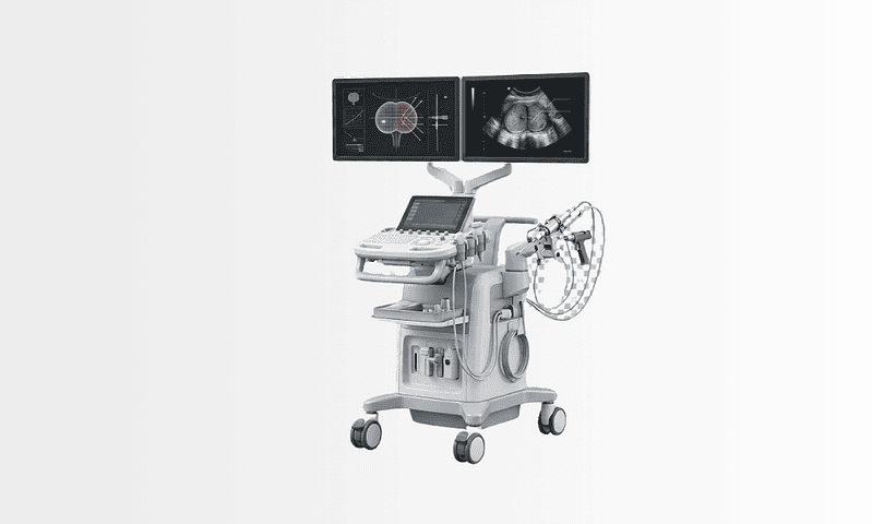
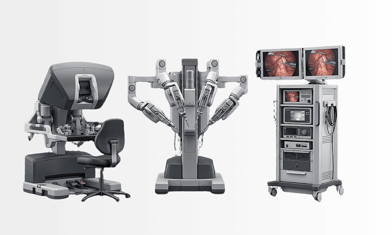
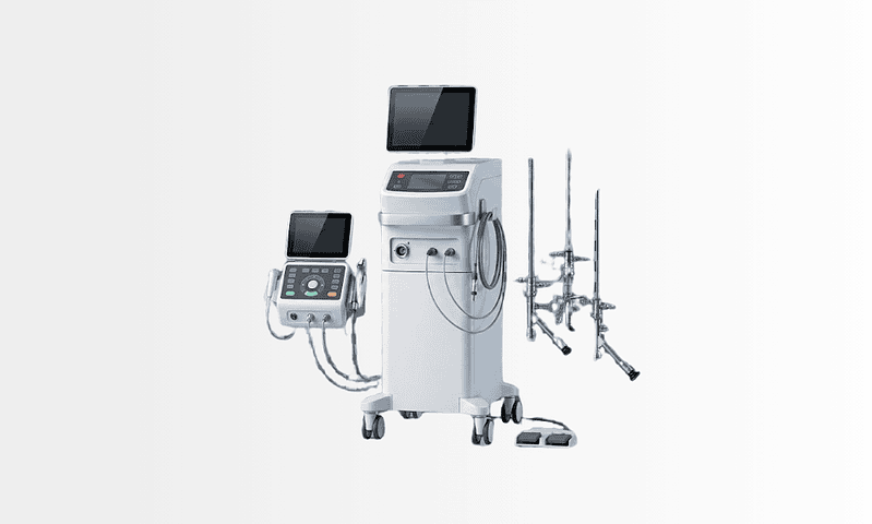
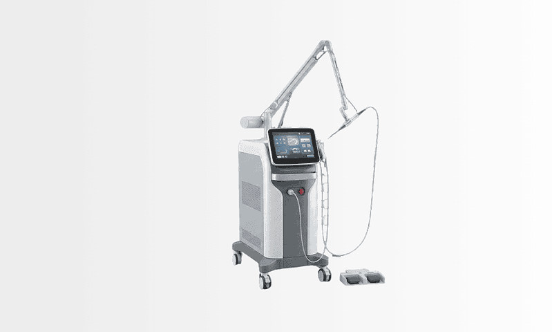
Available at these Hospitals






















What patients say about this unit
★★★★★From 2,400+ verified patient reviews“I was so anxious about my child's developmental neurology review, but the team put us at ease. Prof. Işık answered all our worried questions without making us feel silly. The nursing staff were attentive and kind throughout.”
“Our child's developmental neurology review was explained clearly and managed beautifully. Prof. Dr. Aydinli made our child feel comfortable straight away.”
“The urology department scheduled my tests back to back so the diagnosis took two days, not two months. Laser treatment followed the same week. Discreet, punctual and thorough, everything my previous experience was not.”
“The neurology clinic took my migraine history seriously, imaging to rule out the frightening causes, then a prevention plan that has halved my attacks. Medication choices were explained with alternatives, never dictated.”
Patient stories from this unit
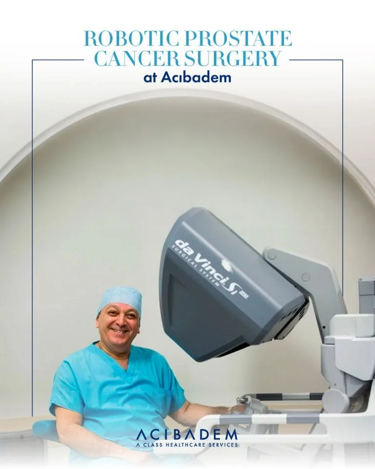 International Robotic Prostatectomy: A Prostate Cancer Recovery Story
 Diagnosed with prostate cancer, he found a minimally invasive path with robotic surgery at Acibadem, and is grateful for the care he received.
Read the story → United Kingdom Deep Brain Stimulation for Parkinson’s: Bani’s Journey from London
 A retired IT architect from London, Bani's tremor, stiffness and slowed movement made daily life a struggle, until deep brain stimulation at Acibadem…
Read the story →Comparison guides for this specialty
Speak with our medical team
Share your case and our international patient team will guide you to the right specialist, free of charge.