Bladder Stones Treatment
Bladder stones are hard mineral deposits that form in the bladder and may cause pain, frequent urination, blood in urine, or urinary blockage. Treatment usually removes stones endoscopically and addresses the underlying…

Quick answer
Bladder stones are hard mineral deposits that form when urine stays in the bladder too long, usually because the bladder does not empty fully. They can cause burning, frequent urination, blood in the urine and repeated infections. Most are removed endoscopically: a surgeon passes a thin scope through the urethra, fragments the stone with laser or ultrasonic energy and washes out the pieces.
Bladder Stones: What They Are and Why They Form
Bladder stones are hard mineral deposits that build up inside the urinary bladder, almost always because urine stays in the bladder longer than it should. When the bladder does not empty completely, minerals in the stagnant urine begin to crystallise; over weeks and months, those crystals can grow into stones that range from fine grit to deposits several centimetres across. Treatment means two things done together: removing the stone, usually through the natural urinary passage without an external incision, and identifying the problem that allowed the stone to form in the first place.
Bladder stones are often confused with kidney stones, and the confusion is understandable, both are urinary stones, and both can cause pain, blood in the urine and infection. They are, however, different problems. Kidney stones form in the kidneys and may travel down the ureter towards the bladder; bladder stones usually form exactly where they are found, because the bladder itself is not emptying properly. The distinction matters, because it changes both the treatment and the plan for preventing the next stone.
Most people first notice a change in urination: a burning sensation, a sudden need to go, urine that looks cloudy or bloody, or pain that becomes worse near the end of urination, when the bladder contracts around the stone. Others have repeated urinary tract infections, an interrupted stream, or the persistent feeling that the bladder never quite empties. Some stones cause nothing at all and are discovered on a scan performed for another reason entirely.
If you are weighing up treatment, your concern is usually twofold. You want the stone removed safely, and you want to understand why it formed, because bladder stones are frequently a signal of something else: prostate enlargement, urinary retention, a urethral narrowing, bladder dysfunction, long-term catheter use or persistent infection. Removing the stone deals with the episode. Finding and treating the cause is what makes the treatment complete.
At Acibadem, urology teams plan evaluation and treatment across the whole pathway of urinary stone disease, from diagnosis and minimally invasive stone removal to management of the underlying cause, so that treatment produces a plan, not just a procedure.
What causes bladder stones?
Bladder stones are caused by urine standing still. A healthy bladder empties almost completely several times a day, flushing out the dissolved minerals before they can settle. When emptying is incomplete, because the outlet is blocked, the bladder muscle is weak, or a catheter changes how urine drains, residual urine becomes concentrated, crystals form, and a stone begins to grow. The most common contributors are:
- Benign prostate enlargement: in men, an enlarged prostate can squeeze the urethra and leave urine behind after every void: the single most frequent background to bladder stones in adults.
- Neurogenic bladder: nerve-related conditions such as spinal injury, multiple sclerosis or diabetes-related nerve changes can stop the bladder contracting effectively.
- Urethral stricture: scar tissue narrowing the urethra slows the stream and traps urine behind the narrowing.
- Long-term catheter use: catheters alter drainage, encourage infection and can act as a surface on which minerals deposit.
- Foreign material in the bladder: rarely, sutures, mesh, stent fragments or other material left from earlier surgery become a nucleus around which a stone grows.
- Recurrent urinary infection: certain bacteria change the chemistry of urine in ways that promote so-called infection stones.
- Bladder diverticula: pouches in the bladder wall hold urine that never fully drains, giving crystals a quiet place to grow.
Stone composition varies. Uric acid stones are common among adult bladder stones, particularly when urine is persistently concentrated. Calcium-based stones occur as well, and struvite stones form in the presence of specific urease-producing bacteria. Composition is worth knowing: fragments retrieved during surgery can be analysed, and the result often shapes the prevention advice you receive afterwards.
Stones in urine bladder: what the phrase describes
Stones in urine bladder is a phrase many people type into a search engine, and it describes precisely what doctors call cystolithiasis: mineral stones sitting inside the urinary bladder rather than in the kidney or the ureter. The wording matters only because it points to the right condition, if your scan report mentions a stone in the bladder itself, this page describes your situation; if the stone is higher in the urinary tract, the treatment pathway is different.
What causes bladder stones in dogs?
Bladder stones in dogs form for much the same reasons as in people: concentrated urine, urinary infection and incomplete emptying, with struvite and calcium oxalate the most common types. Certain breeds are genetically prone, and diet plays a larger role in dogs than in humans. A dog with bladder stones needs a veterinary surgeon, not a hospital urologist, but the overlap is a useful reminder of how universal the mechanism is. Stagnant, concentrated urine grows stones, whatever the species, and prevention in both cases means keeping urine dilute and keeping it moving.
Bladder Stones vs Kidney Stones: How They Differ
Kidney stones and bladder stones share chemistry but not geography, and the difference shapes everything from the symptoms you feel to the operation you might need. A kidney stone forms in the collecting system of the kidney. It may sit there silently for years, or it may begin to travel, and a travelling kidney stone is what produces the notorious colicky pain that sends people to hospital. Once a kidney stone reaches the bladder, it usually passes out through the urethra without difficulty, because the urethra is considerably wider than the ureter it has just navigated. A stone that lingers and grows in the bladder is therefore usually telling you something different: not that a stone travelled, but that the bladder is not emptying as it should.
Stones that lodge in the narrow tube between kidney and bladder are a category of their ownureteral stonesand they behave differently again, often blocking urine drainage from the kidney and causing acute pain. If your imaging shows stones higher in the urinary tract, the treatment pathway is planned differently, with techniques and timing chosen for the kidney and ureter rather than the bladder.
What is the main cause of kidney stones?
The main cause of kidney stones is urine that is too concentrated for too long, most often the result of not drinking enough fluid, so that minerals such as calcium and oxalate reach levels at which they crystallise. Diet, family history, body weight, certain metabolic conditions and some medicines all contribute, and calcium oxalate is the most common stone type. This is a different mechanism from bladder stones, where the dominant problem is retention rather than concentration, which is why the prevention advice for the two conditions is not identical.
What are the symptoms of passing a kidney stone?
Passing a kidney stone typically causes severe pain that arrives in waves, starting in the flank or side below the ribs and radiating down towards the lower abdomen and groin as the stone moves. Nausea, vomiting, blood in the urine and restlessness, the inability to find a comfortable position, are characteristic. As the stone approaches the bladder, many people feel intense urinary urgency and frequency, sometimes mistaken for an infection. Bladder stones rarely produce this pattern; their pain is lower, more positional and tied to urination itself.
Kidney stone symptoms in women
Kidney stone symptoms in women are the same core symptoms, flank pain in waves, blood in the urine, nausea, urgency, but they are more often initially attributed to something else, such as a urinary tract infection, period pain or an ovarian problem, because the anatomy of the female pelvis offers several plausible explanations for lower abdominal pain. Women also develop urinary infections more readily, and an infection sitting alongside an obstructing stone is a combination doctors take seriously. Imaging settles the question, and imaging choices are adapted during pregnancy.
How long does it take to pass a kidney stone?
There is no fixed timetable for passing a kidney stone. Small stones may pass within days; others take several weeks; larger stones, or stones lodged at a narrow point of the ureter, may never pass at all and need to be removed. What determines the outcome is size, position, the anatomy of your urinary tract and whether the kidney is draining adequately in the meantime, which is why a urologist monitors a stone that is being given time to pass, rather than simply waiting indefinitely.
How to get rid of kidney stones fast?
There is no safe shortcut that dissolves a typical kidney stone at home. Drinking plenty of fluid helps small stones move and is the foundation of prevention, and certain uric acid stones can sometimes be shrunk with medical therapy prescribed and monitored by a doctor, but for most stones, the genuinely fast route is a procedure: shock wave lithotripsy or ureteroscopy, chosen by a urologist according to the stone’s size and position. Quick-fix remedies circulating online do not fragment stones, and delaying proper assessment while trying them can allow an obstructed kidney to come to harm.
Can kidney stones cause a bladder infection?
Yes. Any stone that slows or blocks the flow of urine gives bacteria time and territory to multiply, and the rough surface of a stone can shelter bacteria from both the urinary stream and antibiotics. This is true of kidney stones, ureteral stones and bladder stones alike, and it explains why some people cycle through repeated courses of treatment for a bladder infection that keeps returning: as long as the stone remains, the reservoir of bacteria remains with it. An infection combined with an obstructing stone is one of the situations urologists treat with particular urgency.
One final distinction worth making: bladder stones have nothing to do with gallstones. Gallstones form in the gallbladder, a digestive organ that stores bile, and they cause digestive symptoms managed by an entirely different specialty. The shared word “bladder” is the only connection: the organs, the chemistry and the treatments are all different.
Cystolithiasis Symptoms: How a Bladder Stone Announces Itself
Cystolithiasis symptoms, cystolithiasis being the medical term for stones in the urinary bladder, range from nothing at all to severely disruptive, and they overlap with several other urinary conditions, which is why proper evaluation matters more than self-diagnosis. Typical symptoms include:
- Frequent urination, especially small amounts at a time
- An urgent, sudden need to urinate
- Burning or pain during urination
- Lower abdominal or pelvic pain
- Pain at the tip of the penis in men
- An interrupted urine stream or difficulty starting urination
- A feeling that the bladder is not empty after urinating
- Blood in the urine, which may appear pink, red, brown or tea-coloured
- Cloudy or foul-smelling urine
- Recurrent urinary tract infections
- A complete inability to pass urine, acute urinary retention, which doctors treat as an emergency
Symptoms often come and go, because the stone moves within the bladder. Some people notice that pain changes with body position, or that the stream stops abruptly mid-void when the stone rolls across the bladder outlet, then restarts when they shift. Pain at the very end of urination is characteristic: the bladder, nearly empty, contracts directly around the stone.
A minority of bladder stones are silent, producing no symptoms and turning up incidentally on an ultrasound or CT scan done for something else. Silent does not necessarily mean harmless, a stone can still be scraping the bladder lining or harbouring bacteria, so even an incidental finding deserves a considered urological assessment.
How Bladder Stones Are Diagnosed
Diagnosis begins with a medical history and physical examination. The urologist will ask about urinary symptoms, previous stones, infections, prostate problems, catheter use, neurological conditions, medications and any prior pelvic or urinary surgery. For male patients, prostate assessment is usually part of the evaluation, because the prostate is so often part of the story.
Urine testing looks for blood, infection, crystals and other abnormalities. If infection is suspected, a urine culture identifies the bacteria and guides antibiotic selection before any procedure. Blood tests may assess kidney function, infection markers or metabolic factors where clinically relevant, particularly in people who have formed more than one stone.
Imaging confirms the presence, size and number of stones. Ultrasound shows stones within the bladder and, importantly, measures how much urine remains after voiding, which is the single most telling clue to why the stone formed. Plain X-rays detect many stone types. Computed tomography is used when the diagnosis is uncertain, when stones elsewhere in the urinary tract are suspected, or when the surgeon needs detailed anatomy for planning.
Cystoscopy may also be recommended. A narrow camera instrument passed through the urethra lets the urologist inspect the urethra, the prostate area and the bladder directly, confirming the stone, assessing inflammation, ruling out other bladder diseases that can mimic stone symptoms, and detecting structural causes such as a narrowed urethra or obstruction at the bladder outlet.
Where bladder function itself is in question, in neurological conditions, after pelvic surgery, or when the bladder retains urine without an obvious blockage, urinary flow tests and urodynamic studies show how the bladder stores and empties urine. These tests do not treat anything, but they frequently change what the treatment plan should be.
Who May Need Treatment for Bladder Stones
Bladder stones occur in adults and children, though the causes differ. In adults they are most often tied to incomplete bladder emptying; in men, benign prostate enlargement is the common thread. In both men and women, stones accompany neurogenic bladder, bladder outlet obstruction, previous urinary surgery, foreign material in the bladder, recurrent infection or long-term catheter use.
Small stones may pass on their own if nothing obstructs the outlet. Symptomatic stones, larger stones and stones linked to infection usually need active treatment. The common indications are:
- Symptomatic bladder stones: stones causing pain, urgency, frequency, burning, blood in the urine or difficulty urinating.
- Recurrent urinary tract infections: stones harbour bacteria and make infections hard to clear completely.
- Urinary retention: a stone can worsen obstruction or become trapped near the bladder outlet, preventing urination altogether.
- Bladder outlet obstruction: prostate enlargement, urethral stricture or other narrowing causes urine to stagnate and stones to form.
- Neurogenic bladder: nerve-related dysfunction leads to incomplete emptying and, without a strategy, recurrent stones.
- Long-term catheter use: catheters raise the risk of infection and mineral deposits within the bladder.
- Foreign material in the bladder: sutures, mesh, stents or other material acting as a nucleus for stone growth.
- Large or multiple stones: unlikely to pass naturally, and liable to cause progressive symptoms or injury to the bladder lining.
In children, bladder stones may relate to urinary tract abnormalities, infection, dehydration, nutritional factors or metabolic conditions. Paediatric evaluation needs age-appropriate imaging and planning by teams experienced in paediatric urology: the causes and the instruments both differ from adult practice.
In adults, the treatment decision usually turns on whether there is a correctable cause. A man with bladder stones and significant prostate enlargement is better served by a plan that considers both conditions than by repeated stone removal alone, otherwise the same stagnant urine simply grows the next stone.
How a Bladder Stone Is Removed
A bladder stone is most often removed by cystolitholapaxy: the urologist passes a cystoscope, a narrow instrument carrying a camera and working channels, through the urethra into the bladder, sees the stone directly, fragments it with specialised tools and removes the pieces. No external incision is needed for the majority of cases, which is why recovery is usually measured in days rather than weeks.
Different energy sources can do the fragmenting. Laser energy, delivered through a fine fibre, breaks stones precisely into small pieces. Ultrasonic or mechanical instruments suit larger stones or particular compositions. The choice is individual, made from imaging, cystoscopy findings and the surgeon’s assessment of stone hardness and your anatomy.
In some patients, treatment also corrects the underlying problem in the same plan. If an enlarged prostate is preventing the bladder from emptying, prostate treatment may be discussed for the same session or as a staged plan. A urethral stricture may need treatment to restore flow. If the bladder muscle itself is not contracting effectively, the plan may include bladder function testing, a catheter strategy, medication prescribed by the treating doctor, or follow-up in functional urology.
Antibiotics, pain control and supportive care may be part of treatment, especially where infection is present. What medication cannot do is dissolve an established bladder stone: for most symptomatic patients, the definitive step is removal of the stone combined with treatment of the condition that allowed it to form.
Preparation before treatment
Before the procedure, the urology team reviews your history, medications, allergies, prior surgery and imaging. If you are travelling for treatment, previous reports, scans, urine results and any record of urinary infections are all worth bringing: they shorten the diagnostic phase and sharpen the plan. Updated tests are often repeated after arrival to confirm the current picture.
Urine testing carries particular weight here. If bacteria are found, antibiotics may be started before surgery to lower infection risk. If you take blood thinners, antiplatelet medicines or certain supplements, tell the team early: whether and how anything is adjusted before surgery is a decision for the treating doctor, weighed against your heart, vascular and clotting history, never a decision to make on your own.
The anaesthesia team then assesses your general health and discusses the safest option. Many bladder stone procedures are performed under general or spinal anaesthesia; the choice depends on your health, the complexity of the procedure and clinical preference. You will receive clear instructions about fasting, medication timing and arrival at the hospital.
What happens during the procedure
The endoscopic operation follows a consistent sequence:
- Anaesthesia is given and confirmed effective.
- The surgeon gently passes the cystoscope through the urethra into the bladder.
- Sterile fluid fills the bladder enough to allow clear visualisation.
- The stone is identified and assessed, size, shape, number and mobility.
- Laser, ultrasonic or mechanical energy fragments the stone into small pieces.
- Fragments are removed through the scope or flushed out with irrigation, and the surgeon inspects the bladder to confirm significant fragments are cleared and to look for contributing abnormalities.
A temporary urinary catheter may be placed afterwards. It drains urine, rests the bladder and clears small residual debris or blood-tinged urine. In straightforward cases it comes out soon after; the timing depends on the procedure and on the reason the stone formed.
If the stone is very large, if the urethra is too narrow for safe endoscopic access, or if reconstructive work is needed, an alternative approach may be recommended, removal through a small incision into the bladder, or via a minimally invasive suprapubic route. Open surgery is now uncommon for bladder stones, but it remains the right answer for certain complex cases, and it is better chosen deliberately than reached for after a difficult endoscopy.
Technology used in modern bladder stone care
Modern bladder stone treatment rests on accurate imaging, clear endoscopic visualisation and controlled fragmentation. Ultrasound and computed tomography define stone size, number and location before anyone operates. Flexible and rigid cystoscopes give direct sight of the bladder and urethra, and high-resolution camera systems let the surgeon work precisely inside a small space.
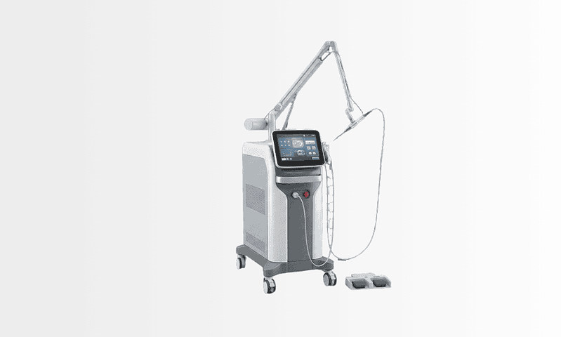
Energy-based fragmentation, laser and ultrasonic systems, is what allows most stones to be treated without external incisions, breaking them into removable pieces while limiting trauma to surrounding tissue when used appropriately. Irrigation systems maintain visibility and clear debris. For patients with suspected obstruction or bladder dysfunction, flow tests, post-void residual measurement and urodynamic evaluation complete the picture of how the bladder stores and empties urine. The technology matters, but it serves clinical judgement rather than replacing it: the same instruments in different hands produce different plans, and the plan is what you are really choosing.
How long does treatment take?
Procedure time varies with stone size, number, hardness and whether another condition is treated in the same session. A straightforward endoscopic bladder stone procedure is relatively short; large or multiple stones take longer to fragment and clear. If prostate surgery or treatment of a urethral narrowing is combined, both the operation and the recovery plan extend accordingly.
Many patients go home the same day or after a short hospital stay. If you have travelled for treatment, you may be advised to remain nearby for a few days afterwards, particularly if you had a catheter, an infection, a larger stone burden or an additional procedure. The medical team gives individual travel guidance based on your recovery, urine clarity, pain control and follow-up needs, so that the journey home is planned rather than improvised.
Recovery after bladder stone removal
After removal, mild burning during urination, increased frequency, urgency and blood-tinged urine are common for a short period. These settle as the bladder lining heals. Drinking adequate fluids, unless another medical condition restricts them, helps flush the bladder and eases the irritation.
Pain is usually manageable with prescribed medication, and antibiotics may continue if infection was present or the physician judges them necessary. Strenuous activity, heavy lifting and sexual activity may be restricted briefly, especially after catheter use or a combined procedure. Before discharge, the team explains which signs, fever, chills, worsening pain, heavy bleeding, clots blocking the stream, or inability to urinate, warrant early medical review, and how to arrange that review wherever you will be recovering.
Follow-up matters as much as the operation. The urologist reviews stone analysis, urine culture results and, where needed, imaging. Prevention may then involve treating prostate enlargement, improving bladder emptying, adjusting catheter care, managing infection risk, increasing hydration or evaluating metabolic factors in people with recurrent stones.
Why Acting Early Matters
Bladder stones often begin as an irritation, but they progress if the cause goes untreated. A stone grows over time, making eventual treatment more complex. It scrapes the bladder lining, causing bleeding and inflammation. It can interfere with emptying, deepening urinary retention and raising infection risk: a slow spiral rather than a stable condition.
Recurrent urinary tract infections are a common reason to treat promptly. Stones give bacteria a surface on which to persist, so infections return even after antibiotics. Repeated infections erode quality of life and, in some patients, can spread towards the kidneys or the bloodstream. People with diabetes, immune suppression, existing kidney disease or urinary catheters carry a higher risk of complications and have the most to gain from early, definitive treatment.
Delay also lets the underlying cause worsen. Untreated bladder outlet obstruction from prostate enlargement gradually changes the bladder muscle itself. Over time the bladder empties less efficiently, retention builds, and pressure can transmit to the upper urinary tract, in severe cases, kidney function suffers.
Early evaluation does not mean immediate surgery. It means understanding the stone, the bladder and the reason the stone formed, and with that understanding, the urologist can recommend the right timing and the least invasive effective treatment for your situation. Watchful waiting is a legitimate plan; drifting is not.
Benefits of Bladder Stone Treatment
When the stone is removed and the underlying cause is addressed, most of what treatment offers falls into a handful of concrete improvements:
| Benefit | What It Means for You |
|---|---|
| Relief from urinary pain and irritation | Removing the stone can reduce burning, pelvic discomfort, bladder spasms and the pain that occurs during or after urination. |
| Improved urine flow | If the stone contributes to blockage or an interrupted stream, removal may help restore more normal emptying, especially when obstruction is treated at the same time. |
| Lower risk of recurrent infection | Stones harbour bacteria. Removing them can make urinary infections easier to clear and reduce repeated episodes when combined with appropriate prevention. |
| Less bleeding and bladder inflammation | Stone movement injures the bladder lining. Treatment can stop ongoing irritation and stone-related blood in the urine. |
| An opportunity to prevent recurrence | Stone analysis, bladder evaluation and treatment of retention or obstruction shape a prevention plan tailored to the actual cause. |
| Minimally invasive options for many patients | Most bladder stones can be treated through the urethra without an external incision, often allowing a shorter recovery than open surgery. |
Recovery Timeline After Bladder Stone Treatment
Recovery varies with stone size, infection status, anaesthesia type and whether another urinary condition was treated at the same session, but many patients follow a broadly similar pattern:
| Time Period | What Patients Can Expect |
|---|---|
| Day 1 | Mild burning, urgency, blood-tinged urine or a temporary catheter are common. The team monitors urination, pain control and signs of infection before discharge. |
| First Week | Urinary irritation improves gradually. Patients are usually advised to drink fluids, avoid heavy lifting and take medications exactly as prescribed. |
| First Month | Most routine activities can usually resume, depending on the procedure. Follow-up may include urine testing, review of stone analysis and assessment of bladder emptying. |
| Longer Term | The focus shifts to preventing recurrence, treating prostate enlargement, infection, catheter-related issues, bladder dysfunction or other contributing factors. |
What Shapes a Good Result
The outcome of bladder stone treatment depends on more than removing the stone. A good result is built from accurate diagnosis, complete stone clearance, infection control and management of whatever caused urine to stagnate. Several factors carry particular weight.
Stone size and number. Small, single stones are generally straightforward to treat endoscopically. Large or multiple stones need longer fragmentation, more careful irrigation and, occasionally, a different surgical approach altogether.
Stone composition. Some stones are harder to fragment; some are bound up with infection. Analysing the retrieved fragments guides prevention, especially in people with a history of recurrent urinary stones.
Bladder outlet obstruction. This is the decisive factor in many men with prostate enlargement. If the obstruction remains, the bladder keeps retaining urine and stones tend to recur, so planning may include medication for prostate symptoms, endoscopic prostate surgery or staged management, depending on overall condition.
Bladder function. Some bladders empty poorly because of nerve-related conditions, spinal injury, diabetes-related nerve changes, prior pelvic surgery or long-standing obstruction. Stone removal relieves symptoms in these patients, but preventing recurrence may require an emptying strategy, intermittent catheterisation, medication or functional urology follow-up.
Infection status. Active infection must be recognised and treated with appropriate antibiotics before definitive surgery; occasionally, drainage or stabilisation comes first. Handling infection carefully reduces the risk of post-operative fever and other complications.
Urethral anatomy. A stricture, prior surgery or anatomical variation can complicate standard endoscopic access. The urologist may treat the narrowing, choose a different instrument or pick an alternative route to protect the urethra.
Overall health. Heart disease, lung disease, diabetes, kidney impairment, bleeding risk and immune suppression are all reviewed before treatment, because they shape anaesthesia planning and recovery. For patients travelling for care, this pre-operative assessment is what keeps the plan realistic for the journey home.
Follow-up adherence. Even after a technically successful procedure, monitoring for recurrent symptoms, infection, incomplete emptying and metabolic contributors is what protects the result. A clear written plan is especially valuable if your follow-up will continue with a physician in your home country.
Bladder Stone Care at Acibadem
Patients who travel for urological care need more than a procedure appointment. They need an accurate diagnosis, a plan that considers the whole urinary system, plain communication and coordinated support before and after treatment. Acibadem’s approach to bladder stone care is organised around that broader pathway rather than around the operation alone.
Urology teams evaluate bladder stones through modern diagnostic pathways, urine testing, imaging, cystoscopy where indicated, and assessment of bladder emptying. The focus is not only on confirming the stone but on understanding why it developed, which matters most for men with prostate enlargement, patients with recurrent infections, people using long-term catheters and those with neurological conditions affecting the bladder.
Treatment plans are individual. Some patients need a straightforward endoscopic cystolitholapaxy; others need combined or staged treatment for prostate obstruction, a urethral stricture, bladder dysfunction or infection. Complex cases draw on collaboration between urologists, anaesthesiologists, radiologists, infectious disease specialists and internal medicine physicians, so the procedure fits the patient’s overall health and travel constraints rather than the other way round.
Advanced endoscopic and imaging technology supports minimally invasive care wherever appropriate, high-quality visualisation, controlled stone-fragmentation systems and contemporary operating room infrastructure. Technology serves clinical judgement, not the reverse: technique is chosen for stone characteristics, anatomy and safety. For international patients, dedicated multilingual services cover medical record coordination, interpretation, appointment planning and admission support, because urology in particular depends on patients genuinely understanding their tests, catheter instructions, prescribed medications and discharge plan.
Experienced teams also recognise that many people want a second opinion before deciding where to be treated. A good second opinion clarifies whether the stone truly needs removal, which approach suits the anatomy, whether another condition should be treated in the same session, and what recovery realistically involves, and it is most useful for people who have been told they need open surgery, who have recurrent stones, or who carry several medical conditions at once.
Preventing the Next Bladder Stone
Bladder stones are treatable, and the symptoms they cause should not be ignored, but the more important point is that they are usually preventable once the cause is understood. Prevention starts with the analysis of the removed stone and the answer to one question: why did urine stand still long enough for this to grow?
If the answer is prostate enlargement, treating the prostate protects the bladder. If it is a stricture, restoring flow does the same. If the bladder muscle itself is weak, a reliable emptying strategy, sometimes intermittent catheterisation, sometimes medication, sometimes scheduled voiding, keeps residual urine low. If catheters are unavoidable, careful catheter care and infection surveillance reduce mineral deposition. And across all causes, adequate hydration keeps urine dilute and less inclined to crystallise, unless another medical condition limits fluid intake.
Timely care relieves discomfort, lowers infection risk and helps protect urinary function over the long term. With careful diagnosis, an appropriate technique and genuine attention to prevention, most people return to daily life with better comfort and, just as valuable, a clear understanding of how to keep their urinary system stone-free.
Preparation
- Evaluation usually includes urine tests, imaging, and cystoscopy when needed to confirm stone size, number, and cause. Blood thinners or certain medications may need adjustment before the procedure. Patients are typically asked to fast for several hours before anesthesia.
Aftercare
- Mild burning during urination, blood-tinged urine, or bladder irritation can occur briefly after treatment. Patients are advised to drink fluids, take prescribed medications, and avoid strenuous activity for a short period. Follow-up helps check healing and manage causes such as prostate enlargement or urinary retention.
Frequently Asked Questions
What affects the cost of bladder stone treatment?
The main factors are stone burden, the removal method, anaesthesia, hospital stay, technology used, preoperative tests, and whether an underlying urinary problem also needs treatment. A personalised quote is prepared after medical records, imaging, and symptoms are reviewed.
How can I get a personalised quote from Acibadem?
You can request a free consultation and share recent imaging, urine test results, previous operation notes, medication details, and a summary of symptoms. The international patient team can help coordinate review by a urology specialist and explain what is included in the proposed package.
Does a package usually include everything I need?
Packages may include consultation, diagnostic review, hospital services, anaesthesia, the procedure, standard medication during admission, and follow-up planning. Items such as additional tests, treatment of unexpected infection, extended stay, or separate procedures may affect the final cost.
Will I need treatment for the cause of the bladder stones?
Possibly. Bladder stones often relate to incomplete emptying, obstruction, infection, catheter use, or bladder function problems. Treating the cause may be recommended to reduce the risk of recurrence, and this can change the treatment plan and cost.
Is bladder stone treatment usually done endoscopically?
Many bladder stones can be treated endoscopically through the urinary tract, but larger or complex stones may require a different approach. The urologist decides suitability based on imaging, stone features, anatomy, infection status, and general health.
Medically reviewed by the Acıbadem International Medical Board September 9, 2026
See our medical review board →
Update history
- PublishedJune 8, 2026
- Medical review approvedSeptember 9, 2026
- Last content updateSeptember 9, 2026
References2
- Bladder stones nhs.uk
- Bladder stones medlineplus.gov
Trusted care for international patients
From the Acibadem Blog
Why Do Bladder Stones Form? Retained Urine, Enlarged Prostate and What Treatment Fixes
Bladder stones usually form when the bladder does not empty fully, so leftover urine sits, concentrates and lets minerals crystallize. In adults the most…