

Gynecology & Obstetrics
Ovarian cysts, fibroids, endometriosis and abnormal bleeding investigated properly, plus gynecologic cancer surgery, urogynecology and high-risk pregnancy care, with free remote review of the scan report you already have.

From a scan report you cannot decode to the operation you may not need
Most women who reach us from abroad arrive with a report, not a diagnosis. The first job is to read it properly, and a great many of the findings that frighten people turn out to need watching rather than surgery.
Cysts, fibroids and bleeding
The benign conditions behind most abnormal scans and most heavy periods, investigated before anything is removed.
Cancer, HPV and surgery
Screening abnormalities and gynecologic cancers, staged and planned by a tumour board rather than a single surgeon.
Pelvic floor and pregnancy
Prolapse and leakage that women live with for years, plus pregnancy follow-up and the conditions that make it high-risk.
The images are read before an operation is offered
A written report is a summary of what one person saw on one day. The images are the evidence, and a second look at them changes the plan more often than people expect: a cyst that was going to be removed turns out to be a normal follicle, or a "fibroid" turns out to be adenomyosis, which is treated entirely differently.
So the sequence is fixed: read the images, time the tests to the right day of the cycle, and only then discuss what surgery would achieve, including the honest answer that for many findings it achieves nothing at all.
What we will not do
- Operate on a simple cyst that ultrasound says will resolve on its own.
- Remove an ovary where the cyst wall can be peeled and healthy tissue kept.
- Diagnose PCOS from a single blood test taken on the wrong day of the cycle.
- Promise that surgery for endometriosis or fibroids will restore fertility.
- Present hysterectomy as the automatic answer to heavy bleeding.
Who would actually read your scan
Gynecology is subspecialised: the surgeon who operates on an ovarian cancer is rarely the one who manages a high-risk pregnancy. Every profile is readable before you decide.
How this works when you are travelling for it
Send the report and the images
The written report is a summary; the images are the evidence. Send both, with your symptoms, cycle pattern and any previous surgery.
Consultant review
What can be settled from the records you already have, and what genuinely needs a scan repeated with our own probe.
Imaging and tests on arrival
Transvaginal ultrasound, MRI where ultrasound cannot settle the question, hormone tests timed to the right day of the cycle.
Surgery and the night after
Most benign laparoscopic and hysteroscopic operations are day-case or one night; cancer surgery and open procedures stay longer.
Follow-up before and after you fly
Pathology explained before departure where it is ready, then review remotely, with flying and recovery advice specific to your operation.
Six things worth knowing first
Sudden one-sided pain is an emergency
Severe pain with nausea or vomiting can mean ovarian torsion, where the ovary twists on its own blood supply. It is saved in hours rather than days, which is why torsion is operated on as an emergency.
Bleeding after menopause
It is usually not cancer. It is still investigated every single time, because it is the earliest and most treatable sign of endometrial disease.
There is no dangerous cyst size
The centimetre figures in a report are follow-up conventions, not danger thresholds. What the cyst contains and your symptoms matter more.
A dermoid is not a reason to panic
A cyst reported to contain fat, hair or a tooth is a mature cystic teratoma, one of the commonest ovarian growths, and almost always benign.
Screening is not diagnosis
An abnormal smear or a high-risk NIPT result is a signal to look further, not an answer. Diagnosis comes from colposcopy, biopsy or a diagnostic test.
Fertility-sparing surgery is conditional
It depends on tumour type, grade, size and imaging, it demands intensive surveillance, and completion surgery may still become necessary.
Quick answer
Gynecology and Obstetrics is the medical unit that cares for women’s reproductive health, pregnancy, and childbirth, covering everything from routine checkups to diagnosis and treatment of gynecologic conditions. At Acibadem in Turkey, this care is provided through coordinated outpatient and hospital-based services, including preventive screening, prenatal follow-up, minimally invasive procedures, delivery planning, and postpartum care.
What our gynecology and obstetrics unit covers, and who it is for
Gynecology and obstetrics is one specialty with two halves that rarely feel connected to the person living through them. One half looks after the reproductive organs, the uterus, ovaries, cervix and pelvic floor, from the first period to long after the last. The other looks after pregnancy and birth. Most women meet the unit somewhere in the middle: a scan report they cannot decode, bleeding that has changed, pain that has been dismissed, an abnormal smear result, or a pregnancy that needs closer watching than the last one.
At Acıbadem International the work is organised into six strands, and knowing which one your problem belongs to is the fastest way to get a useful answer.
- General gynecology and menstrual or hormonal disordersheavy, absent, irregular or unexpected bleeding, polycystic ovary syndrome, endometriosis and adenomyosis, fibroids, cysts, pelvic pain and the investigations that separate them.
- Gynecologic imaging and diagnosishigh-resolution pelvic and transvaginal ultrasound, MRI where ultrasound cannot settle the question, hysteroscopy and endometrial sampling. This is where most international cases begin, because a diagnosis made elsewhere is often the thing being questioned.
- Gynecologic oncologycancers and pre-cancers of the cervix, uterine lining and ovaries, staged and planned with pathology, radiology and the cancer team rather than by a single surgeon.
- Urogynecology and pelvic floorprolapse, urinary leakage and the pelvic floor problems that often follow childbirth or menopause, and that women commonly live with for years before asking.
- Menopause and midlife healthperimenopausal cycle change, symptom control, bone and cardiovascular risk, and the individual risk-benefit conversation about hormone therapy.
- Obstetrics and perinatologyroutine pregnancy follow-up, the scan timeline, bleeding in pregnancy, preeclampsia, fetal movement, high-risk pregnancy, and birth.
Fertility treatment itself, ovulation induction, insemination, IVF and embryo transfer, is a separate unit with its own laboratory and team, and you can read about it under IVF and reproductive health. This page stops at the point where a gynecological problem starts to affect fertility: an endometrioma, a uterine cavity that needs assessing, an ovulation disorder that needs a diagnosis. Those are gynecology’s work, and they are often the step that has to happen before any fertility treatment is worth starting.
Screening is the one part of gynecology that applies to women with no symptoms at all. Cervical screening, a smear test, an HPV test, or both together, usually begins in the early to mid-twenties depending on which test your programme uses, and repeats at intervals that depend on which test is used, your previous results and the programme you follow at home. There is no equivalent screening test for ovarian cancer that works in women at average risk, which is why symptoms and imaging carry so much weight in the sections below.
Who is this unit for? Women who want a diagnosis explained rather than repeated; women told they need surgery who want the reasoning examined before agreeing; women with a complex or previously operated pelvis; and women who are pregnant and want closer follow-up than they are getting. If your question is simply “is what my report says serious?”, the sections that follow were written for you.
Reading your pelvic ultrasound report: anteverted, retroverted and every other word
A pelvic ultrasound report is a description, not a diagnosis. It records what the sound waves showed at one moment, in the vocabulary radiologists use with each other. Read cold, it can sound alarming when it is entirely normal, and occasionally it sounds bland when the important line is buried at the bottom. Here is what those words actually mean.
How the scan was done matters. A transabdominal scan is performed over the lower abdomen with a full bladder, which pushes the bowel out of the way. A transvaginal scan places the probe much closer to the uterus and ovaries and gives far better detail of the endometrium, the ovaries and any cyst. Neither is a “worse” test: they answer different questions, and in many women both are used in the same appointment. If a report says the ovaries were “not well visualised”, that usually reflects bowel gas, body habitus or scan route, not a problem with your ovaries.
Anteverted uterus: what it means on your report
Uterine position: anteverted, anteflexed, retroverted, axial. These words describe which way your uterus is tilted, and nothing more. An anteverted uterus tips forward over the bladder: this is the commonest arrangement and is simply normal anatomy. Anteflexed adds that the body of the uterus is bent forward on the cervix, again a normal variation.
Retroverted uterus, and whether it affects pregnancy
A retroverted (or “tilted”, or “tipped”) uterus leans backwards toward the rectum, and retroflexed means it is bent backwards on itself. Axial or “mid-position” sits between the two. None of these positions is a disease, none needs correcting, and none of them appears on a scan because something has gone wrong.
Two qualifiers do carry meaning. If a report describes the uterus as mobile, it moved freely under the probe, which is reassuring. If it describes it as fixed or retroverted and immobile, or mentions that the ovaries are stuck to the uterus, that suggests adhesions, often from endometriosis, previous surgery or infection, and that finding is worth discussing, because it can explain pain during sex or deep pelvic pain.
Does a retroverted uterus affect pregnancy? On its own, no. A backwards-tilted uterus is not a cause of infertility, and as pregnancy advances the uterus almost always lifts forward out of the pelvis of its own accord. What can matter is the reason behind it: if the tilt is fixed by endometriosis or scarring, it is that underlying condition, not the tilt, that is investigated.
Size and shape. A uterus in the reproductive years is roughly pear-shaped and, in most reports, measured at around seven to nine centimetres in length, with the width and depth given alongside. Those figures move with age, with how many pregnancies you have had, and with the presence of fibroids or adenomyosis; after menopause the uterus becomes noticeably smaller. A single measurement outside a textbook range is not a diagnosis. A uterus described as globular or bulky with asymmetric walls is a different matter: that pattern points toward adenomyosis.
Endometrial thickness. The endometrium is the lining that builds up and sheds each month, so its thickness can only be read against where you are in your cycle. It is thinnest just after a period and considerably thicker in the days before the next one; the same number can be perfectly normal on day 22 and worth explaining on day 5. After menopause the lining should be thin and stay thin. A report saying thickened endometrium is therefore a finding that needs context, cycle day, menopausal status, whether you are bleeding, and whether you take hormones, and not, by itself, a diagnosis of anything.
Myometrium and its echogenicity. The myometrium is the muscular wall. Reports call it homogeneous when the texture is even and heterogeneous when it is not. A heterogeneous myometrium most often reflects adenomyosis or fibroids, sometimes both; radiologists may add specific clues such as small cysts within the muscle, indistinct borders between muscle and lining, or shadowing. Fibroids themselves are usually described by where they sit, pushing into the cavity, within the wall, or bulging outward, and that location predicts symptoms far better than size does.
Ovaries and free fluid. A normal ovary contains several small fluid-filled follicles, and a dominant follicle or a corpus luteum around ovulation is physiology, not pathology. A report that mentions multiple small follicles or “polycystic-appearing ovaries” is describing an appearance, not making a diagnosis of polycystic ovary syndrome, which requires clinical and hormonal criteria as well; the metabolic half of PCOS, insulin resistance, weight and diabetes risk, is co-managed with the endocrinology unit. A small amount of free fluid in the pelvis is common, especially just after ovulation.
When you go through your report with a doctor, the question that gets the most out of the appointment is simple: which of these findings, if any, explains my symptoms, and which are just descriptions of a normal body?
Ovarian cysts: types, symptoms and which sizes actually matter
Ovarian cysts are common, and most are a by-product of ovulation rather than a disease. Understanding which type you have changes everything about what happens next, because the same word covers structures that resolve within weeks and structures that need an operating theatre.
- Follicular cysts form when a follicle grows but does not release its egg. They are thin-walled, fluid-filled, and typically disappear over one or two cycles without treatment.
- Corpus luteum cysts develop after ovulation, when the structure left behind fills with fluid or blood. They can cause one-sided ache in the second half of the cycle and also resolve on their own; in early pregnancy they are an expected finding.
- Haemorrhagic cysts are functional cysts that have bled internally. They can hurt sharply at onset and then settle over several weeks as the blood is reabsorbed.
- Endometriomas are cysts of endometriosis filled with old blood, sometimes called chocolate cysts. They do not resolve spontaneously and are discussed with endometriosis below.
- Dermoid cystsmature cystic teratomas, arise from germ cells and can contain fat, hair and even teeth. They grow slowly, do not disappear, and carry a real torsion risk.
- Cystadenomas grow from the surface cells of the ovary and can reach a considerable size. They are benign but do not go away by themselves.
- Paraovarian and paratubal cysts sit beside the ovary rather than in it, and are usually left alone unless they are large or symptomatic.
What they feel like. Most cysts cause nothing at all and are found during a scan done for another reason. When symptoms do appear, they are usually a dull ache or heaviness on one side, bloating or a sense of pressure, pain during sex, pressure on the bladder causing frequent urination, or pain that comes and goes around ovulation. Pain that is strictly one-sided and matches the side of the cyst is more likely to be caused by it; diffuse central pelvic pain often has another explanation. Sudden severe pain is a different problem altogether (see torsion and rupture).
Which sizes actually matter. There is no single dangerous number, and any source that gives you one is oversimplifying. Three things are weighed together: what is inside the cyst (simple fluid versus solid or vascular components), your menopausal status, and whether it is causing symptoms. A large simple cyst in a woman of thirty may need nothing but a repeat scan; a smaller cyst with solid vascular areas in a woman of sixty-five is taken far more seriously. What size does reliably change is mechanical risk: the larger a cyst grows, the more likely the ovary is to twist on it, and the more likely it is to cause pressure symptoms.
How they are managed. Watchful waiting with a repeat ultrasound is the correct treatment for most functional cysts, and doing nothing is an active decision rather than neglect. Combined hormonal contraception does not shrink a cyst that already exists, but by suppressing ovulation it can reduce how often new functional cysts form: a reasonable option for someone who gets them repeatedly, decided with your doctor and against your own medical history.
Surgery is considered when a cyst persists or grows over successive scans, when it is large enough to cause symptoms or torsion risk, when its features are complex or suspicious, or when it is a type that will not resolve: a dermoid, an endometrioma, a cystadenoma. The default operation is a laparoscopic cystectomy that removes the cyst and leaves the ovary in place, which matters for hormone production and for future fertility. Removing the whole ovary is reserved for situations where the ovary cannot be saved or where the concern is malignancy. Cyst surgery is real surgery: it carries the risks of bleeding, infection, injury to nearby organs and adhesion formation, and removing a cyst wall, particularly a large endometrioma, or a second cyst from the same ovary, can reduce the ovarian reserve that remains. If you may want to become pregnant later, say so before the operation is planned, not after; it changes the surgical approach.
Cysts also recur. A functional cyst removed today does not stop your ovaries from making another one, and endometriomas in particular return in a proportion of women. That is not a sign that the surgery failed: it is the natural history of the condition, and it is part of why an operation is never the automatic answer to a cyst on a scan.
What ovarian cysts look like on ultrasound: simple, anechoic, septated, complex
Ultrasound is the first and usually the best test for an ovarian cyst, and the words used to describe one follow a logic you can learn in five minutes. The whole exercise has one purpose: to sort cysts into those that can be ignored, those that need watching, and the small number that need surgery or a specialist opinion.
A cyst is usually assessed with a transvaginal probe, which sits closest to the ovary and resolves its contents best; a transabdominal view is added when a mass is too large to fit in the transvaginal field, and the two are often combined in one appointment (see how to read the report).
Anechoic. Sound waves pass straight through clear fluid without bouncing back, so pure fluid appears completely black on the screen. That is what anechoic means, no internal echoes. An anechoic cyst with a thin smooth wall, no solid parts and good transmission of sound behind it is the classic simple cyst, and in a woman of reproductive age it is almost always a normal follicle or a functional cyst that will disappear on its own.
Simple versus complex. A cyst stops being simple the moment the report describes anything inside it. The features radiologists look for, roughly in order of significance, are: septations (internal walls dividing the cyst into compartments), how thick those septations are, solid components or papillary projections growing from the wall, irregular or thickened walls, and vascularity on Dopplerblood flow within a solid part. A septated cyst with one thin wall and no blood flow behaves very differently from a cyst with thick irregular septa and a vascular solid nodule, even though both are called “complex”.
Some complex appearances are so characteristic that the ultrasound alone strongly suggests the diagnosis. A haemorrhagic cysta functional cyst that has bled into itself, shows a fine lacy or cobweb pattern of fibrin strands with no blood flow, and typically resolves over a few weeks. An endometrioma shows uniform low-level internal echoes often described as ground glass. A dermoid cyst produces bright echogenic areas with shadowing from fat, hair and calcification. Each of these has its own section further down this page.
How the risk is scored. Radiologists no longer leave this to impression. Two structured systems are in wide use: O-RADS (the Ovarian-Adnexal Reporting and Data System), which assigns a risk category from clearly benign upward, and the IOTA rules and models, which classify masses from standardised ultrasound features. If your report carries an O-RADS number, it is telling you which management pathway the radiologist thinks applies, reassurance, interval follow-up, MRI, or referral to a gynecologic oncologist, and it is a probability statement, not a verdict. No scoring system can prove a cyst is benign from images alone; only pathology after removal can do that.
Does size decide anything? Partly. The figures that follow are follow-up conventions used by radiologists, not danger thresholds, and they differ between guidelines: what a cyst contains, your menopausal status and your symptoms outweigh the measurement. In a woman who has not reached menopause, small simple cysts, up to around five centimetres, are considered normal ovarian physiology and often need no repeat scan at all. Between roughly five and seven centimetres, most centres repeat the ultrasound to confirm the cyst is shrinking or stable. Above about seven centimetres, ultrasound may not capture the whole cyst reliably, MRI is frequently added, and the mechanical risk of torsion rises. After menopause the thresholds tighten, because a new cyst in an ovary that should be quiet deserves more attention, although even then, small simple cysts are common and usually benign.
Timing the follow-up scan. Functional cysts come and go with the cycle, so a repeat scan is usually arranged after six to twelve weeks and, where possible, in the first days after a period, when the ovary is at its quietest. A cyst that has vanished or shrunk needs nothing further. A cyst that is unchanged over months is no longer functional, and the conversation moves on to what it is.
When MRI or a blood test is added. MRI is the usual next step when ultrasound cannot characterise a mass, when a dermoid or endometrioma needs confirming, or when the anatomy is complicated by fibroids or previous surgery. Tumour markers such as CA-125 are sometimes measured, but they are unreliable in isolation: the level rises in endometriosis, fibroids, infection, menstruation and pregnancy, and it can be normal in early ovarian cancer. It is used to refine a picture that imaging has already built, never as a screening test on its own.
Dermoid cyst (mature cystic teratoma): the cyst with hair and teeth
Few lines in a scan report unsettle people more than the one describing an ovarian cyst that appears to contain fat, hair or a tooth. It reads like something has gone badly wrong. It has not. A dermoid cystits proper name is mature cystic teratomais one of the most common ovarian growths found in women in their twenties and thirties, and the great majority are benign.
Most dermoid cysts cause no symptoms at all and are found incidentally on a scan done for another reason. When they do announce themselves, it is usually as one-sided pelvic fullness, a dull ache or pressure on the bladder or bowel as they enlarge; sudden severe pain is a different matter and is dealt with under torsion and rupture.
The explanation is embryological. The ovary contains germ cells, the cells whose job is to become eggs. A germ cell can occasionally begin dividing on its own, and because it keeps the ability to form any tissue in the body, it builds a small, disorganised collection of ordinary tissues: skin, sebaceous glands, hair, fat, sometimes cartilage, thyroid tissue or a tooth. Nothing in a cyst containing teeth and hair is foreign, and it has nothing to do with an absorbed twin, an infection, or anything you did or failed to do.
On ultrasound a dermoid cyst of the ovary usually announces itself with a recognisable signature: a bright, dense nodule inside the cyst that throws a shadow behind it, fine linear echoes where hair sits in fluid, or a fat–fluid level. Because that dense material blocks the sound beam, only the front of the cyst may be visible, radiologists call this the “tip of the iceberg” appearance, and it is one reason a dermoid can be under-measured or missed on a quick scan. Where the picture is unclear, MRI or CT settles it, because both can identify fat directly. Many dermoids are found by accident, during a scan requested for something else entirely.
What decides dermoid cyst treatment is not the word “teratoma” but size, symptoms and your circumstances. These cysts grow slowly and do not disappear on their own the way functional cysts do; no diet, supplement or hormonal treatment dissolves one. A small, quiet dermoid may reasonably be watched with interval scans. Removal is usually advised when the cyst is large, growing, causing pain or pressure, or when it is heavy enough to raise the risk of the ovary twisting, dermoids are among the cysts most likely to cause ovarian torsion. Rupture is uncommon but can spill sebaceous contents into the abdomen and produce an intense inflammatory reaction. Malignant change is rare, and mainly a consideration in older women and in large lesions.
When surgery is chosen, the standard approach is a laparoscopic, ovary-sparing cystectomy: the cyst is peeled away from the healthy ovary and lifted out in a retrieval bag to keep its contents out of the abdomen. Both ovaries are inspected, because dermoids are sometimes present on both sides. Being honest about the trade-offs: any cystectomy removes a little healthy ovarian tissue along with the cyst wall, bleeding, infection and adhesions are recognised risks of any pelvic operation, and a new dermoid can develop later, so a follow-up scan is part of the plan rather than a sign that something went wrong. If a dermoid is found during pregnancy it is often simply monitored, with surgery reserved for complications or for cysts judged risky enough to remove, usually in the second trimester.
Ruptured cyst and ovarian torsion: the two ovarian emergencies
Both conditions in this section are diagnosed and treated in an emergency setting, and in one of them the clock genuinely matters.
A ruptured ovarian cyst is common and usually far less dramatic than it sounds. Every ovulation is, in effect, a small controlled rupture. The signs of a cyst bursting are typically an abrupt, sharp pain on one side, sometimes triggered by exercise or intercourse, often around mid-cycle or just before a period. In most cases the pain eases over hours to a day or two, the free fluid is reabsorbed, and no operation is needed. What changes the picture is bleeding: a haemorrhagic rupture can fill the pelvis with blood. One critical caution: in anyone who could be pregnant, this same picture can be an ectopic pregnancy, which is life-threatening and cannot be told apart from a burst cyst by symptoms alone. A pregnancy test is part of the assessment, always.
Ovarian torsiona twisted ovary, is the true emergency. The ovary rotates on its supporting ligaments, its blood supply is choked off, and the tissue begins to die. It happens most often when a cyst has made the ovary heavy, which is why dermoids and larger cysts are over-represented. The pain is usually sudden, severe, one-sided and colicky rather than steadily settling, and it very commonly comes with nausea and vomiting; some women describe waves of pain over days as the ovary twists and untwists. Ultrasound in torsion may show an enlarged, oedematous ovary displaced from its usual position, sometimes with a twisted vascular pedicle. An important honest limit: Doppler blood flow can still be visible in a torted ovary, so a normal Doppler does not rule torsion out. The diagnosis is made by the whole picture, and if suspicion is high, surgery is both the test and the treatment.
Treatment is urgent laparoscopy. The ovary is untwisted and, even when it looks dark and unpromising, it is usually left in place rather than removed, conserved ovaries very often recover their function. Any responsible cyst is removed at the same time, and in recurrent cases the ovary may be fixed in position. The window for saving the ovary is measured in hours, not days.
Endometriosis and endometrioma (chocolate cyst)
Endometriosis is a chronic condition in which tissue resembling the lining of the uterus grows outside it, on the ovaries, the peritoneum, the ligaments behind the uterus, and in deeper disease on the bowel, bladder or ureters. It responds to the hormonal cycle, bleeds where it should not, and provokes inflammation, scarring and adhesions. That mechanism explains why the pain is so often cyclical at first and more constant later.
The symptoms of endometriosis are broader than period pain: severe cramps that stop you functioning or do not respond to ordinary painkillers, deep pain during or after sex, pain on opening the bowels or passing urine that worsens around your period, cyclical bowel or bladder symptoms, heavy or irregular bleeding, profound fatigue, and difficulty conceiving. Many women are told for years that this is normal. It is not. If pain repeatedly takes you out of work, school or sleep, that alone justifies a proper assessment.
How endometriosis is diagnosed starts with a detailed history and examination, followed by ultrasound performed by someone specifically looking for it, including dynamic signs such as whether the uterus slides freely over the bowel. MRI is added for suspected deep disease, mainly to map it before surgery. Two honest limits belong here: a normal scan does not exclude endometriosis, because superficial peritoneal disease is invisible on imaging, and there is no blood test that diagnoses it. Definitive confirmation still comes from laparoscopy with tissue examination. Staging (I to IV) describes how extensive the disease is anatomically: it does not predict how much pain you will have. Severe pain with minimal-looking disease, and extensive disease with few symptoms, are both common.
Endometriosis is managed rather than cured, and anyone promising otherwise is selling something. Management is built around your priorities, pain, fertility, or both. Options include analgesic strategies, hormonal treatments that suppress the cycle and quieten the lesions (chosen individually with your doctor, and not suitable for everyone, particularly if you are trying to conceive), pelvic floor and pain-management input where the nervous system has become sensitised, and surgery. Endometriosis surgery aims to excise or ablate deposits and free adhesions; for deep disease involving the bowel or urinary tract it belongs in the hands of a team that operates on it regularly, with colorectal and urology colleagues available. The real risks are stated plainly: bleeding, infection, injury to bowel, bladder or ureter, new adhesions, and recurrence of symptoms over time.
An endometrioma, commonly called a chocolate cyst, is endometriosis inside the ovary, filled with old, degraded blood. On ultrasound it has a characteristic homogeneous, low-level “ground glass” appearance with no internal blood flow, recognisable enough that an experienced sonographer will usually name it confidently, while still checking for features that need further assessment. The decision about removing one is genuinely a balance, not a formality: cystectomy can relieve pain and clarify the diagnosis, but stripping the cyst wall also removes ovarian tissue and can reduce ovarian reserve, sometimes substantially in bilateral disease. If pregnancy is part of your plan, that conversation should happen before surgery, together with IVF and reproductive health, so that egg freezing or a treatment sequence can be considered rather than regretted afterwards.
Two frequent questions deserve direct answers. Endometriosis does not directly cause weight gain, but severe bloating, the “endo belly”, pain-driven inactivity and some hormonal treatments can all affect the scales. And while diet, exercise and physiotherapy can genuinely help symptoms for some people, no diet or supplement removes endometriosis.
Adenomyosis, and how it differs from endometriosis
Adenomyosis is often confused with endometriosis, and the distinction is simple once you hear it: in adenomyosis the endometrial-type tissue grows inside the muscular wall of the uterus itself, while in endometriosis it grows outside the uterus. That is the whole of adenomyosis versus endometriosis in one sentence, although the two frequently coexist, and their symptoms overlap enough that many women are living with both.
Because the tissue is embedded in muscle, the uterus becomes bulky, boggy and tender. The classic picture is:
- heavy periods with clots that soak through protection;
- cramping that is deep and dragging rather than sharp;
- pain that often begins days before bleeding starts;
- pressure or fullness low in the abdomen;
- pain during sex;
- a lower belly that looks and feels swollen: the bloating many women nickname “adeno belly”.
Where the bleeding itself is the main problem, it is worth reading this alongside abnormal uterine bleeding, because adenomyosis is one entry in a structured list of causes rather than a diagnosis of exclusion. Adenomyosis is most often recognised in the thirties and forties, frequently in women who have had children, though it is diagnosed in younger women too. Its cause is not fully established; current thinking centres on disruption at the boundary between the lining and the muscle wall, which is why events that breach that boundary are considered contributing factors rather than proven causes.
On ultrasound, an adenomyotic uterus tends to look globular and asymmetrically thickened, one wall noticeably fatter than the other, with small cysts within the muscle, fan-shaped shadowing, and an indistinct border between lining and muscle. This is exactly why a report may say the myometrium is heterogeneous. MRI adds precision by measuring the junctional zone and is useful when fibroids and adenomyosis need to be told apart before planning treatment, since both cause heavy bleeding but respond to different interventions.
Treatment follows what troubles you most. For bleeding and pain, non-hormonal medication and hormonal options, including a hormone-releasing intrauterine device, which is a mainstay for many women, are usually tried first; the specific choice and dosing belong to your doctor, not to a web page. Uterine artery embolisation can reduce bleeding and uterine size in selected cases. Hysterectomy is the only definitive treatment and is a reasonable choice for women with severe symptoms who have completed their families, but it ends fertility permanently and should never be presented as the automatic answer. Where pregnancy is planned, care is individualised. Adenomyosis can affect implantation, and it is associated with a higher chance of preterm birth, of the membranes rupturing early and of problems with the placenta, so decisions about treatment before conception are made jointly with fertility specialists rather than in isolation. This does not mean pregnancy is unlikely or unsafe: many women with adenomyosis conceive and carry to term, and what changes is the closeness of the follow-up rather than the outcome that is expected. Where fertility is the reason for the consultation, the pathway runs with reproductive medicine. Symptoms usually ease after menopause, when the hormonal drive falls away, though they can persist, particularly for women taking systemic hormone therapy.
Uterine fibroids: types, size and when they need treatment
Uterine fibroids (myomas or leiomyomas) are benign muscle growths in the wall of the uterus. They are extremely common, frequently found by accident, and often cause no trouble at all. The single most useful thing to understand about them is that location matters more than sizewhich is the honest answer to the question of which size of fibroid is dangerous. There is no universal centimetre threshold above which a fibroid becomes an emergency and below which it can be ignored.
Fibroids are grouped by where they sit relative to the uterine cavity:
- Submucosalbulging into the cavity, directly under the lining. These are the troublemakers: even a small submucosal fibroid can cause heavy bleeding, prolonged periods, anaemia, and problems with implantation or recurrent miscarriage.
- Intramuralwithin the muscle wall. The most common type; as they enlarge they distort the cavity, increase bleeding and make the uterus feel bulky.
- Subserosalgrowing outwards from the outer surface. These can become very large while causing no bleeding at all, producing pressure symptoms instead: urinary frequency, constipation, back or leg discomfort, a visible swelling.
- Pedunculatedattached by a stalk, inside the cavity or outside the uterus. A stalked fibroid can twist, which causes acute pain and needs urgent assessment.
Typical fibroid symptoms, then, are heavy or prolonged bleeding with clots, pelvic pressure or a feeling of fullness, urinary or bowel pressure, pain during sex, and fatigue from iron deficiency caused by chronic blood loss. Assessment is by pelvic ultrasound, with saline infusion sonography or hysteroscopy used to define fibroids close to the cavity, and MRI where the map needs to be exact, before uterine artery embolisation, before complex myomectomy, or where adenomyosis may also be present. One point of vigilance rather than alarm: fibroids that grow rapidly, or that appear or enlarge after menopause, are always investigated, because a rare malignant tumour of uterine muscle can mimic a fibroid and cannot be reliably distinguished by imaging alone.
Treatment is a decision tree, not a single road. A fibroid causing no symptoms usually needs no treatment, only observation. When bleeding is the problem, medical management, including a hormone-releasing intrauterine device for suitable cavities, and other drug options chosen individually, is often tried first, alongside treating the anaemia. Uterine fibroid embolisation blocks the fibroids’ blood supply through a catheter and can shrink them and control bleeding without surgery, with the caveats that fibroids may regrow, that post-procedure pain is expected, and that its role in women planning pregnancy is debated. Hysteroscopic myomectomy removes submucosal fibroids through the cervix, with no abdominal incision. Laparoscopic, robotic or open myomectomy removes fibroids while preserving the uterus: the right choice for many women who want to keep the possibility of pregnancy, with clearly stated trade-offs: bleeding, adhesion formation, new fibroids developing over the years, a scar in the uterine wall that may influence how a future birth is planned, and the small but real risk of uterine rupture in a later pregnancy. Hysterectomy ends fibroids permanently and is a legitimate choice for women with severe symptoms who do not want future pregnancy; it is discussed in detail alongside the other options under minimally invasive and robotic surgery, never offered as the only one. Whatever is proposed, you are entitled to know why that option, what the alternatives were, and what happens if you simply wait.
Heavy, irregular, absent and unexpected bleeding
Bleeding is the single most common reason women come to a gynecology clinic, and “normal” covers a wide range. A cycle anywhere between 24 and 38 days, bleeding for up to eight days, and some variation from month to month are all within normal limits. What matters is a change from your own pattern, and bleeding that interferes with your life.
Bleeding is worth assessing if it is heavy for you, if you pass clots larger than a coin, flood through protection at night, bleed for more than eight days, or feel breathless, dizzy or exhausted in a way that suggests low iron. Bleeding between periods, bleeding after sex, persistent brown discharge outside your period, and any bleeding at all after menopause also need a proper explanation rather than reassurance over the phone.
Gynecologists group the causes using a framework called PALM-COEIN, which simply separates structural causes from everything else. The structural side covers polyps, adenomyosis, fibroids (leiomyoma) and malignancy or pre-cancerous changethings that can be seen on imaging or under a camera. The non-structural side covers coagulopathy (an inherited or acquired bleeding tendency, often present since your very first periods), ovulatory dysfunction (thyroid disease, polycystic ovary syndrome, perimenopause, significant weight change, heavy exercise, stress), endometrial causes, iatrogenic causes (hormonal contraception, an intrauterine device, blood thinners, some other medicines) and cases not otherwise classified. Two causes can be present at once, which is why “you have a fibroid” is not always the end of the investigation.
A sensible work-up starts with a pregnancy test at any age where pregnancy is possible, because early pregnancy complications are a common cause of unexpected bleeding. It usually continues with a pelvic ultrasound, a full blood count and ferritin to see whether you are anemic, thyroid function, and, depending on your age, your history and what the scan shows, a sample of the womb lining. If you have bled heavily since your teens, or have a family history of bleeding problems, clotting tests belong in the panel too.
Missed or late periods without pregnancy are a different question with the same logic: thyroid disease, raised prolactin, polycystic ovary syndrome, marked weight loss or gain, intensive training and perimenopause are the usual explanations. Three months without a period deserves assessment, and so does a first period that has never arrived by around age 15.
Two honest points. First, we do not recommend home methods to stop heavy bleeding: they delay a diagnosis that is usually straightforward to make, and iron loss is not harmless. Second, bleeding after menopause is not usually cancer, but it is investigated every single time, because it is the earliest and most treatable warning sign of endometrial disease.
PCOS and hormone testing
Polycystic ovary syndrome is diagnosed clinically, not by a single blood test and not by an ultrasound alone. Most clinicians use the Rotterdam criteria: you need two of three features, infrequent or absent ovulation, evidence of raised male-type hormones (either on examination, such as unwanted hair growth, persistent acne or scalp hair thinning, or on blood tests), and ovaries with a high follicle count on ultrasoundafter other causes have been excluded. Thyroid disease, raised prolactin, non-classical congenital adrenal hyperplasia and, rarely, Cushing’s syndrome can all imitate it, so a proper diagnosis includes ruling those out.
This is why the phrase “polycystic ovaries” on a scan report causes so much unnecessary alarm. A large number of small follicles is a common appearance in young women with entirely normal cycles, and on its own it is not a diagnosis and not a disease. In adolescents, ultrasound is generally not used for diagnosis at all, because the appearance is so often seen in healthy teenagers. Equally, you can have PCOS with ovaries that look unremarkable.
A typical assessment covers your cycle history in detail, an examination, and blood tests: total testosterone with SHBG, LH and FSH, prolactin, thyroid function, 17-hydroxyprogesterone, and a metabolic panel, fasting glucose or HbA1c, sometimes a glucose tolerance test, plus lipids and blood pressure. The metabolic side is not an afterthought. Insulin resistance is common in PCOS, independent of body weight, and it is what links the condition to longer-term risks of type 2 diabetes and, in pregnancy, gestational diabetes.
AMH deserves its own sentence, because it is widely misread. It reflects the number of small follicles in your ovaries and is often high in PCOS. It gives an idea of ovarian reserve, but it does not predict whether you can conceive naturally, does not tell you when menopause will arrive, and is not a standalone test for PCOS. It is read alongside your age, cycles and clinical picture, never on its own.
On treatment, we will be direct: PCOS is managed, not cured, and any product or programme promising to reverse it permanently is overpromising. What genuinely helps depends on what is bothering you. If your priority is regular cycles and control of androgen-related symptoms, combined hormonal treatment is usually the first option discussed, and it is not suitable for everyone: migraine with aura, a history of blood clots, uncontrolled high blood pressure and smoking over the age of 35 all change that decision, which belongs with your doctor. If metabolic markers are abnormal, insulin-sensitising medication may be added, prescribed and monitored by a doctor, with the dose set for you, not copied from the internet. If cycles are very infrequent, protecting the womb lining matters, because years of unopposed estrogen raise the risk of endometrial hyperplasia. And if you are trying to conceive, ovulation induction is usually the first step, with IVF and reproductive health involved if it does not succeed. Changes in eating and activity can improve ovulation and insulin sensitivity, and that is said here as a clinical fact, not as a judgement: PCOS makes weight harder to control, and the reverse is also true.
Hysteroscopy, endometrial biopsy and D&C
These three procedures answer questions that imaging cannot. A hysteroscopy looks inside the uterine cavity with a thin telescope passed through the cervix, with saline used to open the cavity gently. An endometrial biopsy takes a sample of the womb lining. A D&C (dilatation and curettage) removes tissue from the lining, and today it is usually performed together with hysteroscopy, under direct vision, rather than blindly.
An office hysteroscopy uses a very fine scope, takes a few minutes, and is done awake in a clinic room with no anaesthetic or a local block. It is enough to inspect the cavity and often to remove a small polyp. A hysteroscopy in the operating theatre, under sedation or general anaesthesia, is chosen when a larger polyp, a submucosal fibroid, a uterine septum or scar tissue (adhesions) needs to be treated, or when the cervix is difficult to pass. Both are day procedures.
Common reasons to do one: abnormal or heavy bleeding that ultrasound has not explained, a polyp or thickened lining seen on a scan, bleeding after menopause, suspected adhesions after a previous procedure, a suspected septum, a lost intrauterine device, or assessment of the cavity before fertility treatment with IVF and reproductive health.
An endometrial biopsy can often be taken in the clinic with a fine plastic catheter, in under a minute. It is recommended when bleeding is abnormal and you are over 45, when bleeding occurs after menopause, when the lining looks thickened, and when risk factors for endometrial disease are present at any age. It is the only way to know what the lining cells are actually doing: a scan can measure thickness but cannot read tissue.
Honestly about how it feels: most women describe strong period-type cramping for the seconds to minutes that the instrument is inside, sometimes with a wave of nausea or faintness afterwards. It is uncomfortable, not unbearable for most, and pain relief taken beforehand helps. If you have had a difficult experience before, or a very tight cervix, say so: that is a good reason to plan sedation instead of pushing through.
The risks are small but real: cramping and spotting for a few days, infection, and rarely a perforation of the uterine wall, injury to the cervix, or fluid absorption during longer operative procedures. Aggressive curettage can itself cause intrauterine adhesions, which is one of the reasons blind D&C has largely given way to hysteroscopic surgery. Most women go home the same day, take simple painkillers, avoid tampons, swimming and sex for the period advised, and receive histology results within one to two weeks.
Cervical screening: pap smear, HPV testing, colposcopy and LEEP
A pap smear and an HPV test are taken from the same swab but answer different questions. Cytology (the pap smear) asks whether the cells currently look abnormal. The HPV test asks whether a high-risk human papillomavirus type is present at all: the virus that causes almost all cervical cancer, usually a decade or more before any cancer develops. Because the virus comes first, many national programmes now use HPV testing as the primary screen.
Intervals differ by country, so follow the programme where you live: broadly, cytology every three years from the early twenties, or HPV-based testing every five years from 25 to 30 onwards, continuing to around 65 if previous results have been normal. Screening continues after the HPV vaccine, and it continues after menopause. If you have had a hysterectomy, whether you still need screening depends on why it was done and whether the cervix was removed, ask, do not assume.
The result vocabulary is worth demystifying. ASC-US means cells of uncertain significance: the mildest abnormal category, usually followed by an HPV test to decide what happens next. LSIL means low-grade change, which very often resolves on its own. ASC-H, HSIL and AGC are higher-grade or glandular abnormalities and lead to colposcopy. None of these words means cancer.
A positive high-risk HPV result also needs plain speaking. HPV is extremely common, most sexually active people encounter it, and the immune system clears the majority of infections within one to two years. A positive test tells you the virus is present now. It does not tell you when you acquired it, it can reflect an infection from many years ago, and it is not evidence about anyone’s fidelity. What matters medically is persistence: infection with the same high-risk type over years, particularly types 16 and 18, is what drives cell change. Genotyping helps decide whether to go straight to colposcopy or repeat the test in a year. And to correct a widespread misunderstanding: the HPV vaccine prevents new infections; it does not treat an infection you already have and does not replace screening.
Colposcopy is an examination, not an operation. The cervix is viewed under magnification after applying dilute acetic acid and sometimes iodine, which make abnormal areas visible. It feels much like a smear and takes several minutes; if a biopsy is taken you may feel a short sharp cramp, and light spotting for a day or two afterwards is normal. Biopsy results are reported as CIN 1, 2 or 3the depth of the abnormal layer, not a cancer stage. CIN 1 is usually monitored, because it commonly regresses. CIN 2 may be monitored in younger women who want to preserve cervical tissue, or treated. CIN 3 is treated.
Treatment is usually a LEEP (also called LLETZ) or a cone biopsy: the affected zone is removed with a fine heated wire loop or a scalpel, under local or general anaesthesia, and the tissue is examined so that treatment and diagnosis happen together. Recovery involves discharge and spotting for two to four weeks, with restrictions on tampons, swimming and sex during that time. The risks to know about: bleeding, infection, narrowing of the cervical canal, and a modest increase in the risk of preterm birth in later pregnancies, greater when the excision is deep or repeated. That is a reason to plan carefully, not to refuse treatment, and a reason to tell your obstetrician about it in any future pregnancy. Follow-up is with an HPV test at around six months, and then a return to regular screening.
Gynecologic cancers: cervix, uterus and ovary
Three cancers dominate this field, and they behave very differently, in how they announce themselves, how they are found, and how they are treated.
Cervical cancer is caused by persistent high-risk HPV and is largely preventable through vaccination and screening. Early disease is usually silent. The symptoms are bleeding after sex, bleeding between periods, bleeding after menopause, watery or foul-smelling discharge, and persistent pelvic pain. Diagnosis is by examination and biopsy, with MRI and often PET-CT to define the extent.
Endometrial (uterine) cancer is the most common of the three and, unusually among cancers, it announces itself early: the overwhelming majority of women present with postmenopausal bleeding. That is why the rule about investigating every episode is worth repeating. Before menopause, heavy or irregular bleeding that persists is the equivalent signal. Risk is raised by long exposure to estrogen without progesterone, obesity, years of anovulatory cycles, estrogen-only therapy, tamoxifen, and by Lynch syndrome, an inherited condition that also raises bowel cancer risk. Endometrial hyperplasia with atypia is the recognised precursor, and finding it is an opportunity, not a diagnosis of cancer.
Ovarian cancer is the hardest of the three, because its symptoms are vague and there is no reliable screening test for women at average risk, neither CA-125 nor routine ultrasound is recommended for screening, because they generate more false alarms and unnecessary surgery than early diagnoses. What helps is pattern recognition: persistent bloating, feeling full quickly, pelvic or abdominal pain, and urinary urgency that are new for you and present on most days for more than a few weeks deserve assessment. Women with a BRCA1, BRCA2 or Lynch-related genetic change are in a different category and are managed with a dedicated risk-reduction plan, including genetic counselling. Uterine sarcomas such as leiomyosarcoma are rare and can be mistaken for a fibroid; rapid growth of a uterine mass, especially after menopause, is a reason for careful imaging, though no scan can distinguish them with certainty before surgery.
Diagnosis rests on tissue: a biopsy of the cervix or lining, or surgical sampling for ovarian masses, interpreted alongside MRI, CT or PET-CT. Staging follows the FIGO system, describing how far disease has spread: it guides treatment and is not a prediction about any individual. Every case is discussed by a multidisciplinary tumor board, so that surgeons, pathologists, medical oncology and radiation oncology agree the sequence before anything begins. Treatment may combine surgery, chemotherapy, radiotherapy with brachytherapy, and, where the tumour’s molecular profile supports it, targeted agents such as PARP inhibitors or immunotherapy.
Fertility-sparing options exist for carefully selected women with early disease: trachelectomy instead of hysterectomy in small early cervical cancers, progestin therapy with close surveillance in some low-grade early endometrial cancers and atypical hyperplasia, and removal of one ovary in certain early ovarian tumours. These are genuine options, and they are also conditional: they depend on tumour type, grade, size and imaging, they require committed follow-up, and they are not appropriate for everyone. They also carry a recognised risk of persistent or recurrent disease compared with standard surgery, they need intensive surveillance, and completion surgery may still become necessary, including after childbirth. If having children matters to you, raise it at the very first consultation, before surgery is planned, not after.
You will not find survival percentages on this page. Published figures describe populations, not people, and they age quickly as treatment changes. What your own team can give you is a plan specific to your tumour, its stage and its biology, and a second opinion on that plan is always a reasonable thing to ask for.
Minimally invasive and robotic gynecologic surgery
Most gynecologic operations that once needed a long abdominal incision are now done through a few keyhole cuts. In a laparoscopy the abdomen is filled with gas, a camera is passed through the navel, and fine instruments work through incisions about the width of a pencil. Laparotomy, open surgery through a larger incision, has not disappeared, and it is still the right choice for a very large fibroid uterus, dense adhesions from earlier operations, and parts of cancer surgery. So the honest comparison is not “keyhole good, open bad”. It is which approach allows the operation to be completed safely and completely in your particular abdomen, with your particular history.
What laparoscopic surgery genuinely changes is recovery: less blood loss, less wound pain, a shorter hospital stay and an earlier return to ordinary life. What it does not change is that this is real surgery under general anaesthesia with real risks, bleeding, infection, injury to the bladder, ureter, bowel or blood vessels, blood clots, hernia at a port site, and adhesions forming later. Sometimes a laparoscopy has to be converted to an open operation partway through, because of bleeding, adhesions or poor visibility. That is a safety decision, and you should be consented for the possibility before the day rather than told about it afterwards.
Hysterectomy is not one operation. A total hysterectomy removes the uterus and the cervix. A supracervical (subtotal) hysterectomy leaves the cervix in place, which means cervical screening must continue afterwards. A radical hysterectomy, used for some cancers, also removes surrounding supporting tissue and the upper vagina. The phrase “partial hysterectomy” is used very loosely in everyday conversation, so ask your surgeon to write down exactly which organs are being removed. Whether the ovaries come out is a separate question from whether the uterus does: removing both ovaries before natural menopause causes immediate surgical menopause, with hot flushes and long-term bone and cardiovascular considerations, so it is discussed deliberately and not bundled in by default. The route, vaginal, laparoscopic, robotic or open, depends on uterine size, previous surgery, whether cancer is suspected, and the surgeon’s experience.
Recovery and the scar. After a laparoscopic hysterectomy most women go home within a day or two and return to desk work in a few weeks; open surgery takes longer. Heavy lifting and penetrative sex are usually avoided until you are reviewed, commonly around six weeks, because the vaginal vault needs to heal. Keyhole scars fade to small pale marks; open surgery usually leaves a low transverse scar along the bikini line. Periods stop permanently and pregnancy is no longer possible: an irreversible consequence that belongs at the centre of the decision, not in a footnote.
Uterus-preserving surgery. A myomectomy removes fibroids and leaves the uterus. Submucosal fibroids bulging into the cavity can often be shaved out through the cervix at hysteroscopic myomectomy, with no abdominal incision at all; intramural and subserosal fibroids are removed laparoscopically, robotically or openly. Two honest limits: fibroids can grow again, because the operation removes the fibroids rather than the tendency to form them; and if the uterine wall was entered deeply, a future pregnancy is monitored more closely and the mode of delivery is planned in advance, since a scarred uterus carries a risk of rupture in labour. Ask for a written operation note describing how deep the repair went.
Ovarian cystectomy peels the cyst away and keeps the ovary, which is what most women want. It is worth knowing that stripping out a cyst wall unavoidably takes a rim of healthy ovarian tissue with it, relevant if you have endometriomas, cysts on both sides, or plans for pregnancy. Sometimes removing the whole ovary is the safer choice, for example when a mass is large or has features that need full pathology. That trade-off is discussed before surgery, not decided silently in theatre.
What robotic surgery adds, and what it does not. In robotic surgery the surgeon controls every movement from a console; the system never acts on its own. Wristed instruments and a magnified three-dimensional view help most in the deep pelvis: severe endometriosis, larger uteruses, higher body weight, dense adhesions and gynecologic oncology, where careful dissection close to the ureter and bowel is the whole job. For a straightforward case, robotic assistance does not automatically produce a better result than conventional laparoscopy in experienced hands. The surgeon’s experience with your specific operation matters more than the platform they use.
Urogynecology: pelvic organ prolapse and urinary incontinence
Pelvic organ prolapse means that the tissues supporting the pelvic organs have stretched or weakened, allowing an organ to drop into or out of the vagina. When the bladder bulges into the front wall it is called a cystocele or bladder prolapse; when the rectum bulges into the back wall, a rectocele; when the uterus itself descends, a prolapsed uterus; and after a hysterectomy the top of the vagina can descend as a vault or vaginal prolapse. Prolapse is graded in stages by how far the leading point has moved relative to the vaginal opening. It is common after childbirth and after menopause, it is not cancer, and it is not caused by anything you did wrong.
The symptoms are usually a feeling of a bulge, heaviness or dragging that is worse by the end of the day, difficulty emptying the bladder or bowel completely, and discomfort with sex. Many women live with mild prolapse without treatment, and that is a legitimate choice, mild prolapse does not have to be repaired simply because it exists. Treatment is aimed at symptoms, not at the picture on examination.
Conservative options come first for most women. Pelvic floor muscle training, taught properly by a physiotherapist rather than guessed at from a leaflet, improves symptoms in mild to moderate prolapse. Managing constipation, chronic cough and heavy lifting takes load off the repair, whether or not you have surgery. A pessary, a silicone device fitted into the vagina to support the walls, is a genuine long-term option, not a stopgap; it needs periodic changing and checking, and can cause discharge or irritation. Vaginal oestrogen may be used alongside these measures after menopause where appropriate.
Surgery is chosen when symptoms outweigh the risks. Repairs may use your own tissue, may suspend the uterus or vaginal vault to a strong ligament, or may use an abdominal approach. Two things should be said plainly: prolapse can come back after any repair, because the underlying tissue quality does not change; and the use of mesh in this field has been restricted or withdrawn in several countries after harm from transvaginal mesh implants, so if mesh is proposed anywhere in your operation you are entitled to know exactly where it goes, why, and what the alternatives are.
Urinary incontinence is treated by type, not by name. Stress incontinence, leaking with coughing, laughing, lifting or exercise, comes from weak support of the urethra. Urge incontinence, a sudden need you cannot defer, comes from an overactive bladder, and it is treated completely differently, with bladder training, fluid and caffeine adjustment, medication or, in resistant cases, bladder injections or nerve stimulation. Many women have both. So the first step is assessment: symptom history, a bladder diary, examination, urine testing to exclude infection, and urodynamic testing in selected cases before surgery.
For stress incontinence, supervised pelvic floor muscle training is the first-line treatment and is worth doing properly for several months before anything surgical is considered. If it is not enough, options include a pessary, urethral bulking agents, a mid-urethral sling such as a TVT or TOT, or a colposuspension using your own tissue. Sling surgery can work well, and it can also cause difficulty emptying the bladder, new urgency, pain or mesh-related complications: the reason it is a shared decision rather than a routine offer. Where bladder function, recurrent infection or suspected fistula is the dominant problem, care is shared with urology, which is a natural border in this field rather than a referral away from us.
Pregnancy care and the scan timeline
Antenatal care is a schedule of appointments, blood tests and scans designed to pick up problems early in a process that usually goes well on its own. In an uncomplicated pregnancy the visits are roughly monthly until around 28 weeks, then more frequent, then weekly near term. At the first visit you will be asked about your medical and obstetric history, medications, family history and previous pregnancies, and blood tests are taken for blood group and antibodies, haemoglobin, immunity and infection screening according to local protocol. If you take regular medication, that conversation belongs at the very start, ideally before conception.
How many ultrasounds you need depends on your pregnancy. There is no single correct number. A typical uncomplicated pregnancy involves a small number of planned scans; a pregnancy with bleeding, twins, a low-lying placenta, growth concerns or a medical condition will involve more, because each extra scan answers a specific question. Scans are a clinical tool, not a keepsake schedule.
Early (dating) scan, roughly 6–9 weeks. This confirms that the pregnancy is inside the uterus, how many embryos there are, whether there is a heartbeat, and the due date, which early measurement dates more accurately than the last period. It is also the scan that identifies an ectopic pregnancy.
11–14 weeks: the NT scan and first-trimester screening. The nuchal translucency measurement, combined with maternal age and a blood test, gives a risk estimate for chromosomal conditions such as Down syndrome. It also checks the number of fetuses, the placenta and the early anatomy. This is a screening test: it produces a probability, not a diagnosis.
NIPT (cell-free DNA). A maternal blood sample analyses placental DNA circulating in your blood, usually from around 10 weeks. It is more accurate than older serum screening for the common trisomies, but it remains a screening test: a high-risk result is confirmed by CVS or amniocentesis before any decision is made, and a low-risk result does not exclude every condition. The panel offered, and what it costs, varies by laboratory and by which conditions are included; ask what is on the panel before you consent. Fetal sex is technically part of the DNA result and can also become visible on ultrasound from mid-pregnancy, but sex determination is not offered here as a service, and it is never a basis for any clinical decision about a pregnancy.
18–22 weeks: the anomaly scan. The 20-week scan is the detailed one, brain, face, spine, heart chambers and outflows, abdomen, kidneys, bladder, limbs, plus placental position, cord and amniotic fluid. It detects many but not all structural problems; some conditions develop later, and some are simply not visible on ultrasound. Body position, fetal position and maternal body habitus can all limit the view, and a repeat scan is sometimes needed for that reason alone.
Third-trimester growth scans and Doppler. These estimate fetal weight against expected growth, measure amniotic fluid and check blood flow in the cord and uterine arteries. Estimated fetal weight carries a margin of error, so a single measurement is never used alone; the trend over time is what matters.
3D and 4D ultrasound uses the same ultrasound energy as a standard scan, rendered as a surface image. It has genuine clinical uses, facial clefts, spine and limb detail, some uterine anomalies. As a photo session it adds nothing medical, and diagnostic scanning is done for a reason, with the shortest exposure that answers the question.
Diagnostic tests. Chorionic villus sampling (around 11–14 weeks) and amniocentesis (usually from about 16 weeks) sample placental tissue or amniotic fluid and give a definitive genetic answer. Both are invasive and carry a small but real risk of pregnancy loss, which is why they follow a high-risk screening result or a specific indication rather than being done routinely. Nobody should feel pushed into them, and nobody should be denied a clear explanation of them.
Bleeding in pregnancy: what it can mean, trimester by trimester
Bleeding during pregnancy is common in the first trimester and much less common after it, and the two situations are investigated in completely different ways. The single most useful thing to understand is that the cause is not decided by how the bleeding looks. Colour, volume, clots and timing overlap almost entirely between a harmless episode and a serious one, which is why bleeding in pregnancy is assessed with an ultrasound scan, a blood test or both, rather than from a description. The sections below set out what each of the possible causes is, how it is separated from the others, and what treatment involves.
Two facts sit behind all of it. First, bleeding that has stopped does not cancel the assessment: the reason for it is what matters, not whether it is still happening. Second, a normal scan does not always settle the question on the day: in very early pregnancy a scan can simply be too early to show what it needs to show, and the answer then comes from repeating a blood test or the scan after a few days. Being asked to come back is usually a sign that the assessment is being done properly, not that something has been found.
Implantation bleeding and early spotting
Implantation bleeding is light spotting that can occur around the time the embryo embeds in the lining of the uterus, roughly when a period would otherwise have been due. It is typically scant, lasts a short time, and is more often brown or pink than red. It is also, in practice, impossible to tell apart from an unusually light period, from spotting caused by the cervix after sex or a smear test, or from a very early pregnancy loss: the descriptions overlap completely, and no chart of colours resolves them. What separates them is a pregnancy test and, where that is positive and the bleeding continues, an ultrasound scan with a blood test for the pregnancy hormone hCG. Spotting later in the first trimester is also common and frequently ends in an entirely normal pregnancy; it is looked into not because it usually means something is wrong, but because the smaller number of cases that do matter cannot be picked out any other way.
Subchorionic hematoma (subchorionic haematoma)
A subchorionic hematoma is a collection of blood between the pregnancy sac and the wall of the uterus, and it is one of the commonest findings when early bleeding is scanned. Sometimes it is found incidentally, in a woman who has had no bleeding at all. Most resolve on their own as the blood is reabsorbed and the pregnancy continues normally. Larger collections, and those found earlier in pregnancy, are followed with repeat scans, because they carry a higher chance of pregnancy loss and, later on, of the membranes rupturing early or the placenta separating. There is no treatment that clears a subchorionic hematoma faster, and strict bed rest has not been shown to change what happens: the plan is observation, and the value of the diagnosis is that it explains the bleeding and sets the follow-up.
Miscarriage: threatened, missed miscarriage and blighted ovum
Early pregnancy loss is far more common than most women are told before it happens to them, and the vocabulary is confusing at exactly the moment clarity matters most. Threatened miscarriage means bleeding in a pregnancy that is still developing normally on the scan, with a closed cervix; most of these pregnancies continue. A missed miscarriagealso called a silent or delayed miscarriage, is one in which the pregnancy has stopped developing but nothing has been felt or seen, so it is discovered at a routine scan, which is part of why it lands so hard. A blighted ovum, or anembryonic pregnancy, is a gestational sac that has developed without an embryo inside it. Incomplete and complete describe whether pregnancy tissue remains in the uterus after a loss has happened.
Once a loss is confirmed, often only after a second scan a week later, because a single early scan cannot always distinguish a very early pregnancy from a failed one: there are three ways forward, and they are genuinely a choice rather than a ranking. Waiting for the pregnancy to pass on its own avoids both medication and surgery, but can take days to weeks and can end in an unplanned hospital visit if bleeding becomes heavy. Medication brings the process on and avoids an operation, but does not always work at the first attempt. Surgical management, emptying the uterus by vacuum aspiration under anaesthesia or sedation, is quick and predictable, and is still an operation with the risks of one. For most women none of the three is medically superior, which means the decision belongs to the woman making it, and it is reasonable to ask for time to make it.
The great majority of early losses are caused by a chromosomal error in that particular pregnancy, arising by chance at conception. They are not caused by working, exercising, lifting, arguing, stress, sex, or the contraception used before. That deserves stating plainly, because the belief that something was done wrong outlasts the loss itself and it is not true. Recurrent pregnancy loss is investigated rather than waved away: the tests look at the shape of the uterus, thyroid and other hormones, blood-clotting antibodies and the chromosomes of both partners. A proportion of couples finish that investigation with no cause identified, which is a frustrating answer to be given, but an unexplained result is not the same as a poor outlook, and it is compatible with a successful next pregnancy.
Ectopic pregnancy symptoms and how it is diagnosed
An ectopic pregnancy is one that has implanted outside the cavity of the uterus, most often in a fallopian tube, less commonly in a caesarean scar, the cervix or the corner of the uterus. It cannot develop into a baby, and it cannot be moved into the uterus; the tissue grows where there is no room for it, and if it ruptures it bleeds internally, which is why it is treated as a life-threatening condition wherever it is diagnosed. The classic ectopic pregnancy symptoms are one-sided lower abdominal pain, light vaginal bleeding, a late or missed period and, where blood has collected in the abdomen, pain felt at the tip of the shoulder, faintness or collapse. The difficulty is that early on it can also produce nothing at all, or exactly the mild symptoms of a normal early pregnancy.
Diagnosis rests on a transvaginal ultrasound scan together with blood tests for hCG, usually repeated after about 48 hours, because the way the level changes differs between a healthy intrauterine pregnancy, a failing one and an ectopic. Where the test is positive but the scan cannot yet see a pregnancy anywhere, the situation is called a pregnancy of unknown location; it is followed closely until it declares itself rather than assumed to be safe. Treatment is chosen from three routes: observation with repeated blood tests, where the pregnancy is already resolving on its own; methotrexate, a medicine given and then monitored by the hospital, which stops the tissue growing; or surgery, usually laparoscopic, either removing the affected tube or opening it and removing the pregnancy from it. Which route fits depends on how unwell the woman is, the hCG level, what the scan shows, and whether the tube has already ruptured. Losing one tube does not, by itself, mean a future pregnancy is unlikely, and having had an ectopic pregnancy is a reason for an early scan in the next one rather than a reason not to try.
Molar pregnancy
A molar pregnancy (hydatidiform mole) is a rare abnormality of the placental tissue rather than of a baby. In a complete mole no fetus develops at all; in a partial mole abnormal placental tissue develops alongside a fetus that cannot survive. It is suspected when a scan shows a characteristic appearance, when the hCG level is much higher than the dates would explain, or when bleeding comes with severe sickness or a uterus larger than expected. Treatment is removal of the tissue by suction evacuation, followed by something that matters as much as the operation: hCG is measured repeatedly afterwards until it falls to zero and stays there. In a minority of cases the tissue persists or becomes invasive and needs chemotherapy, which is highly effective when it is picked up by that follow-up. It is also the reason a further pregnancy is postponed until the monitoring programme is complete, not because the outlook is poor, but because a new pregnancy makes the hCG results impossible to read.
Bleeding later in pregnancy: placental abruption and vasa previa
Bleeding after the middle of pregnancy is much less common, and it is assessed with the baby monitored at the same time as the mother. Some of it is harmless: bleeding from the cervix itself, or a “show” as the cervix begins to change before labour. Two of the causes are not harmless at all.
Placental abruption is separation of the placenta from the wall of the uterus before the baby is born. It typically causes abdominal pain with a uterus that feels hard and tender, usually with bleeding, but the blood can be trapped behind the placenta, so the amount that appears says nothing about how severe the separation is. It reduces the baby’s oxygen supply and can cause heavy maternal bleeding and clotting problems, so it is managed as an obstetric emergency in hospital, frequently by delivering the baby. It is more likely with high blood pressure or preeclampsia, after abdominal trauma such as a fall or a road accident, with smoking or cocaine use, in twin pregnancies, and in women who have had an abruption before.
Vasa previa is rarer and works differently. Unprotected fetal blood vessels run through the membranes across the cervix, below the baby. While the membranes are intact there is no danger at all; when they rupture, those vessels can tear, and because the blood being lost is the baby’s rather than the mother’s, even a small volume is critical. It can be identified before birth on ultrasound with colour Doppler when the scan is specifically looking for it, which is why a low-lying placenta, a velamentous cord insertion or an IVF pregnancy prompts that look. Where it is known in advance, birth is planned by caesarean before labour begins, and that plan is the whole reason the diagnosis is worth making. Placenta previaa placenta lying over or close to the cervix, is described with the other placental problems under high-risk pregnancy.
How bleeding during pregnancy is assessed
The assessment is short, and it follows much the same shape everywhere: the history and the dates; pulse and blood pressure; an abdominal examination and, where it is appropriate, a speculum examination to see whether blood is coming through the cervix and whether the cervix is open; a transvaginal ultrasound scan; and blood tests, hCG, often repeated after about 48 hours, with a full blood count and blood group. After the middle of pregnancy the baby’s heart rate is monitored as well, and a vaginal examination is avoided until a scan has shown where the placenta is.
Blood group matters here more than most people expect. A woman with a rhesus-negative blood group can form antibodies against a rhesus-positive baby’s blood cells if the two mix, which is what bleeding in pregnancy, a miscarriage, an ectopic pregnancy or an invasive test can allow to happen. Those antibodies rarely affect the pregnancy in which they form, but they can seriously affect a later one. An anti-D injection of immunoglobulin prevents that sensitisation; it is given at set points in pregnancy and after bleeding events, with a Kleihauer blood test used after the middle of pregnancy to measure how much fetal blood has crossed. It is one good reason to have every bleeding episode recorded accurately in your notes, even the ones that turn out to be nothing.
High-risk pregnancy and perinatology
“High risk” is not a verdict on how your pregnancy will end. It is a label that changes how closely you are watched. Perinatology, maternal-fetal medicine, is the sub-specialty for pregnancies where the mother has a medical condition, the fetus has a suspected problem, or the obstetric history raises specific concerns. Most high-risk pregnancies still result in a healthy baby; what changes is the number of appointments, the tests, and how far in advance the birth is planned.
You are likely to be offered perinatology input if you have pre-existing diabetes, high blood pressure, thyroid, autoimmune, kidney, cardiac or clotting disorders, epilepsy, or a transplant; if you are carrying twins or more; if a previous pregnancy involved preeclampsia, preterm birth, growth restriction, stillbirth or recurrent loss; if you have had uterine surgery or several previous caesareans; or if a scan or screening test has raised a question about the fetus. Age is part of the picture too. The old term “geriatric pregnancy” for women over 35 is outdated and unhelpful: the great majority of these pregnancies are uneventful; what changes is that certain risks rise gradually, so screening and monitoring are adjusted.
Preeclampsiaraised blood pressure with organ involvement, usually after 20 weeks, is the reason blood pressure and urine are checked at every visit, and a previous episode is one of the strongest reasons for perinatology follow-up in a later pregnancy. Its risk assessment, symptoms, treatment and postnatal course are set out under preeclampsia.
Gestational diabetes is screened for with a glucose test in the second trimester. An abnormal screening result is not the same as a diagnosis; a “failed” one-step screen usually leads to a confirmatory test. When it is confirmed, the first line of treatment is dietary change and home glucose monitoring with a dietitian’s help, along with monitoring of fetal growth and amniotic fluid; some women also need medication, which is prescribed and titrated individually. Well-controlled gestational diabetes usually leads to an ordinary pregnancy and birth, and glucose is rechecked after delivery because the risk of later type 2 diabetes is higher.
Three of the situations that bring a pregnancy into this category are managed by the fetal medicine team rather than here, and are set out in full by perinatology: fetal growth restriction, where serial growth scans and Doppler studies decide the timing of birth; placenta previa and the placenta accreta spectrum, where the position and depth of the placenta determine how and when delivery is planned; and preterm birth risk, where cervical length measurement selects who benefits from progesterone or a cerclage. Where an early delivery is likely, the neonatal team from pediatrics is involved before the birth, so the people who will care for your baby are already part of the plan.
Preeclampsia: how it is diagnosed, monitored and treated
Preeclampsia (pre-eclampsia) is a disorder of pregnancy in which raised blood pressure appears together with evidence that other organs are being affected: the kidneys, the liver, the clotting system, the brain, or the placenta itself and therefore the baby’s growth. It usually begins after 20 weeks, and it can appear for the first time in labour or in the days after birth. The underlying problem lies in the placenta and in the blood vessels that supply it. That is why the only treatment which ends the condition is the birth of the baby and the placenta, and why everything done before that is aimed at keeping mother and baby safe for as long as continuing the pregnancy is the safer of the two options.
Who is at higher risk, and what actually lowers the risk
Risk is assessed at the first antenatal visit, which is why that appointment asks about things that seem to have nothing to do with this pregnancy. The factors carrying most weight are preeclampsia in a previous pregnancy, chronic high blood pressure, chronic kidney disease, type 1 or type 2 diabetes, and autoimmune conditions such as lupus or antiphospholipid syndrome. Others count by accumulating rather than individually: a first pregnancy, a twin or triplet pregnancy, a long gap since the last birth, a family history of preeclampsia, conception through IVF, higher body weight, and older maternal age.
Where that assessment places a woman in the higher-risk group, aspirin at a preventive dose, what most guidance calls low-dose aspirin, started in early pregnancy and continued until near term reduces the chance of preeclampsia developing. It is prescribed, timed and dosed by the doctor who did the risk assessment; aspirin is not suitable for everyone, and it is not something to start independently. Beyond that, nothing has been shown to prevent preeclampsia reliably, not rest, not a particular diet, not the supplements marketed for it. The blood pressure cuff and the urine dipstick at every visit are the screening programme itself, and they work precisely because they find the condition before it can be felt.
Preeclampsia symptoms, and the signs of preeclampsia measured at each visit
The most important thing about preeclampsia symptoms is that early preeclampsia usually produces none. It is normally found on the blood pressure reading and the urine test in a woman who feels completely well, which is the entire reason for the schedule of appointments. When symptoms do appear, they reflect organs being affected: a severe or persistent headache that ordinary painkillers do not settle, visual disturbance such as blurring or flashing lights, pain under the ribs on the right side or high in the abdomen, sudden swelling of the face and hands, breathlessness, vomiting late in pregnancy, or passing much less urine than usual. Swelling on its own, by contrast, is extremely common in ordinary pregnancy and is not by itself a sign of anything.
The signs of preeclampsia that clinicians measure are not the same as the symptoms a woman notices. Blood pressure comes first: a reading at or above 140/90 mmHg, confirmed on a second occasion, is the threshold that opens the assessment, and readings at or above 160/110 mmHg are treated as severe. Protein in the urine is screened by dipstick and then quantified as a protein-to-creatinine ratio on a single sample, which has replaced the 24-hour urine collection in most units. Blood tests follow the platelet count, liver enzymes and kidney function, and an ultrasound scan checks the baby’s growth, the amniotic fluid and the blood flow in the umbilical artery. One change from older teaching still causes confusion and is worth knowing: preeclampsia can now be diagnosed on raised blood pressure with organ involvement even when there is no protein in the urine at all.
HELLP syndrome and eclampsia
HELLP syndrome is a severe form of the same disease process, named for its three components, haemolysis, meaning red blood cells breaking down, elevated liver enzymes, and low platelets. What makes it dangerous is that it can develop with blood pressure that is only modestly raised and with little or no protein in the urine. It is diagnosed on blood tests, while what the woman describes is often nausea, feeling generally unwell, or pain high in the abdomen, nothing that sounds like a blood pressure problem. It is managed in hospital, and delivery is usually part of the treatment rather than something that follows it.
Eclampsia is a seizure occurring in a woman with preeclampsia. It can happen before, during or after birth, and sometimes in a woman whose preeclampsia had not been recognised until that moment. It is an obstetric emergency: the seizure is stopped and further seizures are prevented with intravenous magnesium sulfate given by the maternity team, blood pressure is brought under control, and the baby is delivered once the mother is stable. Magnesium sulfate is also used before any seizure occurs, in severe preeclampsia, for exactly that preventive reason: it is a treatment for the mother rather than for the baby, and it is one of the clearest examples of why severe preeclampsia is managed as an inpatient.
Preeclampsia treatment: what is actually done
Preeclampsia treatment runs on two tracks at once. The first is controlling blood pressure, to protect the mother from stroke and the other consequences of severe hypertension, using medicines chosen because they are established in pregnancy, labetalol, nifedipine and methyldopa are the ones most often used, with the choice, the dose and every adjustment made by the treating team. Lowering blood pressure treats the danger it creates; it does not treat the disorder underneath, which continues regardless. The second track is monitoring: repeated blood tests, fluid balance, and regular assessment of the baby with growth scans, Doppler studies and cardiotocography.
The decision that dominates everything is the timing of birth. Continuing the pregnancy benefits a preterm baby and carries risk for the mother; delivering removes the maternal risk and imposes prematurity on the baby. That balance is reassessed continuously, and it is why women with preeclampsia are often admitted rather than followed from home. Where an early birth looks likely, corticosteroids are given beforehand to mature the baby’s lungs, and the neonatal team from pediatrics joins the plan before the birth rather than after it. At term, from around 37 weeks, birth is normally recommended once preeclampsia is diagnosed, because there is nothing left to gain by waiting. Preeclampsia is not in itself a reason for a caesarean: where the cervix is favourable and neither mother nor baby is unstable, labour is often induced instead, and the route of birth is decided on obstetric grounds like any other.
Postpartum preeclampsia
Delivery removes the cause, but not instantly. Blood pressure commonly rises further over the first days after birth, and postpartum preeclampsia can also appear for the first time after delivery in a woman whose pregnancy was entirely uneventful, usually within the first week, occasionally up to about six weeks later. The symptoms are the same as before birth, and so is the treatment: blood pressure medication compatible with breastfeeding, and magnesium sulfate where the picture is severe. This is why postnatal blood pressure checks are part of the schedule rather than an optional extra, and why a severe headache or visual disturbance after birth is investigated rather than attributed to exhaustion.
There is a long-term dimension that is too often left unsaid. Preeclampsia is recognised as a marker of higher cardiovascular risk decades later: high blood pressure, heart disease and stroke are all more common in women who have had it. That is not a reason for alarm, and it is not a prediction. It is a reason for blood pressure, cholesterol and glucose to be checked periodically for the rest of a woman’s life, and for preeclampsia to stay in her medical history permanently rather than being treated as a pregnancy episode that ended at discharge. It also raises the chance of preeclampsia in a future pregnancy, which is what makes the next booking visit and its aspirin decision matter so much. Where the overall risk profile warrants it, that follow-up is shared with cardiology.
Fetal movement: what is normal, and what reduced fetal movement means
Fetal movement is the one continuous sign of a baby’s condition available between appointments, which is why it is asked about at every visit in the second half of pregnancy. Movements reflect a working nervous system and an adequate oxygen supply, and a change in them can be the earliest indication that the placenta is not delivering what it should, sometimes before a growth scan shows anything at all.
When movements start and how the pattern develops
First movements are usually felt somewhere around 18 to 20 weeks in a first pregnancy, and often several weeks earlier in a later one, when the sensation is already familiar. They begin as flutters easily mistaken for digestion and become unmistakable kicks, rolls and hiccups over the weeks that follow. By the third trimester most babies have a recognisable pattern of active and quiet periods, and that pattern is individual: comparing it with another woman’s pregnancy, or with an app’s average, tells you very little. An anterior placenta, one lying across the front of the uterus, cushions the sensation and can make movements harder to feel without meaning there are fewer of them.
One widely repeated belief is simply wrong, and correcting it directly is worth more than any amount of reassurance. Babies do not move less towards the end of pregnancy because they have run out of room. The character of the movements changes as space gets tighter, more stretching, rolling and squirming, fewer sharp kicks, but the frequency should not fade away as the due date approaches. A baby moving noticeably less in the final weeks is not doing something that is normal for that stage of pregnancy.
Reduced fetal movement: what it means and what is checked
Reduced fetal movement means a fall from what has been usual for that pregnancy, or a change in its pattern, and it is the change rather than any particular number of movements that carries the information. It is a reason for assessment, not a diagnosis in itself: most women who report reduced movements have a completely normal assessment and go on to have a healthy baby. It is taken seriously because the minority in whom something is found are the ones for whom finding it early makes a difference, and because nothing in the description separates the two groups in advance.
The assessment happens in a maternity unit, and it is quick and non-invasive. It begins with the history, when the change was noticed, what the usual pattern had been, how the pregnancy has gone so far, then blood pressure and urine testing, feeling the size and lie of the uterus, and listening to the baby’s heart. Cardiotocography (CTG), also called a non-stress test, records the heart rate over a period and shows how it responds to the baby’s own movements. An ultrasound scan follows where the picture warrants it, measuring growth and amniotic fluid and checking blood flow in the umbilical artery with Doppler. Repeated episodes are followed more closely than a single one, even when each individual assessment has been normal, because the pattern of repetition is itself information.
Kick counts, home dopplers and movement apps
Formal kick countscounting to a fixed number of movements within a set time each day, have been examined in large trials and have not been shown to reduce stillbirth, which is why most guidance has moved away from prescribing a target number and towards a woman knowing her own baby’s pattern. Counting is not harmful in itself, and some women find it steadying. The problem is what a number does to judgement: a target reached slowly can read as a pass when the pattern has in fact changed, and a target missed during one quiet afternoon can cause real distress when nothing is wrong.
Home dopplers deserve a blunter answer. A handheld doppler bought online can pick up the mother’s own pulse or the sound of blood flow in the placenta, either of which is easily mistaken for the baby’s heartbeat. Even a genuine heartbeat says nothing about how well a baby is coping: a heart rate is not the same thing as wellbeing, and the whole point of a proper assessment is that it looks at far more than whether a heart is beating. Movement-tracking apps carry the same limitation in a friendlier interface: they record what is typed into them, and they average across pregnancies that have nothing to do with each other.
Birth: delivery options, C-section and recovery
There is no single best way to give birth. Vaginal birth and caesarean birth each have benefits and risks, and the right choice depends on your medical and obstetric history, how the pregnancy has gone, the baby’s position and wellbeing, and your own informed preference. You should not be pushed toward either one, and a unit that treats one route as the default is not giving you a plan: it is giving you a policy. What you should get is a written birth plan you understand, revisited as the pregnancy develops, with the honest acknowledgement that labour can change the plan.
When a caesarean is medically indicated, the reasons are usually clear: a placenta covering the cervix, a transverse or unstable lie, certain placental or fetal conditions, some maternal medical conditions, previous classical uterine incision, or, during labour, concern about the baby’s condition or a labour that is not progressing safely. A planned caesarean and an emergency caesarean are the same operation performed under different circumstances; the emergency version carries more risk simply because there is less time to prepare.
What a caesarean actually involves. Most are done under regional anaesthesia, a spinal or epidural, so you are awake and your partner can usually be present; general anaesthesia is reserved mainly for genuine emergencies or when regional anaesthesia is unsuitable. The baby is usually delivered within the first several minutes, and the remainder of the operation, closing the uterus and abdominal wall in layers, takes most of the time; the whole procedure commonly takes under an hour. A “gentle” or family-centred caesarean: a clear drape so you can watch the birth, delayed cord clamping where appropriate, immediate skin-to-skin contact in theatre, can be arranged in many planned cases, and is worth asking about in advance.
Caesarean birth is major abdominal surgery, with the risks that go with it: bleeding, infection, blood clots, injury to the bladder or bowel, and effects on future pregnancies, including a higher chance of placental problems and of repeat surgery. That is the honest counterweight to its benefits, and it is why the benefits and risks are weighed for your particular pregnancy, and why a request for caesarean birth is discussed rather than dismissed.
Pain relief in labour ranges from movement, water and breathing techniques through to injectable analgesia and epidural anaesthesia. An epidural is the most effective option available and can usually be topped up if a caesarean becomes necessary. It also has side effects worth knowing: a drop in blood pressure, reduced mobility, difficulty passing urine requiring a catheter, and rarely a headache after a dural puncture. Your anaesthetist should discuss these with you, and you are allowed to change your mind in either direction during labour.
Recovery after a caesarean starts sooner than most people expect: you will usually be helped up within the first day, and early walking reduces clot risk. Hospital stay is commonly a few days. Regular pain relief compatible with breastfeeding is prescribed. Lifting anything heavier than your baby, driving and strenuous exercise wait until you are reviewed, typically around six weeks. The caesarean scar is a low horizontal line that starts firm and red and softens and fades over months; numbness above it is normal and often lasts a long time.
VBACvaginal birth after caesarean, is a reasonable option for many women, and for others it is not. What decides it is the type of your previous uterine incision (a low transverse scar is the usual prerequisite), the reason for the previous caesarean, how many caesareans you have had, how this pregnancy has gone, whether labour starts on its own or needs induction, and whether the unit can perform an immediate caesarean if needed. The specific concern is uterine rupture, which is uncommon but serious, which is why continuous monitoring in labour is recommended and why a planned VBAC belongs in a hospital equipped for it.
The first six weeks matter as much as the birth. Expect bleeding that gradually lightens, uterine cramps, perineal or wound discomfort, and unpredictable sleep. Postpartum haemorrhage, bleeding that soaks a pad within an hour, large clots, dizziness or a racing heart, is an emergency. Persistent low mood, anxiety or intrusive thoughts are treatable and should be raised early rather than endured. Pelvic floor rehabilitation, contraception, and feeding support all belong in the postnatal visit, and your baby’s own care continues with the newborn team in pediatrics.
Menopause and perimenopause
Menopause is a single point in time: the day you have gone twelve consecutive months without a period. Everything before it is perimenopause, and that is the part most women actually experience as difficult. Perimenopause can begin years before the last period, most often somewhere from the mid-forties onwards, and the first sign is usually not hot flushes: it is a change in the rhythm of your cycle. Periods come closer together, then further apart, get heavier or lighter, and skip. Because ovulation still happens some months, pregnancy is still possible during this phase.
The symptoms that bring women in are broader than the ones people talk about: hot flushes and night sweats, broken sleep that is not always explained by sweating, mood changes and anxiety, difficulty concentrating, joint aches, palpitations, changes in skin and hair, vaginal dryness, discomfort during sex, and urinary urgency or repeated urinary infections. The genitourinary symptoms are the ones women most often stay silent about, and also the ones that respond most predictably to treatment. Nothing on that list is something you are expected to simply absorb.
How long it lasts genuinely varies. For some women the vasomotor symptoms fade within a couple of years; for others they continue well into the years after the last period. There is no test that tells you when it will end, and hormone blood tests are unreliable for diagnosing perimenopause in a woman of typical age: the diagnosis is made from your symptoms and cycle pattern. Testing is more useful when menopause seems to be arriving early: periods stopping before 40 is premature ovarian insufficiency and is investigated and managed differently, because the long-term consequences for bone and cardiovascular health are different.
One rule has no exceptions: bleeding that starts after you have completed twelve months without a period is always investigated. Most causes are benign, but this is never a symptom to wait out.
Hormone therapy is the most effective treatment for hot flushes, night sweats and genitourinary symptoms, and for many women it also helps sleep and mood. It is not right for everyone, and the honest framing is a personal risk–benefit conversation rather than a yes-or-no answer. What changes that balance: your age and how long it has been since your last period, whether you still have a uterus (which determines whether progestogen is needed alongside estrogen), the route, patches and gels behave differently from tablets in terms of clot risk, your personal and family history of breast cancer, blood clots, stroke, liver disease and migraine, and how long you intend to continue. Side effects such as breast tenderness, bloating, mood changes and irregular bleeding in the first months are common and are a reason to review the regimen, not necessarily to stop. Local vaginal estrogen for dryness and urinary symptoms is a separate decision from systemic therapy and is suitable for many women who cannot or prefer not to take systemic hormones. Because breast screening is part of that conversation, hormone therapy decisions are made alongside breast health assessment rather than separately from it.
Non-hormonal routes matter too, and not only for women who cannot take hormones: prescription non-hormonal options exist for hot flushes, cognitive behavioural therapy has evidence for flushes and sleep, vaginal moisturisers and lubricants help dryness, pelvic floor training helps urinary symptoms, and weight, alcohol, caffeine and smoking all influence symptom load. Bone and heart health also move to the front of the agenda at this stage, resistance exercise, vitamin D and calcium intake, blood pressure and lipids are part of menopause care, not a separate topic. Any prescribed treatment, hormonal or not, needs an individual assessment and periodic review; nothing on this page is a treatment instruction.
The technology behind diagnosis and surgery
Most gynecological answers still start with an ultrasound probe, and the single most important variable is not the machine: it is who is scanning and how the findings are described. High-resolution transvaginal ultrasound with colour Doppler remains the workhorse: it shows the endometrium in millimetres, the internal structure of an ovarian cyst, blood flow within a solid area, and the sliding of pelvic organs against each other that suggests adhesions. Transabdominal scanning is used alongside it for large masses and in pregnancy.
What makes a report useful is structured reporting. Describing an adnexal mass with IOTA terminology and assigning an O-RADS category means the same cyst attracts the same risk category and the same follow-up recommendation from different readers, and it is the difference between “cyst noted, please follow up” and a report you can act on. The same discipline applies to endometrial measurement and to fibroid mapping.
Beyond ultrasound, the tools are chosen for specific questions:
- 3D ultrasound and saline infusion sonographythe shape of the uterine cavity, congenital uterine anomalies, and polyps or submucosal fibroids that distort the cavity.
- Pelvic MRImapping deep endometriosis before surgery, distinguishing adenomyosis from fibroids, planning myomectomy or embolisation, and characterising an ovarian mass that ultrasound leaves indeterminate.
- Hysterosalpingography or HyCoSyassessment of tubal patency and cavity shape when fertility is part of the picture.
- Office hysteroscopydirect vision inside the uterus, in many cases without general anaesthesia, with the option of treating a polyp or adhesion in the same sitting.
- Laparoscopic and robotic platformsmagnified vision, articulated instruments and advanced energy devices for procedures that would otherwise need an open incision.
- Frozen sectionrapid intra-operative pathology on an ovarian mass so the operation can be adjusted while you are still asleep, with the final histopathology report as the definitive answer.
- HPV testing with genotypingidentifying high-risk types alongside liquid-based cytology, which changes how closely an abnormal screening result is followed.
- Interventional radiologyuterine artery embolisation as a uterus-preserving option for selected fibroids.
Two habits matter more than any single device: a second reading of pathology slides when a diagnosis will change the whole treatment plan, and a multidisciplinary tumour board for every gynecological cancer. Technology narrows uncertainty; it does not remove it. An imaging report gives a probability and a category, not a diagnosis, and any honest report says so.
Risks, recovery and honest limits
Every procedure on this page carries risk, and a consent conversation that skips them is not a consent conversation. Common to gynecological surgery: bleeding, infection, anaesthetic risk, blood clots in the legs or lungs, and adhesions forming afterwards. Specific to laparoscopy: injury to the bladder, bowel, ureter or blood vessels, and the possibility that an operation planned as keyhole has to be converted to an open incision, usually because of dense adhesions or bleeding. Conversion is not a complication of judgement; it is sometimes the safest decision available in the moment.
Then there are the risks particular to each operation, and these are the ones worth understanding before you agree:
- Ovarian cystectomy. Removing a cyst wall inevitably removes some healthy ovarian tissue with it. This matters most for endometriomas, for repeat surgery on the same ovary, and when both ovaries are involved: the effect on ovarian reserve is a real trade-off against symptom relief, and it should be weighed openly if you may want to conceive.
- Endometriosis surgery. Pain often improves substantially, but endometriosis is a condition that is managed rather than cured. Symptoms can return, repeat surgery is sometimes needed, and medical suppression afterwards is frequently part of the plan.
- Myomectomy. New fibroids can grow after the operation, and the uterus is left with a scar. If pregnancy follows, that scar has to be taken into account when planning delivery, because uterine rupture, although uncommon, is a recognised risk depending on how deep the incision went.
- Hysterectomy. It ends periods and fertility permanently. If the ovaries are removed before natural menopause, menopause begins immediately and abruptly, with consequences for bone and cardiovascular health that need a plan; where it is oncologically safe, ovaries are often conserved for exactly this reason. Sex is usually resumed once your surgeon confirms healing, commonly at around six weeks; sensation and libido after hysterectomy vary between women and depend heavily on what was removed and why.
- LEEP and cone biopsy. Effective at removing abnormal cervical tissue, but they do not end your screening obligation: the risk of recurrent abnormality stays higher than average, so follow-up HPV and cytology testing is essential for years afterwards. Removing a larger amount of cervical tissue can also have implications for a future pregnancy, which is why the amount removed is deliberately kept to what is necessary.
The honest limits work in both directions. Surgery is not always the answer. Many simple cysts resolve without any intervention, many fibroids never need treating, and observation, medical management, pelvic floor rehabilitation or an intrauterine hormonal device can be the better route. A recommendation to operate should come with an explanation of what happens if you do nothing, and what the alternative non-surgical route would look like. If that explanation is missing, ask for it.
Planning your care: coordination, timelines and cost
If you are considering care away from home, the process starts long before you travel. The records that matter are: previous ultrasound and MRI reports and, where possible, the images themselves on disc or in DICOM format rather than printed pictures; any pathology reports, with slide or block numbers if a biopsy has been taken; blood results including hormone tests; operative notes from previous surgery; your current medication list; and a short cycle and symptom history. In pregnancy, that list also includes scan reports, dating information and any results already available. Records like these are enough for a specialist to say whether the plan you have been given is reasonable, whether something is missing, and what would need to be repeated.
What happens next follows a predictable order:
- Remote review. A gynecologist with the relevant sub-specialty interest reads your file and responds.
- Written provisional plan. What is likely needed, what it involves, how long you would be here, and what has to be confirmed in person.
- Assessment on arrival. Examination, repeat imaging if the previous study is not adequate, and any pre-operative tests. Until someone examines you, every plan is provisional, and occasionally the plan changes at this point.
- Treatment. The procedure, day-case or with a hospital stay, with your family informed and a named contact throughout.
- Discharge summary and follow-up. A written record of what was done and found, your histopathology result, what to watch for, when you can fly, and a follow-up arrangement that can continue remotely with your own doctor at home.
Timelines depend on what is being treated. A hysteroscopic polyp removal or a laparoscopic cystectomy generally fits into a single short trip. Cancer care, staged surgery or anything requiring pathology before the next decision needs a longer or a second visit. Pregnancy care is different in kind: antenatal care is continuous, airlines restrict travel in late pregnancy, and giving birth away from home is a decision that requires early planning about where you will be for the last weeks, not a booking made at the end.
Practical support is part of the plan, not an extra: interpretation in your language during consultations and consent, a companion accommodated where possible, the option to request a female clinician or a chaperone for examinations, and discretion about your records and your reasons for coming. This is a personal area of medicine and privacy is treated accordingly, who is present in the room and who receives information about your care is your decision.
On cost, we do not publish figures, and any page that quotes a single price for “gynecological surgery” is quoting a number that cannot be honest. What actually moves the total: how much diagnostic work is needed before a decision, whether the procedure is hysteroscopic, laparoscopic, robotic or open, how long you stay in hospital, whether pathology and further imaging are involved, and whether other units, oncology, radiology, neonatology, are part of your care. What you should expect instead is a written, itemised estimate before you commit, including what is not included and what would change the figure.
How to choose your gynecology team: a checklist
Most of this page is about conditions. This part is about the decision you actually have to make, which team to trust. These questions are useful anywhere in the world, and the answers should be given plainly, not deflected.
- Is there a sub-specialty match? Gynecology is wide. Deep endometriosis, gynecological oncology, urogynecology and high-risk obstetrics are distinct areas of expertise. Ask who is leading your care and what their focus is.
- How often does this surgeon perform this specific operation? Not surgery in general: the operation you are being offered.
- What proportion of these cases are done minimally invasively, and would mine be? If open surgery is proposed, the reason should be specific to you.
- Will my case go to a tumour board? For any suspected or confirmed gynecological cancer, a multidisciplinary decision is the standard, not an optional extra.
- Is a second pathology reading available? When a diagnosis determines the whole treatment plan, an independent review of the slides is worth asking for.
- Has fertility been discussed, if it is relevant to me? Ovarian reserve, uterine-preserving alternatives and fertility-sparing options should be raised before surgery, not after.
- What are the alternatives, including doing nothing? A good answer describes what happens if you wait, and how you would know if waiting stopped being safe.
- What are the specific risks for me? Your own history, previous surgery, adhesions, weight, clotting risk, other conditions, changes the risk profile, and the answer should reflect that.
- Who do I contact if something goes wrong after I go home, and how fast? A named contact and a defined route matter more than a phone number on a leaflet.
- Do I have this in writing? The diagnosis, the proposed procedure, the alternatives, the risks and the follow-up plan, in a document you can read again later, and show to another doctor.
If a team is unwilling to answer these calmly, that is information. And if you leave a consultation not understanding what is wrong with you or why the recommended treatment is the right one, the problem is the explanation, not you, ask again, or ask someone else.
Frequently Asked Questions
What does “anteverted uterus” mean on my ultrasound report?
Anteverted simply describes the direction your uterus tilts, forward, towards your bladder. It is the most common position and it is an anatomical description, not a diagnosis. It does not cause pain, does not affect your periods, does not reduce fertility and needs no treatment. Radiologists record it because it helps whoever scans or examines you next, and because the position changes how instruments are angled during a procedure. If your report says anteverted and nothing else is flagged, there is nothing here for you to act on.
Is a retroverted or tilted uterus a problem for getting pregnant?
A backward-tilting, or retroverted, uterus is a normal variant found in around a quarter of women and is not by itself a cause of infertility. In most pregnancies the uterus lifts and rotates forward as it grows, usually by the end of the first trimester. What matters more is whether the tilt is fixed in place by scar tissue from endometriosis, infection or previous surgery, because that can cause deep pelvic pain or pain with sex. In that situation it is the underlying cause that is assessed, not the tilt.
What is a normal uterus size and endometrial thickness?
There is no single number that fits everyone. Uterine measurements change with age, with pregnancies and after menopause, and the endometrium changes across your cycle, thin just after a period, considerably thicker before the next one. That is why one measurement read on its own is not a diagnosis. After menopause the lining is expected to be thin, and a thicker measurement is interpreted differently, particularly if you have had any bleeding. Your report should always be read next to your age, your cycle day and your symptoms.
What does “heterogeneous myometrium” mean?
It means the muscular wall of your uterus does not look uniform on ultrasound, brighter and darker areas mixed together rather than an even texture. The common explanations are adenomyosis and fibroids, both benign, and sometimes it reflects nothing more than image quality or the angle of the scan. On its own the phrase does not mean cancer. What guides the next step is whether you have heavy periods, painful periods or pressure symptoms; if you do, an MRI or a repeat scan by a gynecologist may be suggested.
My report says “anechoic cyst”, is that good or bad?
Anechoic means the area sends back no echoes, so the scanner is reading clear fluid rather than tissue. A round, thin-walled, anechoic ovarian cyst is the classic description of a simple cyst, and most simple cysts are functional: they form with ovulation and settle on their own over one to three cycles. Larger ones are usually rechecked after a few months rather than removed. Imaging describes probability rather than certainty, so the wording is always read together with your age, your symptoms and any previous scans.
What is a septated or complex cyst, and does it mean cancer?
Septated means the scan sees thin walls dividing the cyst into compartments; complex means it contains solid areas, thick septations or blood flow as well as fluid. These features raise the question of something other than a simple cyst, but most such cysts are still benign, endometriomas, dermoids and haemorrhagic cysts all look complex. Radiologists use structured systems such as O-RADS or IOTA to place a cyst in a risk category, and that category guides whether you need repeat imaging, an MRI, blood tests or surgery.
What size of ovarian cyst is dangerous?
There is no single cut-off. Size matters, larger cysts are more likely to twist the ovary and to cause pressure symptoms, but content, your age and your symptoms matter at least as much. A large simple cyst in a young woman and a small solid mass after menopause are handled very differently. Decisions here are individual, and planned follow-up rather than immediate surgery is often a perfectly legitimate answer.
Can birth control pills shrink an ovarian cyst?
Combined hormonal contraception does not shrink a cyst you already have. What it can do, by suppressing ovulation, is reduce how often new functional cysts form, which is why it is sometimes offered to women who get them repeatedly. An existing simple cyst is usually watched instead, because most resolve without any treatment at all. Whether the pill suits you depends on your medical history, migraine pattern, blood pressure, smoking and clotting risk, so this is a decision to make with a doctor rather than online.
Why does a dermoid cyst contain hair and teeth, and how is it treated?
A dermoid cyst, or mature cystic teratoma, develops from a germ cell that has kept the ability to form several tissue types, so it can contain skin, greasy sebaceous material, hair and occasionally tooth-like structures. It is almost always benign and is typically found in younger women, often by chance on a scan done for another reason. Small, quiet dermoids can be monitored; growing or symptomatic ones are usually removed laparoscopically, taking the cyst while preserving as much healthy ovary as possible. They can recur.
What are the signs of a cyst bursting, and when is it an emergency?
A ruptured cyst usually announces itself as sudden, sharp, one-sided pelvic pain, sometimes after sex or exercise, and many settle with rest and simple pain relief. What changes the picture is severe or worsening pain with nausea and vomiting, fever, dizziness, fainting or a racing heart, which can mean significant internal bleeding or ovarian torsion, where the ovary twists and loses its blood supply. Both are emergencies, diagnosed and treated in hospital.
How is endometriosis diagnosed?
Diagnosis starts with your history: the pattern, timing and severity of pain, bleeding, bowel or bladder symptoms and pain with sex, followed by examination and a detailed pelvic ultrasound performed by someone specifically looking for endometriosis. MRI is added in selected cases, particularly for deep disease. A normal scan does not rule the condition out, because superficial disease is often invisible on imaging, which is why laparoscopy with tissue sampling remains the definitive answer. Diagnostic delays of several years are common, so persistence on your part is reasonable.
Does endometriosis cause weight gain?
Endometriosis does not directly make you gain weight. What it commonly causes is bloating, sometimes severe enough that your abdomen visibly swells and clothes stop fitting, often called endo belly, which is inflammation and fluid rather than fat. Chronic pain also limits how much you move, and some hormonal treatments used to control the disease can affect appetite or fluid balance. If your weight has changed noticeably, it is worth checking thyroid function and other explanations rather than assuming endometriosis accounts for it.
What is a chocolate cyst (endometrioma)?
An endometrioma is a cyst on the ovary filled with old, degraded blood, which gives it the dark brown colour behind the nickname chocolate cyst. On ultrasound it has a fairly characteristic ground-glass appearance, though the finding is confirmed alongside your symptoms rather than from the image alone. Treatment is a balance: removing it can relieve pain, but ovarian surgery takes healthy tissue with it and can reduce ovarian reserve. Smaller cysts in women planning pregnancy are often monitored, and that trade-off should be discussed openly.
What is adenomyosis?
Adenomyosis is a condition in which endometrial-type tissue grows inside the muscular wall of the uterus, so the wall thickens and the uterus becomes bulky and tender. It typically causes heavy periods with clots, deep dragging cramps that start days before bleeding, low abdominal pressure and pain during sex, and it is most often recognised in the thirties and forties. It is benign, it is common, and it is diagnosed with ultrasound and, where the distinction from fibroids matters, MRI, full detail is in the adenomyosis section.
Can adenomyosis be cured?
Adenomyosis is managed rather than cured, and any treatment promising to reverse it permanently is overpromising. Medical and hormonal options, including a hormone-releasing intrauterine device, control bleeding and pain for many women, and uterine artery embolisation helps in selected cases; symptoms usually ease after menopause, though they can persist with systemic hormone therapy. The only definitive treatment is hysterectomy, which ends fertility permanently and is a decision made with your own doctor when symptoms are severe and childbearing is complete.
Adenomyosis vs endometriosis, what is the difference?
In adenomyosis, endometrial-type tissue sits inside the muscular wall of the uterus itself; in endometriosis it grows outside the uterus, on the ovaries, ligaments, bowel or pelvic lining. The symptoms overlap, painful, heavy periods and pelvic pain, but adenomyosis more often produces heavy bleeding and a bulky, tender uterus, while endometriosis more often produces pain with sex, bowel symptoms and adhesions. The two frequently coexist. They are investigated differently, respond to different treatments, and both are managed over the long term rather than cured.
Which size of fibroid is dangerous, and what do submucosal, intramural and subserosal mean?
Position often matters more than size. Submucosal fibroids sit just under the lining and can cause heavy bleeding, or interfere with implantation, even when they are only a centimetre or two across. Intramural fibroids grow within the muscle wall and cause bleeding and pressure once they enlarge. Subserosal fibroids bulge outwards and can reach a considerable size while staying silent. There is no diameter above which a fibroid automatically needs surgery; the real questions are your symptoms, the location, and whether you are planning pregnancy.
Why is my period so heavy and full of clots?
Passing clots and soaking through protection is common, but it is not something you simply have to accept. The usual causes are fibroids, adenomyosis, polyps, hormonal changes around perimenopause, thyroid disease and, less often, an inherited bleeding disorder. Bleeding that soaks a pad or tampon every hour for several hours, lasts longer than a week, or leaves you breathless and exhausted needs assessment, including a blood count for anaemia and a pelvic ultrasound. Home remedies to stop heavy bleeding are no substitute for finding the cause.
Is bleeding after menopause always cancer?
No. Most postmenopausal bleeding turns out to be benign, thinning of the vaginal and uterine lining, a polyp, or an effect of hormone therapy. But cancer of the uterine lining is the one diagnosis that has to be excluded, and it is far more treatable when it is found early, so the rule is simple: any bleeding after twelve months without periods should be investigated, even a single episode of spotting. Assessment usually means a transvaginal ultrasound and, depending on the lining thickness, a sample of the endometrium.
My period is late but I’m not pregnant, what else could it be?
Once pregnancy has been excluded, the usual explanations are stress, significant weight loss or gain, intense exercise, thyroid disease, polycystic ovary syndrome, raised prolactin, certain medications, recent illness, and perimenopause if you are in your forties. Cramping without bleeding is common in these situations and can also come from the bowel or bladder rather than the uterus. An occasional late period is not alarming; cycles that are consistently longer than 35 days, or three months with no period at all, deserve assessment.
How do I get tested for PCOS, and does it cause weight gain?
There is no single PCOS test. Diagnosis rests on a combination: irregular or absent ovulation, signs of excess androgens either clinically or on blood tests, and the polycystic appearance of the ovaries on ultrasound, with other causes such as thyroid disease and raised prolactin excluded first. Many women with PCOS also have insulin resistance, which makes weight harder to control and is worth addressing in its own right. Be cautious with anything promising a permanent cure: PCOS is managed, often very effectively, rather than cured.
What does an AMH test actually tell me?
AMH reflects the size of your remaining pool of ovarian follicles, so it gives an indication of ovarian reserve and helps predict how your ovaries might respond to stimulation in fertility treatment. What it does not do is tell you whether you will conceive naturally, or how good your eggs are, women with low AMH do conceive, and a reassuring AMH is not a guarantee. Read in isolation it causes needless alarm; read alongside your age, cycle history and scan, it is genuinely useful.
What does an abnormal pap smear (ASCUS, LSIL) mean?
An abnormal smear means some cervical cells looked different from normal: it is not a cancer diagnosis. ASCUS means the changes are minor and uncertain; LSIL usually reflects a recent HPV infection; HSIL means more significant changes that need closer assessment. Most low-grade changes clear on their own as the immune system clears the virus, which is why repeat testing is often preferred to immediate treatment. What happens next depends on your HPV result and your age, and usually means either a timed repeat test or colposcopy.
Does a colposcopy hurt?
For most women it feels much like a smear: a speculum examination lasting several minutes while the cervix is viewed through a magnifying scope that never touches you. A vinegar-like solution is applied and can sting briefly. If a biopsy is taken you may feel a short, sharp cramp similar to period pain, and light bleeding or dark discharge can follow for a few days. Local anaesthetic is used for some procedures and often is not needed for a simple biopsy. Say if you are anxious or uncomfortable.
What do CIN 1, CIN 2 and CIN 3 mean?
CIN stands for cervical intraepithelial neoplasia and grades how much of the surface layer of the cervix contains abnormal cells, none of the grades is cancer. CIN 1 changes are mild and usually regress without treatment, so surveillance is common. CIN 2 sits in between and may be monitored in younger women or treated. CIN 3 involves the full thickness and is normally treated, most often with LEEP or a cone biopsy, to prevent progression. Follow-up testing afterwards matters as much as the treatment itself.
How long is recovery after a hysterectomy, and what about sex afterwards?
Recovery depends on the route. After laparoscopic or robotic surgery many women go home within a day or two and return to light activity in two to three weeks; open surgery takes longer. Heavy lifting and sex are usually avoided for around six weeks, until the vaginal vault has healed, and your surgeon’s timing takes priority over any general figure. Sex is not lost after hysterectomy: some women find it easier once bleeding and pain have gone, while others need time, and dryness can be treated.
Can pelvic floor exercises fix leaking urine, or do I need surgery?
For stress incontinence, leaking with coughing, laughing or exercise, properly taught pelvic floor muscle training is the first-line treatment and works for many women, but it needs supervision and around three months of consistent practice before you judge it. If leaking persists, options include a pessary or a sling procedure, each with its own risks that should be explained to you in detail beforehand. Urge incontinence, where the need is sudden and overwhelming, is treated differently, so establishing the type comes first.
What happens at the 12-week and 20-week scans?
The scan between 11 and 14 weeks confirms dating and the number of babies, checks early anatomy and measures nuchal translucency, which is combined with blood tests to estimate the chance of certain chromosomal conditions. The scan at around 18 to 22 weeks is a detailed anatomical survey covering the brain, heart, spine, abdomen, limbs, placenta and fluid volume. Both are screening rather than certainty: findings can be missed, and a flagged finding often turns out to be normal, which is why further tests are offered.
How accurate is NIPT, including for gender?
NIPT analyses fragments of placental DNA in your blood and is a highly accurate screening test for the common trisomies, particularly Down syndrome, but it is not a diagnostic test. A high-risk result needs confirmation with CVS or amniocentesis before any decision is taken, and false positives do occur, more often for rarer conditions. It also reports chromosomal sex reliably from around ten weeks. That information is part of the medical result; it is not offered as a basis for selecting or choosing.
What is a “geriatric pregnancy”, am I high risk at 35?
It is an outdated label for pregnancy at 35 or older, and many clinicians have dropped it in favour of advanced maternal age, or nothing at all. Most pregnancies in this group proceed entirely normally. What changes is the background likelihood of chromosomal conditions, gestational diabetes, raised blood pressure and growth problems, so you may be offered more frequent monitoring, additional screening and growth scans. Age alone is not a reason for any particular delivery method; that decision rests on how your pregnancy actually unfolds.
How long does C-section recovery take, and can I have a VBAC next time?
Most women stand and walk within the first day, leave hospital after a few days, and feel substantially better by six weeks, though numbness or twinges around the scar can last months. Lifting and driving restrictions are usually lifted at around six weeks, but the instruction from your own team takes priority. Vaginal birth after caesarean is a reasonable option for many women, particularly with a low transverse incision, one previous caesarean and no recurring indication, but uterine rupture, while uncommon, is a real risk, so it needs a hospital equipped for immediate surgery and a decision made together.
How long does menopause last and what signals the end?
Perimenopause: the years of fluctuating hormones, irregular cycles and symptoms before periods stop, commonly lasts several years and varies widely between women. Menopause itself is defined looking backwards: twelve consecutive months without a period. Hot flushes often continue for some years afterwards and, for a minority, considerably longer, so there is no fixed endpoint that applies to everyone. Vaginal dryness tends to persist rather than resolve, and it is treatable. Any bleeding after those twelve months is not part of the process and must be assessed.
Conditions We Treat
- Adenomyosis
- Amenorrhea
- Asherman Syndrome
- Bacterial Vaginosis
- Bartholin Cyst
- Cervical Dysplasia
- Cervical Polyps
- Ectopic Pregnancy
- Endometrial Hyperplasia
- Endometrial Polyps
- Endometriosis
- Female Infertility
- Genital Warts
- HPV
- Hyperemesis Gravidarum
- Lichen Sclerosus
- Menopause
- Menorrhagia
- Menstrual Irregularities
- Molar Pregnancy
- Ovarian Cysts
- Ovarian Torsion
- Painful Intercourse (Dyspareunia)
- Painful Menstruation
- Pelvic Inflammatory Disease
- Pelvic Organ Prolapse
- Placenta Previa
- Polycystic Ovary Syndrome
- Postpartum Fever
- Postpartum Hemorrhage
- Preeclampsia
- Premature Ovarian Insufficiency
- Premenstrual Syndrome
- Primary Fallopian Tube Carcinoma
- Recurrent Pregnancy Loss
- Stress Urinary Incontinence
- Uterine Fibroids
- Uterine Polyps
- Uterine Prolapse
- Uterine Septum
- Vaginal Atrophy
- Vaginal Infection
- Vaginal Yeast Infection
- Vaginismus
- Vulvodynia
No condition matches your search.
Select a condition to read its guide
All conditions A–ZMedically reviewed by the Acıbadem International Medical Board September 26, 2026
See our medical review board →
Update history
- PublishedJune 7, 2026
- Medical review approvedSeptember 26, 2026
- Last content updateSeptember 25, 2026
References9
- Ovarian cysts before the menopause rcog.org.uk
- Endometriosis acog.org
- Cervical Cancer Screening cancer.gov
- Cesarean Birth acog.org
- Bleeding During Pregnancy acog.org
- Early Pregnancy Loss acog.org
- Ectopic pregnancy rcog.org.uk
- Preeclampsia and High Blood Pressure During Pregnancy acog.org
- Your baby’s movements in pregnancy rcog.org.uk
Add us as a Preferred Source to see more of our trusted health content across Google Search, AI Overviews and Discover.
Treatments in Gynecology & Obstetrics
Specialists in this Unit























Medical Technologies Used
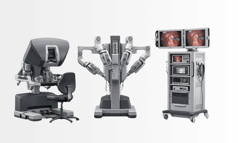
Available at these Hospitals























What patients say about this unit
★★★★★From 2,400+ verified patient reviews“I chose to have my gynecology operation abroad and the language support made it feel local: a translator at every appointment including anesthesia consent. Recovery ward was calm and the follow-up plan was emailed to my own doctor.”
“The gynecology department investigated symptoms my local doctors had dismissed for two years. Ultrasound, hysteroscopy and a diagnosis in one week, with every step explained to me first and my consent treated as a real question.”
Patient stories from this unit

Comparison guides for this specialty
Speak with our medical team
Share your case and our international patient team will guide you to the right specialist, free of charge.