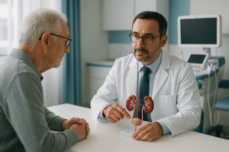
Ureter Diseases
Learn about ureter diseases, including stones, strictures, reflux, and tumors: common symptoms, causes, how doctors diagnose them, and treatment options.

Quick answer
Ureter diseases are conditions affecting the two tubes that carry urine from the kidneys to the bladder. They include stones lodged in the ureter, narrowing from scar tissue, congenital blockages or backward urine flow, injury, and rarely cancer. Symptoms often include flank pain and blood in the urine; untreated blockage can harm kidney function.
What is ureter diseases?
The ureters are two thin, muscular tubes, each roughly 25 to 30 centimeters long, that carry urine from the kidneys down to the bladder. The term ureter diseases covers any condition that blocks, narrows, damages, or otherwise affects these tubes. Because the ureters are the only route for urine to leave the kidneys, a problem in a ureter can quickly affect kidney function if it is not recognized and managed.
Ureter diseases are not a single illness. They include several different conditions, such as:
- Ureteral stoneskidney stones that have moved into the ureter and become stuck.
- Ureteral stricturea narrowing of the ureter caused by scar tissue.
- Ureteropelvic junction (UPJ) obstructiona blockage where the kidney joins the ureter, often present from birth.
- Vesicoureteral reflux (VUR)urine flowing backward from the bladder into the ureter, most often seen in children.
- Ureteral cancera tumor in the ureter lining, usually a type called urothelial carcinoma.
- Congenital anomaliesdifferences present from birth, such as a duplicated ureter, a ureterocele (a balloon-like swelling at the lower end of the ureter), or a megaureter (an abnormally wide ureter).
- Ureteral injurydamage from an accident or, less commonly, during pelvic or abdominal surgery.
Ureter diseases can affect people of any age. Congenital conditions are usually identified in infancy or childhood, sometimes even before birth on a prenatal ultrasound. Stones are most common in adults, and ureteral cancer is rare and typically occurs in older adults, particularly those with a history of smoking. In hospital settings these conditions are generally managed by a urology department, working with nephrology (kidney medicine), radiology, and, when needed, oncology.
Symptoms of ureter diseases
Ureter diseases symptoms depend on the type of problem, how quickly it developed, and whether one or both ureters are involved. Some conditions cause sudden, severe pain, while others cause no symptoms at all for a long time and are found by chance on imaging done for another reason.
Common symptoms include:
- Pain in the flank (the side of the body between the ribs and hip), which may spread to the lower abdomen or groin
- Pain that comes in waves, often called renal colic
- Blood in the urine (hematuria), which may be visible or detected only on a urine test
- Nausea and vomiting, especially with severe pain
- A frequent or urgent need to urinate
- Burning or discomfort when urinating
- Fever and chills, which may signal an infection behind a blockage
- Reduced urine output, or no urine at all if both ureters are blocked or a person has only one working kidney
- In infants and young children: recurrent urinary tract infections, poor feeding, irritability, or a swollen abdomen
How symptoms differ by type:
- Ureteral stones usually cause sudden, intense flank pain that shifts downward as the stone moves. Blood in the urine is common.
- Strictures and UPJ obstruction often develop gradually. Pain may be dull and intermittent, sometimes worse after drinking large amounts of fluid. Some people have no pain, and the condition is discovered when a swollen kidney (hydronephrosis) is seen on a scan.
- Vesicoureteral reflux in children is often silent, with repeated urinary infections being the main clue.
- Ureteral cancer most often presents with painless blood in the urine. Flank pain and weight loss can occur in later stages.
- Ureteral injury may cause pain, fever, urine leaking into the abdomen, or a fluid collection noticed days after surgery or trauma.
Because these symptoms overlap with other kidney, bladder, and abdominal conditions, they do not by themselves confirm a ureter problem. A medical evaluation is needed to find the cause.
Causes and risk factors
Ureter diseases causes vary widely, and in some cases more than one factor is involved.
Stones form when minerals in the urine, most often calcium, crystallize in the kidney. A stone that travels into the ureter can become lodged at one of its naturally narrow points. Low fluid intake, a diet high in salt or animal protein, certain metabolic conditions, some medications, and a family history of stones all raise the risk.
Strictures develop when the ureter wall is injured and heals with scar tissue. Common triggers include stones that have sat in one place for a long time, previous ureteral surgery or instrumentation, radiation therapy to the pelvis, and infection. Rarely, a condition called retroperitoneal fibrosis, in which scar tissue forms around the ureters from outside, can squeeze them shut.
Congenital conditions such as UPJ obstruction, duplicated ureters, ureteroceles, megaureter, and vesicoureteral reflux arise during fetal development. The exact cause is often unknown, though some have a genetic component and may run in families.
Ureteral cancer shares risk factors with bladder cancer because the same type of lining cell is involved. Smoking is the most important known risk factor. Others include long-term exposure to certain industrial chemicals, prior bladder cancer, chronic irritation or infection, and rare inherited conditions such as Lynch syndrome.
Ureteral injury most often occurs during pelvic or abdominal surgery, such as gynecologic, colorectal, or vascular operations, because the ureters lie close to these organs. Blunt or penetrating trauma is a less common cause.
General risk factors for ureter diseases include:
- A personal or family history of kidney stones
- Previous urinary tract surgery or procedures
- Recurrent urinary tract infections
- Smoking
- Radiation therapy to the pelvis
- Certain congenital kidney or urinary tract differences
- Older age, particularly for ureteral cancer
Diagnosis of ureter diseases
Ureter diseases diagnosis starts with a careful medical history and a physical examination. Your doctor will ask about the type and location of pain, changes in urination, any fever, previous stones or surgeries, and family history. From there, a combination of laboratory tests and imaging is used to locate the problem and judge how much it is affecting the kidney.
Urine tests. A urinalysis checks for blood, infection, and crystals. A urine culture identifies bacteria if infection is suspected. In cases where cancer is a concern, urine cytology (examining cells in the urine under a microscope) may be requested.
Blood tests. Measures of kidney function, such as creatinine and estimated glomerular filtration rate (eGFR), show whether a blockage is affecting how well the kidneys filter waste. Blood counts and inflammatory markers help detect infection.
Ultrasound. This is often the first imaging test because it uses no radiation and can quickly show hydronephrosis, the swelling of a kidney that occurs when urine cannot drain. It is the main tool for diagnosing congenital conditions in babies and children.
CT scan. A non-contrast CT of the abdomen and pelvis is the most accurate test for finding ureteral stones and showing their size and location. CT urography, which uses contrast dye to outline the urinary tract, is commonly used to look for strictures, tumors, and injuries.
MRI. Magnetic resonance urography may be chosen when radiation should be avoided, such as in pregnancy or in children, or when CT contrast cannot be used.
Nuclear medicine renal scan. This test measures how well each kidney is working and how quickly urine drains. It is frequently used to decide whether a UPJ obstruction or a stricture is significant enough to need treatment.
Voiding cystourethrogram (VCUG). In children with suspected vesicoureteral reflux, contrast is placed in the bladder through a catheter and X-rays are taken during urination to see whether urine flows backward toward the kidneys.
Ureteroscopy. A thin, flexible telescope is passed through the urethra and bladder into the ureter under anesthesia. This allows the doctor to see the inside of the ureter directly, take a biopsy (a small tissue sample) if a tumor is suspected, and often treat a stone or stricture during the same procedure.
The specific tests used depend on the suspected condition. Not everyone needs every test, and your doctor will explain which are appropriate in your situation.
Treatment options for ureter diseases
Ureter diseases treatment options range from watchful waiting to major surgery. The choice depends on the type of condition, its severity, how much it is affecting kidney function, whether infection is present, and your overall health.
Observation and supportive care. Small ureteral stones, generally those under about 5 millimeters, often pass on their own with time, adequate fluids, and pain relief. Mild vesicoureteral reflux in children frequently resolves as the child grows, and doctors may simply monitor it with periodic ultrasounds. Some mild congenital obstructions that are not harming the kidney may also be watched rather than operated on.
Medications. Pain relievers, including nonsteroidal anti-inflammatory drugs where appropriate, are used for renal colic. A class of drugs called alpha-blockers may be prescribed to relax the ureter and help a stone pass. Antibiotics are used to treat urinary infections and are sometimes given long term at low doses to children with reflux to prevent kidney infections. For ureteral cancer, chemotherapy or immunotherapy may be part of the treatment plan.
Urgent drainage. When a blocked ureter is combined with infection or with failing kidney function, the first priority is to relieve the pressure. This is usually done by placing a ureteral stent (a thin hollow tube that runs from the kidney to the bladder and keeps the ureter open) or a nephrostomy tube (a drain placed through the skin of the back directly into the kidney). Definitive treatment of the underlying problem follows once the person is stable.
Minimally invasive procedures.
- Ureteroscopy with laser lithotripsya scope is passed to the stone and a laser breaks it into fragments that are removed or allowed to pass.
- Shock wave lithotripsy (SWL)sound waves delivered from outside the body break up a stone, mainly used for stones in the upper ureter.
- Balloon dilation or endoscopic incisiona short stricture may be stretched with a balloon or cut from the inside, often followed by a temporary stent.
- Endoscopic injection for refluxin children, a bulking material is injected at the ureter opening in the bladder to stop backward flow.
- Endoscopic tumor removalsmall, low-grade ureteral tumors may sometimes be treated through a scope with laser ablation, with close follow-up.
Surgery. When a stricture is long, an obstruction is significant, or a tumor is more advanced, surgery is usually needed. Options include pyeloplasty (reshaping the junction between kidney and ureter for UPJ obstruction), ureteral reimplantation (reattaching the ureter to the bladder, used for reflux and lower strictures), ureteroureterostomy (removing a damaged segment and joining the healthy ends), and, in some cases, using a segment of bowel or a flap of bladder to replace a long missing portion. For most ureteral cancers, the standard operation is nephroureterectomy, which removes the kidney, the entire ureter, and a small cuff of bladder on that side. Many of these operations can now be done laparoscopically or with robotic assistance, which often means smaller incisions and shorter recovery, though the best approach varies by case.
Recovery and follow-up. After stone treatment, doctors often analyze the stone and may recommend dietary or medication changes to lower the chance of new stones. After stricture repair or pyeloplasty, imaging is repeated over months to confirm urine drains well. People treated for ureteral cancer need regular surveillance of the bladder and remaining urinary tract because the same type of cancer can appear elsewhere in the lining.
Living with ureter diseases and outlook
The outlook for ureter diseases depends heavily on the specific condition and how early it is addressed. Most ureteral stones pass or are removed without lasting harm, although people who have had one stone are at higher risk of another and may benefit from long-term prevention measures such as increased fluid intake and dietary adjustments guided by a doctor.
Congenital conditions such as UPJ obstruction and vesicoureteral reflux generally have a good long-term outlook when monitored and treated appropriately, and many children grow up with normal kidney function. Strictures can recur after treatment, so ongoing follow-up imaging is common, and some people need more than one procedure.
A ureter that has been blocked for a long time may leave a kidney with permanently reduced function even after the blockage is relieved. This is why prompt evaluation of persistent flank pain, recurrent infections, or blood in the urine matters. If one kidney has lost function, the other kidney can often maintain overall health, but regular monitoring of blood pressure and kidney function is usually advised.
For ureteral cancer, outcomes vary widely with the stage and grade of the tumor at diagnosis. Cancers found early, while confined to the lining, are generally more treatable than those that have grown through the ureter wall or spread. Because these cancers can recur in the bladder, lifelong surveillance is typically part of care. Stopping smoking is one of the few modifiable factors that may reduce the risk of recurrence.
Living with a ureteral stent, if one is needed for a period of time, can involve side effects such as urinary urgency, mild discomfort, and blood-tinged urine. These usually settle after the stent is removed. Your care team can advise on managing these effects and on when the stent should be exchanged or taken out.
Frequently asked questions
What is ureter diseases in simple terms?
Ureter diseases are conditions that affect the two tubes carrying urine from the kidneys to the bladder. They include stones stuck in the tube, narrowings from scar tissue, blockages or backward urine flow present from birth, injuries, and, rarely, cancer. What they share is the potential to disrupt urine drainage and, over time, to affect kidney health.
What are the most common ureter diseases symptoms?
The most frequent symptoms are flank pain that may spread toward the groin, blood in the urine, nausea, and changes in urination such as urgency or burning. Fever with flank pain can indicate an infection behind a blockage and needs prompt attention. Some ureter conditions, particularly congenital ones and early cancers, cause few or no symptoms and are found on imaging or urine tests.
What are the main ureter diseases causes?
Causes depend on the type of disease. Stones result from minerals crystallizing in urine. Strictures usually follow injury, infection, previous procedures, or radiation. Congenital conditions arise during development before birth. Ureteral cancer is strongly linked to smoking and certain chemical exposures. Surgical injury is a recognized cause in people who have had pelvic or abdominal operations.
How is ureter diseases diagnosis usually made?
Doctors typically combine urine and blood tests with imaging. Ultrasound is often the first step and can show a swollen kidney. CT scans are highly accurate for stones and can outline strictures or tumors when contrast is used. A nuclear renal scan measures how well each kidney drains and functions. Ureteroscopy allows direct viewing of the inside of the ureter and biopsy when cancer is a concern.
What ureter diseases treatment options exist without surgery?
Many small stones pass on their own with fluids, pain control, and sometimes an alpha-blocker medication. Mild reflux in children is often observed and may resolve with growth. Antibiotics treat associated infections. Short strictures can sometimes be treated through a scope with balloon dilation rather than open surgery. Whether a non-surgical approach is suitable depends on the individual case and on how the kidney is coping.
Can ureter diseases damage the kidney permanently?
They can if a blockage or reflux goes untreated for a long time, because ongoing pressure or repeated infection can scar the kidney. Early recognition and relief of obstruction often allow the kidney to recover, though some loss of function may be permanent in prolonged cases. Regular follow-up after treatment helps detect any recurrence before it causes further harm.
Is ureteral cancer common?
No. Cancer of the ureter is rare compared with bladder or kidney cancer, and it usually occurs in older adults, especially those who smoke or have smoked. Its most common sign is painless blood in the urine, which should always be evaluated by a doctor even if it happens only once.
When to see a doctor
Persistent flank pain, blood in the urine, or repeated urinary tract infections should be evaluated by a doctor, even if the symptoms come and go. Children with recurrent urinary infections or unexplained fevers should also be assessed, as these can be signs of reflux or obstruction.
Seek urgent medical care if you experience any of the following red-flag warning signs:
- Severe flank or abdominal pain that does not ease with rest or usual pain relief
- Fever, chills, or shaking together with flank pain, which may indicate an infected, blocked kidney
- Inability to pass urine, or a sudden marked drop in urine output
- Persistent vomiting that prevents you from keeping fluids down
- Heavy or continuous blood in the urine, or urine with clots
- Confusion, rapid heartbeat, or feeling faint alongside urinary symptoms
- Pain, fever, or fluid leakage in the days or weeks after pelvic or abdominal surgery
- Any of the above in a person with a single kidney, a kidney transplant, or during pregnancy
These situations can progress quickly and may require emergency drainage of the kidney or treatment of infection. Timely assessment gives the best chance of protecting kidney function.
Medically reviewed by the Acıbadem International Medical Board September 9, 2026
See our medical review board →
Update history
- PublishedSeptember 9, 2026
- Medical review approvedSeptember 9, 2026
- Last content updateSeptember 9, 2026