Ureteral Stones
Learn what ureteral stones are, common ureteral stones symptoms, causes, how doctors confirm the diagnosis, and the treatment options that may be used.

Quick answer
Ureteral stones are kidney stones that have moved into the ureter, the narrow tube carrying urine from the kidney to the bladder. They often cause sudden, severe side or back pain, blood in the urine, and nausea. Small stones frequently pass on their own; larger stones may need lithotripsy or ureteroscopy.
What is ureteral stones?
Ureteral stones are hard mineral deposits that have moved out of the kidney and become lodged in the ureter. The ureter is the narrow tube that carries urine from each kidney down to the bladder. Most ureteral stones start as kidney stones (medically called renal calculi), which form when substances in the urine such as calcium, oxalate, and uric acid become concentrated enough to crystallize. When a stone leaves the kidney and travels into the ureter, it may pass on its own or it may get stuck, partly or fully blocking the flow of urine.
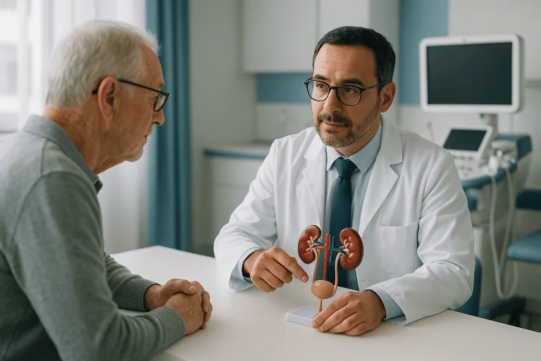
Because the ureter is only a few millimeters wide, even a small stone can cause significant pain. The condition is common and affects adults of all ages, although it is seen most often in people between their twenties and fifties. Men have historically been affected more often than women, although the gap appears to be narrowing. People who have had one stone have a meaningful chance of forming another one later in life. Ureteral stones are usually managed by a urologist, a doctor who specializes in the urinary tract and the male reproductive organs. In the hospital setting, this care is typically coordinated through the urology department.
Ureteral stones symptoms
Ureteral stones symptoms can range from none at all to severe, sudden pain. Many people first learn they have a stone when it starts to move and causes an episode of pain known as renal colic. Common ureteral stones symptoms include:
- Sharp, cramping pain in the side and back, below the ribs, often coming in waves
- Pain that spreads to the lower abdomen, groin, or genitals as the stone moves down the ureter
- Blood in the urine (hematuria), which may look pink, red, or brown, or may only be visible under a microscope
- Nausea and vomiting, often triggered by the intensity of the pain
- Frequent or urgent need to urinate, especially when the stone is near the bladder
- Burning or pain when urinating
- Passing only small amounts of urine
- Fever and chills, which can signal an infection behind the blockage
The location of the stone within the ureter often shapes the symptoms. A stone high in the ureter, near the kidney, tends to cause flank pain (pain in the side between the ribs and hip). As the stone travels lower, the pain frequently shifts toward the lower abdomen and groin. When the stone reaches the lowest part of the ureter near the bladder, people may notice urinary urgency, frequency, and a feeling of incomplete emptying that can resemble a urinary tract infection.
The pain of a ureteral stone is often described as one of the most intense a person can experience. It typically comes in waves lasting minutes at a time, as the ureter contracts against the stone, and people often find it hard to stay still. Some stones, however, cause little or no pain, particularly when they do not fully block the ureter. A stone that causes no symptoms is sometimes discovered by chance on imaging done for another reason.
Causes and risk factors
The main cause of ureteral stones is the formation of stones in the kidney, which then migrate into the ureter. Kidney stones form when urine contains more crystal-forming substances than the fluid can dilute, or when urine lacks substances that normally prevent crystals from sticking together. Ureteral stones causes are therefore closely tied to the chemistry of the urine and to how much fluid a person drinks.
Several types of stones exist, and each has somewhat different causes:
- Calcium stones are the most common type. They are usually made of calcium oxalate and, less often, calcium phosphate. High levels of calcium or oxalate in the urine contribute to their formation.
- Uric acid stones form when urine is persistently acidic. They are more common in people who eat a diet high in animal protein, who have gout, or who lose a lot of fluid.
- Struvite stones are linked to certain urinary tract infections and can grow quickly and become large.
- Cystine stones are rare and result from an inherited disorder called cystinuria, in which the kidneys leak too much of an amino acid called cystine into the urine.
Risk factors that make stone formation more likely include:
- Not drinking enough fluid, which produces concentrated urine
- A personal or family history of stones
- Diet high in sodium, animal protein, or foods rich in oxalate, and low in calcium from food
- Obesity and metabolic conditions such as type 2 diabetes
- Digestive conditions such as inflammatory bowel disease, chronic diarrhea, or a history of gastric bypass surgery, which change how the body absorbs calcium and water
- Certain medical conditions, including hyperparathyroidism (overactive parathyroid glands that raise blood calcium), gout, and recurrent urinary tract infections
- Some medications and supplements, including high doses of vitamin C, certain diuretics, and some antacids containing calcium
- Living in a hot climate or working in hot conditions, which increases fluid loss through sweat
Having one or more of these factors does not mean a person will definitely form stones, but it does raise the likelihood. In many cases, several factors act together.
Ureteral stones diagnosis
Ureteral stones diagnosis usually begins with a description of the symptoms and a physical examination. Doctors will ask about the location and pattern of the pain, any blood in the urine, previous stones, family history, diet, and medications. Because renal colic can be confused with other conditions such as appendicitis, gallbladder disease, ovarian problems, or a blood vessel emergency, testing is used to confirm the diagnosis and rule out other causes.
Common tests and methods include:
- Urinalysis: a urine sample is checked for blood, crystals, signs of infection, and acidity. Blood in the urine is common with stones but is not present in every case.
- Blood tests: these can measure kidney function, calcium, uric acid, and markers of infection. They also help doctors judge how urgently treatment is needed.
- Non-contrast CT scan: a computed tomography scan of the abdomen and pelvis without contrast dye is widely considered the most accurate imaging test for finding ureteral stones. It shows the size and exact location of the stone and the degree of blockage.
- Ultrasound: this test uses sound waves and no radiation. It is often the first choice for pregnant women and children, and for people who need repeated imaging. It may not show small stones in the ureter directly but can reveal swelling of the kidney caused by a blockage (hydronephrosis).
- Plain X-ray of the kidneys, ureters, and bladder (KUB): this can show some stones and is sometimes used to track a known stone over time, but it misses stones that do not contain calcium.
Two features of the stone matter most for planning treatment: its size and its position in the ureter. Smaller stones located in the lower ureter are more likely to pass on their own, while larger stones and those higher up are less likely to do so. Doctors also look for signs of infection or severely reduced kidney function, which change the urgency of care. Once a stone has passed or been removed, it may be sent to a laboratory to determine its chemical composition, which helps guide prevention.
Ureteral stones treatment options
Ureteral stones treatment options depend on the stone’s size and location, how much pain and blockage it is causing, whether infection is present, the person’s overall health, and personal preference. The goals of treatment are to relieve pain, protect the kidney, and clear the stone.
Observation and supportive care. Many small ureteral stones pass on their own within days to a few weeks. In these cases, doctors often recommend watchful waiting with pain relief and regular fluid intake. Nonsteroidal anti-inflammatory drugs (NSAIDs) such as ibuprofen are commonly used for pain, provided the person can take them safely; stronger medication may be needed for severe episodes. People are usually asked to strain their urine to catch the stone for analysis and to return for follow-up imaging to confirm the stone has passed.
Medical expulsive therapy. Your doctor may prescribe a medication called an alpha-blocker, such as tamsulosin, which relaxes the muscle in the wall of the ureter. This may make it easier for a stone, particularly one in the lower ureter, to pass and may shorten the time it takes. These drugs are used off-label for this purpose in many countries, and the benefit appears to be greatest for somewhat larger stones rather than very small ones.
Shock wave lithotripsy (SWL). This procedure uses focused sound waves delivered from outside the body to break the stone into smaller fragments that can then pass in the urine. It is non-invasive and is often used for stones in the upper ureter or kidney. It may be less effective for very hard stones, very large stones, or stones in people with a higher body weight, and more than one session is sometimes needed.
Ureteroscopy. A thin, flexible or rigid telescope called a ureteroscope is passed through the urethra and bladder into the ureter, without any incision. The stone can be removed with a small basket or broken up with a laser and the pieces extracted. Ureteroscopy is used for stones anywhere in the ureter and is frequently chosen for stones in the lower ureter or when SWL has not worked. A temporary internal tube called a ureteral stent is sometimes left in place afterward to keep urine draining while swelling settles.
Percutaneous nephrolithotomy (PCNL). For very large stones, or stones that have moved back into the kidney, a surgeon may make a small incision in the back and pass instruments directly into the kidney to remove the stone. This is more invasive than the options above and usually requires a short hospital stay.
Emergency drainage. If a stone is blocking the ureter and there is also infection, or if the kidney is at risk, doctors may first insert a ureteral stent or a nephrostomy tube (a drain placed through the skin into the kidney) to relieve the pressure and treat the infection. The stone itself is then dealt with once the person is stable.
Open surgery is rarely needed today and is reserved for unusual situations where other methods are not possible.
After treatment, follow-up imaging is commonly used to confirm the ureter is clear. Depending on the stone type and the person’s history, a doctor may also recommend a metabolic evaluation, which includes blood and 24-hour urine tests, to identify why stones are forming and to guide longer-term prevention.
Living with ureteral stones and outlook
For most people, a single ureteral stone is a painful but temporary event. Once the stone passes or is removed, symptoms usually resolve, and the kidney typically recovers fully if the blockage was relieved in a reasonable time. Prolonged obstruction, especially when combined with infection, can damage the kidney, which is why timely evaluation matters.
The main long-term concern is recurrence. A significant proportion of people who have had one stone will form another within several years, although the exact risk varies from person to person. Lifestyle changes can lower the chance of new stones in many cases. These commonly include drinking enough fluid to produce pale-colored urine throughout the day, limiting salt, moderating animal protein, avoiding excessive intake of oxalate-rich foods if advised, and maintaining normal calcium intake from food rather than cutting it out. Depending on the type of stone and the results of a metabolic evaluation, a doctor may also prescribe medication such as thiazide diuretics, potassium citrate, or allopurinol to change the urine chemistry.
People who form stones repeatedly, who have a single kidney, who have underlying metabolic or digestive disease, or whose stones are cystine or struvite may need ongoing follow-up with a urologist or nephrologist (a kidney specialist). Living well with a tendency to form stones is largely a matter of consistent hydration, sensible dietary habits, taking any prescribed medication, and reporting new symptoms promptly.
Frequently asked questions
What is ureteral stones and how is it different from kidney stones?
The term describes a stone that has moved from the kidney into the ureter, the tube connecting the kidney to the bladder. Kidney stones and ureteral stones are made of the same material; the difference is simply location. Because the ureter is narrow, a stone lodged there is more likely to cause pain and blockage than one sitting quietly in the kidney.
What are the most common ureteral stones symptoms?
The classic symptom is severe, wave-like pain in the side and back that may spread to the lower abdomen and groin. Blood in the urine, nausea, vomiting, and a frequent urge to urinate are also common. Some stones cause no symptoms until they move. Fever with these symptoms is a warning sign of infection and should not be ignored.
What are the main ureteral stones causes?
Ureteral stones form in the kidney when the urine becomes too concentrated with minerals such as calcium, oxalate, or uric acid. Low fluid intake, diet, family history, obesity, certain digestive and metabolic conditions, and some medications all raise the risk. In many people several factors contribute at once.
How is ureteral stones diagnosis confirmed?
Doctors usually combine a history and examination with a urine test, blood tests, and imaging. A non-contrast CT scan is the most accurate way to see the stone’s size and position. Ultrasound is often used instead for pregnant women, children, and people who need repeated scans, because it avoids radiation.
Can a ureteral stone pass on its own?
Many small stones, particularly those in the lower part of the ureter, pass without a procedure, often within days to a few weeks. Larger stones and stones higher in the ureter are less likely to pass by themselves. Your doctor may suggest a trial of watchful waiting with pain relief, and sometimes an alpha-blocker medication, followed by imaging to confirm the stone has gone.
What are the ureteral stones treatment options if the stone does not pass?
The most common procedures are shock wave lithotripsy, which breaks the stone from outside the body, and ureteroscopy, in which a thin telescope is passed up the urinary tract to remove or laser the stone. Very large stones may need percutaneous nephrolithotomy. The choice depends on stone size, location, hardness, and individual factors, and your doctor will discuss which option suits your situation.
How can I reduce the chance of getting another stone?
Drinking enough fluid to keep urine pale throughout the day is the single most widely recommended step. Limiting salt and excess animal protein, keeping a normal dietary calcium intake, and following any specific dietary advice based on your stone type may also help. If a metabolic evaluation shows an underlying abnormality, medication may be prescribed to lower the risk.
When to see a doctor
Anyone who experiences sudden, severe pain in the side or back with blood in the urine should be evaluated, because a stone needs to be confirmed and other serious conditions ruled out. Seek urgent or emergency medical care if you have any of the following red-flag signs:
- Fever or chills together with flank or abdominal pain, which may indicate an infected, blocked kidney
- Pain so severe that you cannot sit still, find a comfortable position, or control it with recommended pain relief
- Persistent nausea and vomiting that prevents you from keeping fluids down
- Inability to pass urine, or passing very little urine
- Blood in the urine with clots or heavy bleeding
- Known stone in a person with only one kidney, a kidney transplant, or existing kidney disease
- Symptoms during pregnancy
- Confusion, rapid heartbeat, or feeling faint, which can be signs of a serious infection spreading in the body
If you have been diagnosed with a ureteral stone and are waiting for it to pass, contact your doctor if the pain changes character, lasts longer than expected, or if follow-up imaging has not confirmed that the stone is gone. Timely care protects the kidney and allows treatment to be adjusted when needed.
Medically reviewed by the Acıbadem International Medical Board September 9, 2026
See our medical review board →
Update history
- PublishedSeptember 9, 2026
- Medical review approvedSeptember 9, 2026
- Last content updateSeptember 9, 2026