Laser Lithotripsy
Laser lithotripsy is a minimally invasive endoscopic procedure that uses laser energy to fragment kidney or ureter stones so they can pass or be removed safely.
Quick answer
Laser lithotripsy is a minimally invasive procedure that breaks kidney and ureter stones using pulses of laser energy delivered through a thin telescope passed along the natural urinary passages, usually without an incision. Under anaesthesia, the surgeon fragments the stone into fine dust or retrievable pieces; a temporary stent is sometimes placed afterwards, and many patients go home the same day.
Kidney Stones Treatment: When a Stone Will Not Pass on Its Own
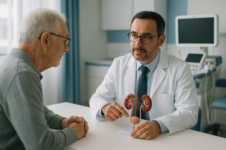
Laser lithotripsy is a minimally invasive kidney stones treatment that breaks stones in the kidney or ureter into fragments small enough to remove or to pass naturally. It is performed through the natural urinary passages, usually without any external incision, using a thin telescope and a very fine laser fibre. It is typically offered when a stone is too large, too painful, infected or too obstructive to be managed safely with observation alone.
A stone rarely announces itself politely. For many people it begins as a sudden, severe pain in the back or side, arriving in waves and refusing to settle. For others, the stone is silent, discovered on a scan ordered for an entirely different reason. Either way, the uncertainty is often as distressing as the pain itself. Will the stone pass on its own? Could it damage the kidney? Is a procedure really necessary? And if you are travelling for care, there are practical questions layered on top: timing, communication, recovery, and how soon you can fly home.
Treatment matters because a stone can block the flow of urine, trigger recurrent infections, cause repeated attacks of pain and, in some situations, impair kidney function. Not every stone needs an intervention. But when a stone is unlikely to pass safely, or is already causing complications, timely treatment protects the kidney and reduces the risk of emergency episodes. At Acibadem, evaluation and treatment are planned by experienced urology teams using modern imaging, endoscopic techniques and individualised care pathways for both local and international patients. Good kidney stones treatment is as much about judgement as technique, knowing when to operate, when to drain the kidney first, and when the wisest move is simply to wait and watch.
What Is Laser Lithotripsy?
Laser lithotripsy is an endoscopic stone treatment that uses laser energy to fragment stones lodged in the kidney or the ureter. The ureter is the narrow tube that carries urine from the kidney down to the bladder: a passage only a few millimetres wide, which is precisely why stones become stuck there. Stones may lodge anywhere along this tube or remain within the collecting system of the kidney itself. When a stone is too large, too painful, infected or obstructive to manage conservatively, laser lithotripsy is one of the treatments most commonly recommended.
What does lithotripsy mean?
Lithotripsy simply means breaking stones: the word combines the Greek terms for “stone” and “crushing”. It is an umbrella term, and this is where confusion often starts. There are two broad families of stone-breaking treatment: shock wave lithotripsy, which delivers energy from outside the body, and laser lithotripsy, which delivers energy from inside the urinary tract under direct vision. They share a name but differ substantially in how they reach the stone, what they can treat and what recovery looks like. When your doctor recommends “lithotripsy”, it is worth asking which type is meant.
How does laser lithotripsy work?
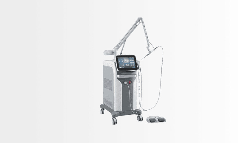
Laser lithotripsy works by delivering short, controlled pulses of light energy directly onto the surface of the stone through a flexible glass fibre thinner than a strand of spaghetti. The energy is absorbed by the stone, creating tiny vapour bubbles and micro-fractures that break it apart. Holmium laser systems are the workhorse of modern stone surgery, and the surgeon adjusts the pulse energy and frequency depending on the effect required. Two techniques dominate. In dusting, the settings are chosen to grind the stone into particles fine enough to wash out with the urine. In fragmentation, the stone is broken into slightly larger pieces that are then captured with a small wire basket and physically removed. The choice depends on the stone’s location, size, hardness and the patient’s anatomy, and surgeons frequently combine both approaches in a single procedure.
How is laser lithotripsy different from shock wave lithotripsy?
The defining difference is access. Laser lithotripsy is performed internally, through the natural urinary channels, with the surgeon watching the stone on a screen as it breaks. Shock wave lithotripsy uses focused sound waves generated outside the body, aimed at the stone using imaging: the surgeon never sees the stone directly, and fragments must find their own way out. Laser lithotripsy is also different from percutaneous stone surgery, in which a small access tract is created through the skin of the back directly into the kidney; that route is generally reserved for larger or more complex stone burdens. Laser lithotripsy tends to be selected when direct visualisation and precise, controlled fragmentation matter, for example, with hard stones, stones in awkward positions, or patients in whom shock waves are unsuitable.
For many patients, laser lithotripsy offers a workable balance between effectiveness and recovery time. It can treat ureter stones and many kidney stones in a single session, although large or complex stone burdens sometimes require staged procedures: an honest surgeon will tell you this beforehand rather than promise everything in one sitting. The goal is not only to relieve the immediate problem, but also to reduce the likelihood of future obstruction, infection and repeat attacks.
Who May Need Laser Lithotripsy?
You may be considered for laser lithotripsy when a stone is causing symptoms, is unlikely to pass on its own, or presents a risk to the health of the kidney. The decision rests on a careful assessment of stone size, location, degree of obstruction, infection risk, kidney function, your prior stone history and your overall medical condition, never on the stone alone.
Typical symptoms of a kidney or ureter stone include:
- Intense pain in the side or back, often arriving in waves
- Pain radiating toward the lower abdomen or groin
- Nausea and vomiting
- Urinary urgency or burning during urination
- Blood in the urine, visible or detected on testing
- Repeated episodes of discomfort that settle and return
Some patients develop fever or chills alongside these symptoms. In urology, fever with a blocked kidney is treated as an emergency in every department in the world, because it can signal infection trapped behind the obstruction. Other patients have mild symptoms or none at all, with the stone found on ultrasound, X-ray or computed tomography performed for another reason.
When do kidney stones hurt the most?
Kidney stones typically hurt most when they move, specifically, when a stone drops out of the kidney and lodges in the ureter, blocking the flow of urine. Pressure builds behind the blockage, stretching the kidney’s collecting system, and this stretch is what produces the wave-like pain known as renal colic. Counter-intuitively, a small moving stone often hurts far more than a large stationary one; sizeable stones can sit quietly in the kidney for years while a stone a few millimetres across causes agony on its journey down the ureter. Pain also tends to shift location as the stone travels, moving from the flank toward the groin as the stone approaches the bladder.
Can a kidney stone go away on its own?
Yes, many stones pass without any procedure at all. Whether yours will depends chiefly on its size and position: smaller stones, particularly those already low in the ureter, stand a reasonable chance of passing naturally, while larger stones and those stuck high in the ureter are much less likely to. When passage seems plausible and there is no infection, severe obstruction or uncontrolled pain, urologists often allow a supervised period of waiting, with pain management and follow-up imaging to confirm the stone is actually moving. What matters is that the waiting is monitored: a stone that silently blocks a kidney for weeks can do harm even when the pain has faded.
Diagnosis begins with a detailed history and physical examination, but imaging is central to planning. Ultrasound can identify swelling of the kidney and some stones. Low-dose CT imaging provides more detailed information about stone size, density, location and the degree of obstruction, density in particular hints at how hard the stone will be to break. In selected cases, contrast studies help define the anatomy of the urinary tract. Urine testing checks for blood, crystals and infection, and blood tests evaluate kidney function, inflammation and metabolic factors such as calcium or uric acid levels.
Laser lithotripsy tends to enter the conversation when a ureter stone is stuck and causing persistent pain, when the stone is too large to pass comfortably, when conservative treatment has been tried and failed, or when the patient has only one functioning kidney and cannot afford to gamble. It also suits recurrent stone formers, stones associated with infection once that infection has been brought under control, patients who need to return to work or travel promptly, and stones poorly suited to shock wave treatment because of their position, density or patient-specific factors.
Some patients need stabilising before any definitive stone treatment. If a blocked kidney is infected, the priority is urgent drainage, with a ureteral stent or a nephrostomy tube, together with antibiotics. Laser lithotripsy is performed later, once the infection has settled. This staged approach is a core safety principle of modern stone care, and a team that proposes it is exercising caution, not delay.
Conditions Treated With Laser Lithotripsy
Laser lithotripsy is used across the urinary tract. It is especially valuable for ureteral stones, where obstruction produces severe pain and puts direct pressure on the kidney. It also treats stones within the kidney itself, in the renal pelvis and the calyces, when size and anatomy suit an endoscopic approach, and laser fragmentation is used for bladder stones as well. One point of frequent confusion: kidney stones are unrelated to gallstones, which form in the gallbladder, are made of different material and are treated by entirely different specialists.
Common indications include symptomatic ureter stones, kidney stones causing recurrent pain or bleeding, stones that keep growing on serial imaging, stones associated with repeated urinary tract infections, stones blocking urine flow, and residual fragments left behind after previous stone treatment. Patients who have not responded to medication and observation are also candidates.
The procedure handles a range of stone compositions, calcium-based stones, uric acid stones and rarer types. Hardness matters practically: a dense stone takes longer to fragment, and this influences whether everything can be cleared in one session. Where fragments are retrieved, they can be sent for analysis, and for anyone with a history of recurrent stones this analysis is genuinely useful: it identifies what the stones are made of and therefore how to make the next one less likely.
Not every stone is best treated with a laser. Very large kidney stones, complex branching “staghorn” stones and stones behind challenging anatomy are often better served by percutaneous techniques or a planned staged approach. Small, non-obstructing, symptom-free stones may simply be monitored. The right plan emerges from imaging review, symptom assessment and an honest discussion of your goals, travel constraints and medical risks, not from a default preference for any single tool.
4 Methods for Kidney Stone Removal
Most kidney stones treatments fall into one of four approaches, and understanding all of them makes the recommendation for any one of them easier to weigh.
- 1. Observation and medical therapy. Small stones with a fair chance of passing are managed with supervised waiting, pain control and, where a doctor prescribes them, medications that relax the ureter to ease the stone’s passage. Certain uric acid stones can sometimes be dissolved over time with prescription treatment that changes the chemistry of the urine: a decision that belongs entirely to the treating physician, guided by imaging and blood results.
- 2. Shock wave lithotripsy (ESWL). Focused sound waves generated outside the body break the stone without any instrument entering the urinary tract. It is the least invasive option and often done as a day case, but it works best for softer, moderately sized stones in favourable positions, fragments must pass on their own, and more than one session is sometimes needed.
- 3. Ureteroscopy with laser lithotripsy. The subject of this page: a telescope passed through the natural passages, the stone broken under direct vision, fragments dusted or retrieved. It offers precision and the ability to confirm, there and then, what has been achieved.
- 4. Percutaneous nephrolithotomy (PCNL). A keyhole tract through the skin of the back directly into the kidney, used for large or complex stone burdens that endoscopic and external methods cannot clear efficiently. It is the most invasive of the four and usually involves a longer hospital stay.
What is the best thing to treat kidney stones?
There is no single best treatment, only the best treatment for a specific stone in a specific patient. Size, location, density, the shape of your urinary tract, the presence of infection, your kidney function, your medications and even your travel plans all move the answer. A small lower-ureter stone in a healthy patient and a hard lower-pole kidney stone in a patient with one functioning kidney are different problems demanding different tools. Be wary of any clinic that offers one method for every stone; a well-equipped stone service matches the technique to the case, and is willing to explain why.
How to get rid of kidney stones without a procedure
If you are researching how to get rid of kidney stones without surgery, the honest answer is that it is sometimes possible and sometimes not. A small stone may pass with time, generous fluid intake as advised by your doctor, and medication prescribed to ease its journey. What no amount of water can do is shift a stone that is too large for the ureter, dissolve a calcium stone that has already formed, or make a safely ignorable problem out of an obstructed or infected kidney. Conservative management is a legitimate strategy precisely because it is supervised, with imaging to confirm progress and a plan B agreed in advance.
What foods help clear kidney stones?
No food dissolves an existing stone, and it is worth being clear about that before spending money on remedies that claim otherwise. Fluids are the genuinely useful part of the equation: drinking enough, as advised for your individual health situation, dilutes the urine and helps small fragments wash through. Citrus fruits contribute citrate, a natural inhibitor of calcium stone formation, which is why lemon water appears in so much prevention advice. For the longer term, sensible measures include moderating salt, keeping dietary calcium at normal levels rather than cutting it out, moderating animal protein and being aware of high-oxalate foods, but effective dietary advice should follow stone analysis and metabolic testing, because the right diet depends on what your stones are made of.
How Is Laser Lithotripsy Done?
Preparation before the procedure
Preparation begins with confirming the diagnosis and planning the safest route to the stone. Your urologist reviews the imaging to pin down the stone’s exact position, size, density and the anatomy of the kidney and ureter. Urine is tested for infection; if bacteria are found, antibiotics are given first, and in some cases the operation is postponed until the infection is controlled, operating through infected urine is a risk no careful surgeon takes willingly. Blood tests assess kidney function, blood count and clotting.
You will be asked about your medications, allergies, previous anaesthetics, prior stone procedures and conditions such as diabetes, heart disease, bleeding disorders or pregnancy. Decisions about blood-thinning medication belong to your treating doctor and anaesthetist: they will tell you exactly what to do and when. You will be instructed not to eat or drink for a set period before anaesthesia. For international patients, the pre-operative evaluation is usually coordinated in advance, with imaging and laboratory results reviewed by the care team before travel wherever possible, so that surprises on arrival are kept to a minimum.
What is ureteroscopy?
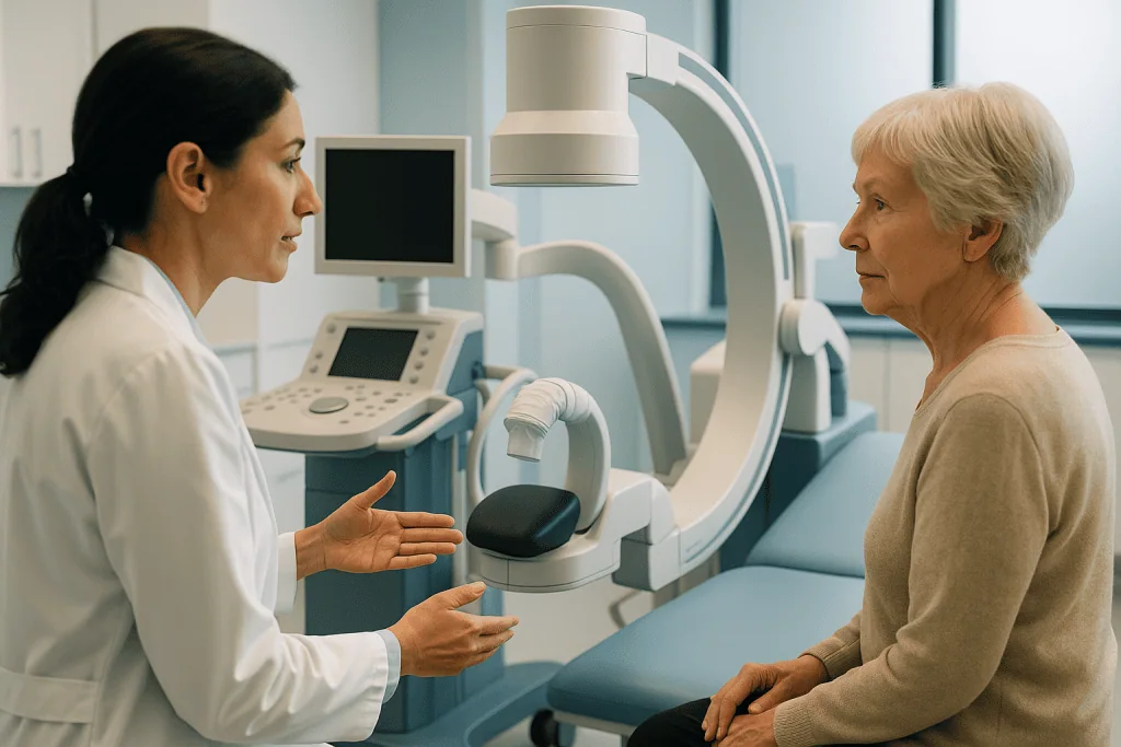
Ureteroscopy is the passage of a thin telescope through the urethra and bladder into the ureter and, when needed, up into the kidney itself: the delivery route for the laser. The procedure is most often performed under general anaesthesia, so you are asleep throughout; regional anaesthesia is an option in selected cases. Modern ureteroscopes come in two forms: semi-rigid instruments for stones in the lower and middle ureter, and flexible instruments that can navigate up into the kidney and steer into its individual calyces. Each scope carries a camera and a working channel for delicate instruments. Irrigation fluid keeps the view clear, and fluoroscopy, real-time X-ray, is used when needed to confirm position and anatomy. Nothing is cut; the entire journey uses passages your body already has.
Fragmenting and removing the stone
Once the stone is in view, the laser fibre is advanced through the scope and positioned against it. The surgeon applies controlled pulses, adjusting the settings for the effect required, dusting the stone into fine particles that will pass with the urine, or fragmenting it into pieces that are captured with small retrieval baskets and taken out. Impacted stones, hard stones and multiple stones each demand slightly different tactics, and the surgeon adapts in real time because, unlike external treatments, everything happens under direct vision.
The technology surrounding the laser exists to improve precision and safety. High-resolution endoscopic imaging lets the urologist work accurately in spaces a few millimetres wide. Flexible instruments reach areas of the kidney that were once accessible only through open surgery. The laser delivers its energy in controlled bursts designed to act on the stone while minimising effect on surrounding tissue. The specific equipment chosen depends on your anatomy and your stone, not the other way round.
Will I need a ureteral stent?
Possibly, and it helps to know why before you wake up with one. A stent is a soft, flexible tube placed temporarily inside the ureter to keep it open, support drainage from kidney to bladder and prevent swelling from blocking the passage after treatment. Not every patient needs one, but stenting is common after ureteroscopy, particularly if the stone was impacted, the ureter was narrow, the procedure involved significant manipulation, or fragments are expected to keep passing. Stents cause noticeable symptoms in many people (urgency, frequency, a dragging discomfort), which is unwelcome but temporary. Removal is a brief procedure, often done in the outpatient clinic, and you will leave hospital with a clear plan for when it happens.
How long does a laser lithotripsy take?
Many laser lithotripsy procedures are completed in less than two hours, though the time varies with stone size, number, hardness, location, your anatomy and whether a stent is placed. Complex stone burdens may take longer or be deliberately planned in stages: a decision made for safety, since very long procedures carry their own risks. Some patients go home the same day once they have recovered from anaesthesia; others stay overnight for observation, pain control, antibiotics or monitoring of kidney function. Before discharge, the team walks you through expected symptoms, medications, fluid intake, activity limits and, if you have a stent, the removal plan.
Recovery After Laser Lithotripsy
Recovery is generally quicker than after more invasive stone surgery, but it is not symptom-free, and pretending otherwise sets patients up for worry. Expect some burning during urination, urinary frequency, bladder irritation, mild flank ache and blood-tinged urine for several days. These symptoms are usually more noticeable when a stent is in place. Medication may be prescribed to manage pain, bladder spasm or infection risk, take it as directed by your team.
Most patients return to light daily activity within a short time, depending on how they recover from anaesthesia and how they feel. Strenuous exercise, heavy lifting and long journeys may be restricted briefly, particularly with a stent in place or when fragments are still passing. Drinking fluids as your team advises helps flush fine particles through, though fluid targets differ for patients with heart, kidney or other conditions, which is why the advice is individual rather than universal.
Follow-up imaging is often recommended to confirm the stone has cleared and the kidney is draining well. Retrieved fragments can be analysed, and recurrent stone formers are usually offered a metabolic evaluation, urine and blood testing, dietary review and, where a doctor judges it appropriate, preventive medication.
| Time Period | What Patients Can Expect |
|---|---|
| Day 1 | Recovery from anaesthesia; fluids as advised. Burning with urination, urgency, mild flank discomfort and blood-tinged urine are common and expected. |
| First Week | Light activity is usually possible. Stent discomfort, urinary frequency and intermittent pain may come and go; medication helps control symptoms. |
| First Month | Irritation typically settles after stent removal and passage of fine fragments. Follow-up imaging or urine testing confirms drainage and stone clearance. |
| Longer Term | Focus shifts to prevention: stone analysis, metabolic evaluation, hydration planning, dietary guidance and medication where a doctor prescribes it. |
Benefits of Laser Lithotripsy
The benefits depend on your stone and your circumstances, but laser lithotripsy is often chosen because it pairs direct, visible stone treatment with a comparatively short recovery.
| Benefit | What It Means for You |
|---|---|
| Minimally invasive access | The stone is treated through the natural urinary passages, usually without any external incision. |
| Direct visualisation | The surgeon sees the stone throughout, allowing controlled fragmentation and removal rather than working blind. |
| Suits many ureter and kidney stones | Effective in locations and for stone densities where external shock wave therapy performs poorly. |
| Relief of obstruction and symptoms | Clearing the stone restores urine drainage, reduces pain episodes and lowers the risk of obstruction-related complications. |
| Relatively quick return to routine | Light activity usually resumes soon after treatment, though recovery varies and stent symptoms are possible. |
| Stone analysis | Retrieved fragments can be tested to explain why your stones form: the starting point for genuine prevention. |
Can Laser Lithotripsy Damage the Kidney?
Serious kidney damage from laser lithotripsy is uncommon. The laser is applied under direct vision, its energy penetrates tissue only very shallowly, and the settings are chosen to act on the stone rather than the kidney around it. The more relevant comparison is usually the other way round: an obstructing stone left untreated poses a greater long-term threat to the kidney than the procedure designed to remove it, because sustained blockage, especially with infection, can erode kidney function over time.
That said, no procedure is free of risk, and a fair account includes the possible complications:
- Irritation or, rarely, injury to the ureter, which occasionally requires longer stenting to heal
- Urinary tract infection or, uncommonly, more serious infection needing antibiotics and observation
- Bleeding, usually minor and self-limiting
- Stent-related discomfort while the stent remains in place
- Residual fragments that need time to pass, or occasionally a further procedure
- Rarely, later narrowing of the ureter (stricture), which follow-up is designed to detect
Your surgeon should discuss these with you in the context of your own anatomy, stone and health: the risk profile of a straightforward lower-ureter stone and a complex kidney stone burden are not the same, and generic reassurance serves nobody.
Factors That Influence Outcomes
Stone size is among the most important factors. Smaller stones are usually fragmented and cleared in a single procedure; larger burdens may need longer treatment, deliberate staging or a different technique altogether. The number of stones matters too, several stones scattered through different parts of the kidney present a more complex task than a single stone stuck in the ureter.
Location shapes both access and clearance. Lower-ureter stones are reached with semi-rigid instruments; stones inside the kidney require flexible ureteroscopy. Some regions, the lower-pole calyces in particular, are awkward for fragments to leave naturally, because gravity and anatomy work against them. A narrow or tortuous ureter can also determine whether the scope passes safely on the first attempt or whether a stent must be placed beforehand to let the ureter widen, with the lithotripsy performed at a second visit.
Composition and hardness matter practically. Some stones crumble readily; dense calcium-based stones demand more laser time. Uric acid stones can sometimes be managed with prescription therapy that alkalinises the urine, depending on size, symptoms and obstruction. Infection-related stones require rigorous infection control before, during and after treatment, plus a prevention plan, because they tend to return if the underlying problem is ignored.
Patient factors round out the picture: baseline kidney function, particularly relevant for anyone with chronic kidney diseasehydration, active infection, previous surgery, pregnancy, bleeding risk, body build and other medical conditions. Age changes the calculus as well; stone care in older kidneys is planned with extra attention to anaesthetic fitness and medication interactions. A patient with fever and obstruction needs drainage before definitive treatment; a patient on anticoagulants or with significant heart disease needs coordination with other specialists before anaesthesia.
A good result is more than a broken stone. It depends on safe perioperative care, sensible stent management, follow-up imaging that actually happens, and prevention. Stone disease is recurrent by nature: treat the episode but not the cause, and another episode is likely. This is why metabolic evaluation, dietary counselling and individualised prevention are built into comprehensive stone care rather than bolted on.
You have a role too. Recovery goes better for patients who take medications as directed, attend stent removal and follow-up appointments, and keep up fluid intake as their team advises. Before discharge, the care team will spell out which symptoms they want to hear about promptly, typically high fever, chills, inability to urinate, severe uncontrolled pain, heavy bleeding or persistent vomiting, so that small problems are caught before they become large ones.
Why Acting Early Matters
A stone that causes no obstruction and no symptoms can often be monitored safely. A symptomatic or obstructing stone is a different matter, and urologists treat it as a problem to assess promptly rather than one to endure. When urine flow is blocked, pressure builds inside the kidney; if the obstruction persists, kidney function can suffer. The stakes rise further with infection, a single functioning kidney, pre-existing kidney disease or a complete, unrelenting blockage.
The costs of delay are predictable: repeated emergency visits, escalating pain, dehydration from nausea and vomiting, worsening obstruction and infection. An infected, obstructed kidney is a genuine medical emergency in every health system, because bacteria trapped under pressure can spread into the bloodstream. In that scenario, the correct first step is drainage and antibiotics, stone fragmentation waits until it is safe.
Early evaluation does not automatically mean early surgery. It means making a safe, informed decision on the basis of imaging, symptoms, urine findings, kidney function and your circumstances, including, for international patients, the practical realities of travel timing, stent management, follow-up imaging and fitness to fly, all of which are far easier to plan in advance than to improvise afterwards.
Preventing the Next Stone
Prevention is the quiet half of kidney stones treatment, and the half most often neglected once the pain has gone. Because stone disease recurs, the weeks after a successful procedure are precisely the time to ask why the stone formed. Fragment analysis identifies the stone’s composition. A metabolic evaluation, urine collection, blood testing, dietary review, identifies the chemistry driving it. From there, prevention becomes specific rather than generic: hydration targets suited to your health, dietary adjustments matched to your stone type, and preventive medication where your doctor judges it worthwhile.
The habits are unglamorous but effective over years: consistent fluid intake spread through the day, moderated salt, normal rather than restricted dietary calcium, moderated animal protein and attention to individual risk factors identified in testing. Periodic imaging can catch a new stone while it is still small, silent and easy to deal with, which is a far better position than discovering it in an emergency department at three in the morning.
Laser Lithotripsy at Acibadem
For anyone considering treatment away from home, the quality of the decision-making matters as much as the procedure. Laser lithotripsy sounds straightforward; the judgement behind it is not. The best plan requires careful reading of imaging, an understanding of how stones behave, and the discipline to know when to treat, when to drain first and when a different approach is safer.
At Acibadem, stone care is delivered by urology teams experienced in the endoscopic management of kidney and ureter stones, working to evidence-based treatment principles with attention to patient safety, infection control, anaesthetic evaluation and follow-up. Complex cases draw on specialists across disciplines, urology, nephrology, radiology, anaesthesiology, infectious diseases and internal medicine, which matters most for patients with recurrent stones, reduced kidney function, infection, multiple medical conditions or previous procedures. Diagnostic pathways include ultrasound, CT-based stone mapping and laboratory assessment of kidney function, infection and metabolic risk; during treatment, endoscopic visualisation, flexible instruments, laser systems and imaging guidance support precise access and fragmentation. The working principle is to select the right method for the individual patient rather than to apply one technique to every stone.
International patients are supported by dedicated coordination services covering appointment planning, hospital admission processes, interpretation in multiple languages and follow-up arrangements, so that the practical side of travelling for care is organised before arrival rather than negotiated during recovery. Patients weighing up their options, observation, medical therapy, shock wave treatment, ureteroscopy with laser lithotripsy or percutaneous surgery, often find a second specialist opinion clarifying before committing to a single pathway.
Moving Forward With Clarity
Kidney and ureter stones disrupt life suddenly, and pain has a way of making every decision feel urgent. Good decisions start with a precise diagnosis and a clear-eyed view of the options. Laser lithotripsy is a minimally invasive endoscopic procedure that treats many stones effectively by fragmenting them under direct vision, relieving obstruction, easing symptoms and helping protect kidney function when intervention is genuinely needed.
Understanding how the procedure works, what recovery honestly involves, where its limits lie and how prevention follows treatment puts you in a far stronger position, whether your stone ultimately passes on its own, responds to a single session of laser treatment, or calls for a staged plan built around your anatomy, your health and your life.
Preparation
- Before laser lithotripsy, patients usually have urine tests, blood tests, and imaging such as ultrasound or CT to confirm stone size and location. Blood thinners may need to be stopped under medical guidance. Fasting is typically required for several hours before anesthesia.
Aftercare
- Mild burning, blood in the urine, or frequent urination can occur for a few days, especially if a ureteral stent is placed. Patients are encouraged to drink fluids and take prescribed pain relief or antibiotics as directed. Follow-up imaging may be scheduled to confirm stone clearance and stent removal if needed.
Frequently Asked Questions
What affects the cost of laser lithotripsy?
The main factors are stone location, stone burden, the endoscopic technique, anaesthesia, hospital stay, need for a stent, imaging, laboratory tests and follow-up care. Hospital accreditation, surgeon experience and international patient services can also influence the final quote.
How can I get a personalised quote from Acibadem?
You can request a free consultation and share your recent scans, reports, blood and urine test results if available. A urology specialist can review your case and the international patient team can prepare a personalised treatment plan and cost estimate.
Is laser lithotripsy always the best option for stones?
No. Laser lithotripsy is suitable for many kidney and ureter stones, but the best option depends on the stone characteristics, anatomy, infection risk, kidney function and overall health. A specialist decides suitability after medical evaluation.
What is usually included in an international patient package?
Packages may include consultation, review of imaging, the procedure, anaesthesia, hospital services, nursing care and follow-up planning. Inclusions vary by case, so the quotation should clearly state what is covered and what may be billed separately.
Could additional costs occur after the procedure?
Additional costs may arise if extra imaging, laboratory tests, stent removal, medication, longer hospital care or staged treatment is required. Your care team should explain likely scenarios before treatment.
Is this information medical or financial advice?
No. This is general educational information and not a substitute for medical or financial advice. A free consultation with a specialist is recommended for a personalised assessment and quote.
Medically reviewed by the Acıbadem International Medical Board September 13, 2026
See our medical review board →
Update history
- PublishedJune 8, 2026
- Medical review approvedSeptember 13, 2026
- Last content updateSeptember 12, 2026
Trusted care for international patients
From the Acibadem Blog

Laser Lithotripsy Recovery Day by Day: Fragments, Stent Discomfort and the Follow-Up Scan
Laser lithotripsy recovery time is usually short in the sense that most people go home the same day and return to light routines within…