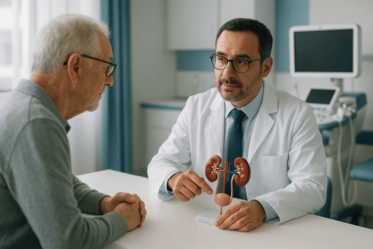
Ureter Diseases Treatment
Ureter diseases affect the tubes carrying urine from the kidneys to the bladder and may involve stones, narrowing, obstruction, reflux, infection or tumors. Diagnosis and treatment depend on cause, severity and kidney…

Quick answer
Ureter diseases are conditions affecting the two tubes that carry urine from the kidneys to the bladder, most commonly stones, strictures, reflux, injuries and tumours. Treatment ranges from observation and medication to endoscopic stone removal, stent placement and reconstructive surgery. The aim is always the same: restore normal urine drainage and protect the kidney before pressure or infection causes lasting damage.
Ureter Diseases: Understanding the Problem
Ureter diseases are conditions that affect the ureters: the two thin, muscular tubes that carry urine from each kidney down to the bladder. They include stones lodged in the tube, narrowed segments called strictures, blockages at the junctions where the ureter meets the kidney or the bladder, backward flow of urine known as reflux, injuries and tumours. When any of these stops urine draining normally, pressure builds in the kidney, and the aim of treatment is to restore drainage before that pressure causes lasting harm.
These conditions can be unsettling because they announce themselves in very different ways. You may feel sudden, severe pain in your side and feel sick: the classic picture of a stone. You may develop repeated urinary infections because urine is pooling above a blockage. You may learn, during a scan ordered for something else entirely, that one of your kidneys is swollen because urine is not draining from it. In other cases, blood in the urine raises concern for a tumour, or a child or an adult is found to have urine flowing backward from the bladder toward the kidneys.
Because “ureter disease” is not a single diagnosis, there is no single treatment. The right approach depends on the cause, the exact location of the problem, the severity of the obstruction and how well the affected kidney is still working. A small stone, a scar-related stricture and an invasive tumour can all produce the same swollen kidney on a scan, yet each demands a different plan. That is why careful diagnosis comes before every treatment decision in ureter care, and why a structured evaluation matters more than a quick answer.
As you weigh up treatment options, the questions tend to be practical ones. Is surgery truly necessary? Can the problem be treated through a minimally invasive approach? Is the kidney still working well enough to be worth saving? How quickly does treatment need to happen? This page explains how these conditions are assessed and treated, what each option involves, and which factors genuinely change the outcome, so that you can put those questions to any urologist with a clear picture of what the honest answers should cover.
What are the symptoms of ureter problems?
The most common symptoms of ureter problems are flank or back pain, blood in the urine, recurrent urinary tract infections, fever, nausea during episodes of colic, and changes in how often or how easily you pass urine. Pain from a stone is typically sudden and intense, radiating from the side toward the lower abdomen or groin. Pain from a slowly developing obstruction may be dull, intermittent or absent altogether. Some ureter conditions produce no symptoms at all and are discovered only when an ultrasound, CT or MRI shows a swollen kidney. The absence of pain is not proof that the kidney is safe: a partially blocked ureter can damage kidney tissue quietly over months or years while the other kidney compensates and blood tests stay normal.
What the Ureters Do
Ureter function is active, not passive. Each ureter is roughly 25 to 30 centimetres long and only a few millimetres wide. Waves of muscular contraction, called peristalsis, push urine downward from the kidney in small spurts, even when you are lying flat. At its lower end, the ureter passes through the bladder wall at an angle, creating a natural one-way valve that stops urine flowing back toward the kidney when the bladder fills and empties.
This anatomy explains most ureter problems. The tube has natural narrow points, where it leaves the kidney, where it crosses the pelvic blood vessels and where it enters the bladder, and stones tend to lodge at exactly these points. Scar tissue anywhere along the tube can narrow it enough to slow drainage. A faulty valve mechanism at the bladder allows reflux. And because the ureter’s blood supply is delicate and segmental, both injuries and surgical repairs demand careful technique if the tissue is to heal without new scarring.
What Treatment for Ureter Conditions Means
Treatment for ureter diseases covers the medical, endoscopic, minimally invasive and reconstructive procedures used to restore normal urine flow, remove obstruction, treat infection, manage reflux or address tumours involving the ureter. Because the underlying causes are so varied, treatment is personalised after careful evaluation rather than chosen from a fixed menu. The same finding on a scan can lead one patient to observation and another to major reconstruction, depending on what is driving it.
Some ureter conditions can be managed with medication, observation and follow-up imaging. Others require urgent drainage of the kidney, especially when obstruction is combined with infection, fever or falling kidney function. Ureter stones may pass on their own if they are small and not causing complications, but larger or impacted stones often need endoscopic removal or fragmentation. Narrowing of the ureter, a ureteral stricture, may need dilation, internal incision, stenting or reconstructive surgery. Vesicoureteral reflux, in which urine flows backward from the bladder toward the kidneys, may be treated medically, endoscopically or surgically depending on age, severity and the risk to the kidney. Ureter tumours require precise staging and may involve endoscopic management, segmental surgery, removal of the kidney and ureter in selected cases, and coordinated oncological care.
Modern ureter care puts kidney preservation first wherever that is realistic. In practice, this often means a staged approach: relieve the blockage with a temporary ureteral stent or a nephrostomy tube, let infection settle, clarify the anatomy with imaging, and only then carry out definitive treatment. Many procedures are performed entirely through the natural urinary passages using fine endoscopic instruments, which avoids external incisions and can shorten recovery. More complex strictures, injuries and tumours may require laparoscopic, robot-assisted or open reconstructive surgery. Neither route is automatically better; the right one depends on the length and position of the diseased segment, the quality of the surrounding tissue and the condition of the kidney above it.
Who Needs Evaluation for a Ureter Disease?
You may need evaluation for a ureter problem when symptoms, laboratory tests or imaging suggest that urine is not draining normally from a kidney to the bladder. The trigger can be dramatic, an episode of renal colic severe enough to bring you to hospital, or entirely incidental, such as hydronephrosis noticed on a routine ultrasound. Between those extremes sit recurring infections, unexplained blood in the urine and slowly changing kidney function on blood tests. All of these deserve a proper explanation, because each can be the first sign of a treatable drainage problem.
Common symptoms and findings that lead to evaluation include:
- Flank or back pain, either sudden and severe or persistent and dull
- Blood in the urine, visible or detected on urinalysis
- Recurrent urinary tract infections or kidney infections
- Fever, chills or feeling generally unwell alongside urinary symptoms
- Nausea and vomiting during episodes of kidney colic
- Difficulty passing urine or changes in urinary frequency
- Hydronephrosis found on ultrasound, CT or prenatal imaging
- Reduced kidney function on blood tests
- A history of pelvic surgery, radiation therapy, endometriosis, cancer or previous stone disease
How ureter diseases are diagnosed
Diagnosis begins with your medical history, a physical examination, urine tests and blood tests to assess infection, bleeding and kidney function. Imaging then does most of the work. Ultrasound can identify kidney swelling and some stones without any radiation. Computed tomography shows stones, the level of an obstruction, inflammation, tumours and the surrounding anatomy in detail. In selected cases, CT urography, MR urography, nuclear renal scans, retrograde pyelography, cystoscopy or diagnostic ureteroscopy are used to map the ureter more precisely. A nuclear renal scan is particularly useful when doctors need to measure how much each kidney contributes to overall function and whether an apparent obstruction is genuinely impairing drainage: a distinction that can change the entire treatment plan.
Part of the diagnostic task is ruling out look-alikes. Problems inside the kidney tissue itself, such as glomerular diseases or tubulointerstitial diseases, can also reduce kidney function but are managed medically by nephrologists rather than by relieving a blockage. Conditions of the bladder can mimic or coexist with ureter problems, since the two share the same lining and the same drainage pathway. Distinguishing a drainage problem from a tissue problem is one of the first questions the diagnostic pathway has to answer.
Whenever a second opinion is sought, prior records carry real weight. Imaging discs, radiology reports, operative notes, pathology reports, laboratory results and medication lists let a treating team see what has already been done and which decisions are still open. A second opinion built on complete records can clarify whether immediate intervention, watchful follow-up or a more complex reconstructive plan is the appropriate next step, and can spare you repeat tests that add delay without adding information.
Conditions and Indications Treated
Ureter diseases may involve obstruction, inflammation, reflux, injury, stones or cancer. The treatment pathway is shaped by the underlying condition and, above all, by whether the kidney is threatened. The most frequent indications are described below.
Ureteral stones form when mineral deposits develop in the urinary tract and lodge in the ureter. They can block urine flow abruptly and cause severe pain. Small stones in a favourable position may pass on their own under observation. If a stone does not pass, or if there is infection, persistent obstruction or risk to the kidney, treatment may include ureteroscopy with laser fragmentation, stent placement or other stone removal techniques. Patients who form stones repeatedly may also need a metabolic work-up to understand why.
Ureteral strictures are narrowed segments of the ureter. They develop after stones, surgery, radiation, infection, inflammation or injury, and they can damage the kidney slowly if drainage stays impaired. Treatment options range from endoscopic dilation and endoureterotomy to stenting and reconstructive surgery that removes or bypasses the narrowed segment. The length of the stricture and the health of the surrounding tissue largely decide which option is realistic.
Ureteropelvic junction obstruction is a blockage where the kidney’s drainage system meets the ureter. It may be congenital or acquired and affects both children and adults. When it causes pain, infection, stones or falling kidney function, surgery to reconstruct the junction, pyeloplasty, is often recommended.
Ureterovesical junction obstruction occurs where the ureter enters the bladder. It can cause hydronephrosis and recurrent infection. Management depends on severity and may involve observation, endoscopic procedures or reimplanting the ureter into the bladder.
Vesicoureteral reflux is the backward flow of urine from the bladder into the ureter and sometimes up to the kidney. It is most often diagnosed in children but is also relevant in adults with recurrent infection or previous urinary tract abnormalities. Treatment, medical, endoscopic or surgical, aims to reduce infection risk and protect kidney tissue, and the threshold for intervening depends on age, severity and how much the kidneys have already been affected.
Extrinsic compression is pressure on the ureter from outside the tube itself, from pregnancy, endometriosis, retroperitoneal fibrosis, enlarged lymph nodes, vascular abnormalities or pelvic masses. Because the ureter itself may be healthy, treatment focuses on the underlying condition, often with a temporary stent protecting drainage while that condition is addressed.
Ureteral tumours are usually urothelial cancers, arising from the same lining that also covers the renal pelvis and the bladder, which is why patients treated for ureter tumours need ongoing surveillance of the whole urinary tract, and why the field overlaps with bladder diseases. Diagnosis and treatment require careful staging, pathology review and usually multidisciplinary discussion. Options range from endoscopic treatment for highly selected low-risk lesions to more extensive surgery and systemic therapy for higher-risk disease.
Ureteral injury can follow pelvic surgery, trauma or radiation therapy. Early recognition matters, because a fresh injury and a scarred, months-old injury are repaired very differently. Treatment may involve stenting, drainage, direct repair or formal reconstruction, depending on the timing, location and extent of the damage.
Can you live with hydronephrosis?
Yes, many people live with mild hydronephrosis, particularly when the cause has been identified, the kidney is draining adequately and function is stable on follow-up tests. Hydronephrosis simply means the kidney is swollen with urine; it is a finding, not a diagnosis, and its seriousness depends entirely on what is causing it and how severe the pressure is. Mild swelling with good drainage may need nothing more than periodic imaging. Significant or worsening obstruction is a different matter: left untreated, it can lead to irreversible loss of function in that kidney, and infection developing behind the blockage adds real danger. The honest answer is therefore conditional, living with hydronephrosis is safe only once a doctor has established why it is there, confirmed that the kidney is not deteriorating, and set up a follow-up plan to catch any change.
What are the 10 diseases of the urinary system?
There is no official list of ten, but the urinary system conditions most often cited are:
- Urinary tract infections, including kidney infections
- Kidney stones
- Ureteral stones and ureteral obstruction
- Hydronephrosis
- Ureteral strictures
- Vesicoureteral reflux
- Cystitis and other bladder conditions
- Urinary incontinence
- Chronic kidney disease
- Cancers of the urinary tract, including bladder and urothelial cancers of the ureter and renal pelvis
Several of these overlap. A stone can cause obstruction, obstruction causes hydronephrosis, and stagnant urine above a blockage invites infection. That interconnection is why a full urinary tract assessment, rather than treatment of a single finding in isolation, is the standard of good care.
How Ureter Disease Treatment Is Performed
Treatment starts with defining the exact problem. A ureter stone, a scar-related stricture and a ureter tumour can all cause hydronephrosis, but they require very different strategies. A disciplined diagnostic phase protects you from both directions of error: undertreatment that lets a kidney deteriorate, and unnecessary procedures on a problem that could have been observed.
Preparation and diagnostic planning
Before any procedure, the care team reviews your symptoms, kidney function, infection status, imaging and overall health. Urinalysis and urine culture identify infection. Blood tests assess kidney function, inflammation, anaemia and fitness for anaesthesia. Imaging is reviewed to locate the obstruction, grade the hydronephrosis and map the relationship of the ureter to nearby organs and blood vessels, details that decide whether an endoscopic route is feasible or reconstruction will be needed.
If a tumour is suspected, additional imaging and endoscopic evaluation follow. Urine cytology and ureteroscopic biopsy can provide the tissue diagnosis that guides treatment. Complex cases are typically discussed in specialist meetings, urology-radiology conferences for difficult anatomy, multidisciplinary tumour boards when cancer is suspected. This structure exists for a reason: decisions that balance kidney preservation against cancer control, or endoscopic simplicity against reconstructive durability, are stronger when several specialists weigh in.
If infection is present behind an obstructed ureter, drainage becomes the first priority and everything else waits. Clinicians treat this combination as an emergency because infected urine trapped above a blockage often cannot be controlled by antibiotics alone; the pressure must be relieved first. Definitive stone removal or reconstruction is then deferred until fever, infection and inflammation have settled. This staged approach lowers risk and makes the definitive procedure safer. Where infections are complicated or recurrent, input from an infectious diseases specialist is part of the pathway.
Temporary drainage: ureteral stent or nephrostomy
When urine cannot drain, doctors place either a ureteral stent or a nephrostomy tube. A ureteral stent is a thin, flexible tube positioned inside the ureter, usually through the bladder with a cystoscope, that lets urine flow from kidney to bladder past an obstruction or after a procedure. A nephrostomy tube is placed through the skin directly into the kidney under image guidance, draining urine externally into a collection bag. The choice depends on the cause of the blockage, the anatomy, the severity of infection and what is technically feasible at that moment.
Temporary drainage relieves pressure, protects kidney function and helps bring infection under control. It is worth knowing the trade-offs in advance: stents commonly cause bladder irritation, urinary frequency or flank discomfort, especially in the first days. These symptoms are usually manageable, but they matter for planning, particularly if you will be living with a stent in place for a period while waiting for a second-stage procedure.
Endoscopic treatment through the urinary tract
Many ureter conditions can be treated endoscopically. During ureteroscopy, the urologist passes a very small camera through the urethra and bladder into the ureter, usually with no external incision at all. This is the standard approach for most ureter stones and for selected strictures and tumours.
For stones, fine instruments and laser energy fragment the stone into pieces small enough to pass or to be removed with a tiny basket. A temporary stent is often left afterwards to prevent swelling from blocking the ureter while it heals. For strictures, endoscopic dilation or an internal incision (endoureterotomy) may be appropriate in selected cases, typically short strictures with favourable anatomy and healthy surrounding tissue. For carefully selected low-risk ureter tumours, endoscopic biopsy, ablation or removal can be considered, always paired with close surveillance because urothelial tumours can recur.
Endoscopic work in the ureter depends on high-quality imaging, precise optics, controlled irrigation and instruments fine enough for a tube only millimetres wide. The advantage for you is treatment through the natural urinary passages, generally with a shorter hospital stay and a faster return to routine activity than open surgery. The honest limit is that endoscopy cannot fix everything: long strictures, dense scarring, complex injuries and aggressive tumours usually need more than an endoscope can offer, and choosing endoscopy in those situations only postpones the definitive operation.
How long does it take to recover from a ureteroscopy procedure?
Most people return to routine activities within a few days of ureteroscopy, and many procedures are done as day surgery or with a single overnight stay. Mild burning on urination, urinary frequency and a small amount of blood in the urine are common in the first days and usually settle on their own. If a stent was placed, some discomfort typically continues until it is removed: a matter of days to weeks depending on the reason it was inserted. Your surgeon will set the exact timeline, because it depends on what was treated, how inflamed the ureter was and whether further procedures are planned. Recovery from ureteroscopy is quick compared with reconstructive surgery, but the follow-up appointment for stent removal and post-treatment imaging is not optional; a forgotten stent can itself cause blockage and stones.
Minimally invasive and reconstructive surgery
When the ureter needs rebuilding rather than clearing, surgery may be laparoscopic, robot-assisted or open, depending on the complexity of the problem and the surgeon’s judgement. Reconstructive options include removing a narrowed segment and joining the healthy ends, reimplanting the ureter into the bladder, creating a flap from bladder tissue, using a segment of bowel in selected complex cases, or reconstructing the ureteropelvic junction.
For ureteropelvic junction obstruction, pyeloplasty is the standard reconstructive operation: the narrowed connection is removed or reshaped so urine drains freely from the kidney into the ureter. For lower ureter strictures and injuries, ureteral reimplantation connects the ureter to a new position in the bladder; where a gap has to be bridged, techniques such as a psoas hitch or a Boari flap bring bladder and ureter together without tension. These operations demand careful planning because the ureter’s blood supply is fragile, durable healing depends on tension-free reconstruction, healthy tissue and reliable drainage while the repair matures.
For ureter cancer, the operation depends on tumour grade, stage, location, kidney function and your general health. Some patients need removal of the kidney and ureter on the affected side. Others, particularly those with a solitary kidney, reduced kidney function or a low-risk localised lesion, may be candidates for kidney-sparing approaches. These decisions are individualised and usually made in a multidisciplinary setting, because the trade-off between cancer control and kidney preservation has no one-size answer.
Typical duration, hospital stay and recovery
Treatment length varies widely. A diagnostic cystoscopy or ureteroscopy is a short procedure; complex reconstruction or cancer surgery takes considerably longer and requires inpatient recovery. Endoscopic stone procedures are often day surgery or involve a short stay. Reconstructive operations need more monitoring, for pain control, urinary drainage, kidney function and early healing, before discharge is sensible.
Afterwards, you may carry a ureteral stent for days to weeks depending on the procedure. Follow-up imaging confirms that urine is draining well, and urine tests, kidney function tests and symptom review track your recovery. For tumour-related disease, ongoing surveillance is essential, because urothelial tumours can recur in the ureter, the kidney’s drainage system or the bladder long after the original treatment.
Hydration, activity levels, prescribed medicines and the timing of each follow-up test all belong in a proper discharge plan, and that advice should be specific to your procedure rather than a generic rule. Before leaving hospital, it is worth confirming when any stent comes out, when imaging is due and which results the care team will review with you.
Why Acting Early Matters
Ureter diseases can progress quietly. When urine flow is partially blocked, the kidney keeps producing urine while pressure gradually rises. Pain may come and go. Blood tests may stay normal for a long time if the other kidney is working well. All of this can create a false sense of safety, yet persistent obstruction can cause irreversible loss of function in the affected kidney.
Infection behind an obstructed ureter is the scenario clinicians take most seriously, and it is managed as an emergency in practice. Antibiotics alone are often not enough, because infected urine trapped above a blockage behaves like an abscess under pressure: it usually has to be drained before the infection can be controlled. This is why obstruction plus fever changes the pace of everything in ureter care.
Delay also complicates later treatment. A stone that stays impacted causes inflammation and scarring at the point where it sits. A stricture can lengthen and become more fibrotic, turning an endoscopic candidate into a reconstructive one. Repeated reflux-related infections scar kidney tissue in ways that cannot be undone. A tumour can advance past the stage where kidney-sparing treatment is reasonable. Early evaluation does not automatically mean early surgery, often it means the opposite, a confident decision to observe, but it is what separates the problems that can safely wait from the ones that cannot.
Benefits of Treatment
The benefits depend on the diagnosis, but the central aim is constant: restore safe urine drainage and protect the long-term health of the urinary tract.
| Benefit | What It Means for You |
|---|---|
| Relief of obstruction | Urine drains more normally from kidney to bladder, reducing pressure on the kidney and helping preserve its function. |
| Pain and symptom control | Treating stones, strictures or infection can reduce flank pain, nausea, urinary discomfort and repeated emergency visits. |
| Reduced infection risk | Correcting reflux or obstruction can lower the likelihood of recurrent urinary tract or kidney infections in appropriately selected patients. |
| Kidney preservation | Timely drainage and reconstruction may protect kidney tissue, especially when the affected kidney still has meaningful function. |
| More precise cancer management | For ureter tumours, accurate staging and tailored treatment balance cancer control against kidney preservation where clinically appropriate. |
| Clear long-term planning | A firm diagnosis lets your care team set out a defined plan for imaging, kidney tests, stent removal or tumour surveillance. |
Recovery Timeline After Ureter Treatment
Recovery depends on the procedure performed, the severity of the disease and whether infection, stents or reconstruction are involved. The pattern below is typical, not universal.
| Time Period | What You Can Expect |
|---|---|
| Day 1 | After endoscopic procedures, most patients walk the same day; burning on urination, frequency and mild blood in the urine are common. After major reconstruction, monitored hospital recovery is expected. |
| First week | Discomfort eases gradually. Your doctor may prescribe antibiotics, pain relief or bladder-relaxing medicines. Hydration and activity instructions are individualised. |
| First month | Many patients are back to routine activities after endoscopic treatment; reconstructive recovery continues for longer. Stent removal or follow-up imaging is often scheduled in this period. |
| Longer term | Kidney function tests and imaging confirm drainage. Patients treated for reflux, strictures, stones or tumours need periodic follow-up to watch for recurrence and protect kidney health. |
What Influences Outcomes and a Good Result?
A good result is not defined only by technical success on the operating table. It means the kidney drains adequately, infection risk stays controlled, symptoms improve and the plan still makes sense a year later. Several factors genuinely move the needle, and it is worth understanding them before consenting to any procedure.
The underlying cause matters most. A small stone in an otherwise healthy ureter carries a different outlook from a long radiation-related stricture or an invasive tumour. Benign and malignant conditions demand different follow-up and carry different risks, and no comparison between two patients’ experiences is meaningful without knowing what each was treated for.
Duration and severity of obstruction shape what is recoverable. A kidney blocked for a long time may not regain function even after perfect drainage is restored. Imaging shows swelling, but functional tests are sometimes needed to establish how much that kidney actually contributes. If the answer is very little, the honest discussion shifts from reconstruction to symptom control, infection prevention or, in selected cases, removal.
Infection status governs timing and safety. Procedures performed during uncontrolled infection carry higher risk. When obstruction and infection coincide, urgent drainage followed by staged definitive treatment is usually the safer path, even if it means two procedures instead of one.
Stricture location and length decide the technique. Short strictures may respond to endoscopic approaches in selected patients; longer or recurrent ones usually need reconstruction. Prior surgery, radiation therapy, endometriosis, inflammatory disease and pelvic tumours all make reconstruction more demanding, and a surgeon who has seen those situations before is better placed to judge what will actually last.
Stone size, hardness and position determine whether a stone is likely to pass and how easily it can be treated. Stones lodged in the ureter for a long time provoke swelling and scarring around themselves. If you form stones repeatedly, a metabolic evaluation can identify causes that diet, hydration and, where your doctor considers it indicated, medication can address, reducing the odds of going through this again.
For ureter tumours, grade, stage, location, multifocality and kidney function guide the plan. A kidney-sparing approach may be reasonable for selected low-risk tumours; higher-risk disease usually requires more extensive surgery and oncological treatment. Regular surveillance afterwards is not an optional extra but a core part of the treatment itself.
Your own circumstancesage, general health, diabetes, blood-thinning treatment, existing kidney disease, previous operations and your realistic ability to attend follow-up, all influence planning. A technically elegant operation is the wrong operation if its follow-up requirements do not fit your circumstances, your medical support at home or your capacity for long-term monitoring. Good surgeons plan around the patient they have, not the ideal patient.
How Ureter Care Is Organised at Acibadem
At Acibadem, ureter conditions are assessed through a structured diagnostic pathway rather than a single consultation. Urologists work with radiologists, nephrologists, anaesthesiologists, infectious disease specialists, oncologists and pathologists as each case requires, and patients with suspected ureter cancer are reviewed in multidisciplinary tumour boards where imaging, pathology and treatment options are weighed together. The purpose is straightforward: to identify the cause accurately and choose a plan that protects kidney function while avoiding unnecessary intervention.
Diagnostic depth matters in ureter care because small anatomical differences change the treatment. High-resolution cross-sectional imaging, ultrasound, endoscopic visualisation, functional kidney testing and image-guided drainage techniques are used to define the problem and plan the safest sequence. During procedures, modern endoscopic systems, laser technologies for stones, minimally invasive surgical platforms and image guidance are applied according to your condition and the hospital’s clinical protocols: the technique follows the diagnosis, not the other way round.
Experience carries particular weight in recurrent strictures, ureter injuries, congenital abnormalities, complex stones and tumours. These conditions demand judgement as much as technique. The real question is rarely whether a ureter can be opened or repaired; it is which approach will prove durable, which preserves kidney function, and what follow-up will be needed afterwards.
Planning Treatment and Follow-Up
If you have been told you have hydronephrosis, a ureteral stone, a narrowing of the ureter, reflux, an injury or a suspected tumour, the most important first step is usually not choosing an operation. It is understanding the cause, the condition of the kidney and the safest sequence of treatment. Sometimes the right plan involves urgent drainage; sometimes it is a scheduled procedure weeks later; sometimes it is structured surveillance with no procedure at all. Each of these is a legitimate outcome of a good evaluation.
Sequencing deserves particular attention. A staged plan, drainage first, definitive treatment after infection settles, means more than one procedure, and each stage carries its own preparation and recovery. A stent placed during treatment sets a removal date that has to be honoured, and a forgotten stent creates new problems of its own. Reconstructive surgery adds a longer recovery period during which strenuous activity may be inadvisable. None of this should be discovered after the fact: a realistic treatment plan states the expected hospital stay, the follow-up schedule and who is responsible for each step of surveillance.
Finally, keep every report. Operative notes, stone analysis results, pathology reports and imaging form the baseline against which all future findings are judged. These are, in many cases, conditions you manage over years rather than events you put behind you in a week, and patients who hold a complete record of their own care consistently get better-informed decisions from every doctor they see afterwards.
Preparation
- Patients usually have a urology consultation, urine tests, blood tests and imaging such as ultrasound or CT when needed. Bring previous scans, laboratory results and medication lists. Fasting or anesthesia preparation is only required if an endoscopic or surgical procedure is planned.
Aftercare
- Follow-up depends on the diagnosis and may include medication, stone management, stent care or surgery. Patients should drink fluids as advised, monitor fever, pain or changes in urination, and attend imaging or laboratory follow-ups. Urgent care is needed for severe pain, high fever or reduced urine output.
Frequently Asked Questions
What affects the cost of ureter disease treatment?
Cost depends on the diagnosis, the severity of obstruction or infection, kidney function, required imaging, procedure type, hospital stay, devices such as stents, pathology needs and follow-up. A personalised quote is only possible after a specialist reviews your medical records.
How can I get a personalised quote from Acibadem?
You can request a free consultation by sharing your reports, imaging, urine and blood test results, previous procedure notes and current symptoms. The international patient team can then coordinate a urology review and provide a case-based estimate.
Does a package usually include all ureter disease costs?
Packages may include selected consultations, tests, the planned procedure, hospital stay and standard coordination, but inclusions vary by diagnosis and treatment plan. Additional imaging, emergency care, pathology, extended stay or staged treatment may change the final cost.
Why might the final cost change after arrival?
The treatment plan may change if new tests show infection, reduced kidney function, a different cause of obstruction, a tumour suspicion or the need for drainage before definitive treatment. Your specialist will explain medically necessary changes before proceeding.
Is treatment in Turkey suitable for international patients with ureter diseases?
Turkey can be a practical option for international patients when care is coordinated through experienced urology teams and international patient services. Suitability depends on your diagnosis, urgency, travel safety and the specialist’s assessment.
Medically reviewed by the Acıbadem International Medical Board September 13, 2026
See our medical review board →
Update history
- PublishedJune 8, 2026
- Medical review approvedSeptember 13, 2026
- Last content updateSeptember 12, 2026
References2
- Ureteral Disorders medlineplus.gov
- Hydronephrosis my.clevelandclinic.org
Trusted care for international patients
From the Acibadem Blog

Which Scans Show a Ureter Problem? Ultrasound, CT and Kidney Function Tests Explained
A non-contrast CT scan is the most accurate way to find a stone or blockage in a ureter, while ultrasound is often the first…