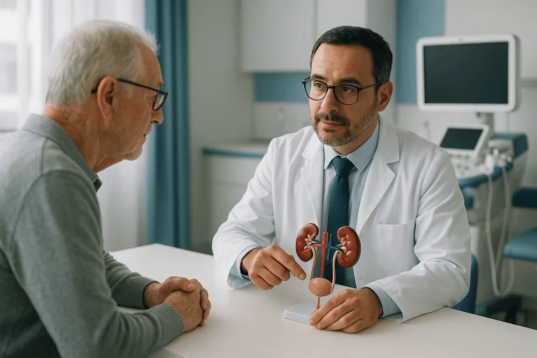
Bladder Tumor Resection Treatment
Bladder tumor resection, usually TURBT, removes visible bladder tumors through the urethra for diagnosis, staging, and treatment while preserving the bladder when appropriate.
Quick answer
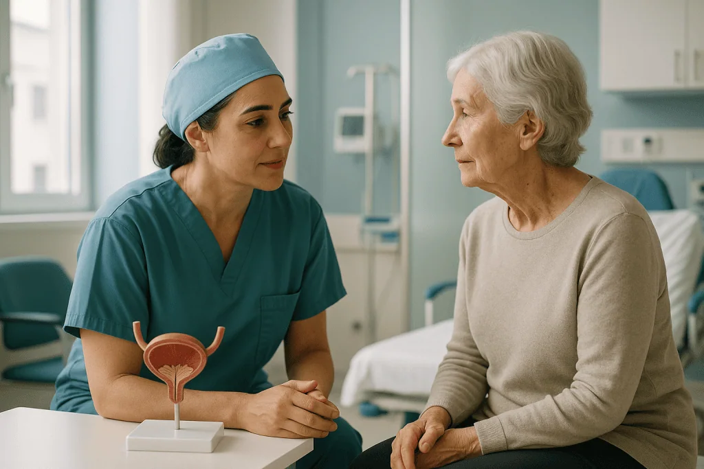
Bladder tumor resection, usually performed as transurethral resection of bladder tumor (TURBT), removes abnormal growths from the bladder lining using an instrument passed through the urethra, with no external incision. It serves two purposes: it removes visible tumour tissue, and it provides the specimen pathologists examine to identify the tumour type, grade and depth: the findings that determine all further treatment.
Bladder Tumor Resection: What It Is and What It Involves
Bladder tumor resection is an endoscopic procedure that removes abnormal growths from the inner lining of the bladder. It is most often performed as transurethral resection of bladder tumor, known as TURBT, in which the surgeon works through the urethra, the natural passage that carries urine out of the body, so there is no external incision. The procedure does two jobs at once. It removes the visible tumour, and it provides the tissue pathologists need to establish exactly what the growth is and how deeply it reaches into the bladder wall. Those two answers, taken together, decide everything that follows.
Most people who undergo bladder tumor resection have a suspected or confirmed bladder cancer. Some arrive at the procedure after blood appeared in their urine and a camera examination found a growth. Others are in follow-up after earlier treatment and a new lesion has been spotted during surveillance. A smaller group has abnormal urine test results without an obvious mass on first inspection. In every case the goal is the same: remove what can be seen safely, and find out precisely what it is.
A bladder tumour diagnosis raises immediate, practical questions. Is it cancer? Can the bladder be kept? How uncomfortable is the procedure, and how long does recovery take? This page works through those questions in order, what the operation is, who needs it, how it is performed, what recovery looks like, and how the pathology results steer the next steps.
What is a bladder tumor resection?
A bladder tumor resection is the surgical removal of a growth from inside the bladder, carried out with instruments passed through the urethra rather than through a cut in the abdomen. It is both diagnostic and therapeutic. It treats the condition by physically removing visible abnormal tissue from the bladder lining, and it diagnoses the condition by supplying that tissue for microscopic examination. For many people with disease confined to the bladder lining, the resection itself may be the main treatment. For people whose tumour has grown deeper, it provides the staging information on which every further decision rests.
The pathology report answers the critical questions. Is the tumour cancerous? Is it low grade or high grade? Has it stayed within the bladder lining, or has it invaded the muscle layer beneath? Are there features that point to a higher risk of recurrence or progression? These details shape what happens next: surveillance cystoscopy at set intervals, medication placed directly inside the bladder, a planned second resection, or a discussion about more extensive treatment. TURBT sits at the centre of bladder cancer decision-making because it produces the information that every later decision depends on.
What is transurethral resection of bladder tumor?
Transurethral resection of bladder tumor, abbreviated TURBT, is the standard technique for this operation worldwide. “Transurethral” simply means through the urethra. The urologist passes a thin surgical instrument called a resectoscope along the urethra into the bladder, fills the bladder gently with sterile fluid to open it up and improve visibility, and removes the tumour piece by piece using a fine electrical loop. Small blood vessels are sealed as the surgeon works to limit bleeding. Samples are usually taken from the base of the tumour, and sometimes from nearby areas of lining, to determine whether cancer cells have moved into the deeper layers of the bladder wall.
Is a laparoscope used for bladder tumor resection?
No. TURBT does not use a laparoscope. A laparoscope is a camera inserted through small incisions in the abdominal wall: the “keyhole” approach used for operations such as gallbladder surgery. TURBT uses a resectoscope passed along the urethra instead, so no incisions or ports are needed at all. Laparoscopic and robotic techniques do have a place in bladder cancer care, but for a different operation: removal of the entire bladder, called cystectomy, which is considered for selected patients with muscle-invasive disease. Removing a tumour from inside an intact bladder is always an endoscopic, transurethral procedure.
Who May Need Bladder Tumor Resection?
You may be advised to have the procedure when a camera examination or imaging shows a suspicious growth inside the bladder. The most common symptom that starts the whole process is blood in the urine, known medically as haematuria. The urine may look red, pink, tea-coloured or cola-coloured, or the blood may be detectable only on a laboratory test. Even painless bleeding that comes and goes deserves proper evaluation, particularly in adults and in people with recognised risk factors such as smoking or long-term occupational exposure to certain industrial chemicals. Intermittent bleeding is a feature of bladder tumours, not evidence against one.
Other symptoms include frequent urination, urgency, burning with urination, pelvic discomfort, or waking repeatedly at night to pass urine. These overlap heavily with common non-cancerous conditions. A bladder infection, bladder stones, an overactive bladder, prostate enlargement and various other bladder diseases can all produce very similar complaints. That is exactly why diagnosis relies on a structured evaluation rather than assumptions: a medical history, physical examination, urine testing, imaging where appropriate, and a direct look inside the bladder.
How do a cystoscopy and a transurethral resection fit together?
They are two separate steps that often follow one another. A diagnostic cystoscopy is a look, not an operation: the urologist passes a slim flexible camera into the bladder, usually in a clinic setting with local anaesthetic gel, to inspect the lining. If a tumour or suspicious area is seen, the next step is normally a TURBT: a formal operation in a theatre, under general or regional anaesthesia, using a larger rigid instrument capable of cutting tissue and controlling bleeding. In other words, cystoscopy finds the problem and TURBT removes it. Imaging studies such as ultrasound, CT urography or MRI may be added at either stage to assess the rest of the urinary tract, check kidney drainage, look for additional lesions and help plan treatment.
Several typical situations lead to a recommendation for TURBT:
- A newly discovered bladder mass found during investigation of symptoms or on imaging done for another reason.
- A recurrence detected during scheduled follow-up after previous bladder cancer treatment.
- Abnormal urine cytology, meaning laboratory tests have found suspicious cells in the urine even when a tumour is not immediately obvious on inspection.
- A planned repeat resection after an initial TURBT, recommended particularly when the first tumour was high grade, large or multiple, or when the first pathology specimen did not include enough of the muscle layer to stage the disease reliably.
A repeat resection deserves a word of reassurance, because patients sometimes read it as a failure of the first operation. It is not. In selected higher-risk situations, a planned second resection is standard, evidence-based practice: it confirms the staging, removes any residual tumour at the original site, and gives the pathologist a second, deeper specimen to examine. Many treatment guidelines around the world specifically recommend it.
Conditions and Indications Treated With Bladder Tumor Resection
Bladder tumor resection is most often used for suspected or confirmed bladder cancer arising from the bladder lining. The most common type by far is urothelial carcinoma, previously called transitional cell carcinoma. The same lining extends up through the ureters towards the kidneys, which is why patients investigated for bladder tumours are sometimes also assessed for ureteral tumors. TURBT is also used to evaluate less common bladder tumours and abnormal lesions that cannot be diagnosed safely by appearance alone.
The procedure is especially important in non-muscle-invasive bladder cancertumours confined to the inner lining or to the thin layer of connective tissue just beneath it. Depending on their appearance and depth, these are categorised as Ta (confined to the lining), T1 (into the connective tissue but not the muscle) or carcinoma in situ (a flat, high-grade change within the lining itself). For many of these tumours, TURBT removes the visible disease and, together with the pathology findings, determines whether further treatment inside the bladder is needed.
Two words come up constantly in these conversations, and it helps to keep them apart. Grade describes how abnormal the cells look under the microscope: low-grade cells still resemble normal bladder lining and tend to behave indolently, while high-grade cells look disorganised and carry a genuine capacity to invade. Stage describes depth, how far the tumour has physically grown into or through the bladder wall. A tumour can be low stage but high grade, and that combination is treated with particular respect, because the cells have aggressive potential even though they have not yet travelled far. The pathology report always addresses both.
In muscle-invasive bladder cancer, where the tumour has grown into the muscular wall, TURBT is usually not the definitive treatment on its own. It remains essential nonetheless, because the tissue it provides is what proves the invasion and stages the disease. Those findings anchor the discussion of the larger options: bladder removal surgery, bladder-preserving protocols that combine resection with radiation and systemic therapy, chemotherapy, immunotherapy, or combinations of these.
TURBT is also the standard response to recurrent tumours found during surveillance. Bladder cancer has a well-recognised tendency to come back in the lining, which is why structured monitoring is part of standard care rather than a sign of pessimism. When a new lesion appears, resecting it establishes whether it resembles the previous tumour or whether its grade or stage has changed: a distinction that can alter the whole treatment plan.
Finally, in selected cases the procedure is used to remove benign growths, inflammatory lesions that mimic tumours, or areas of tissue change that simply cannot be identified with confidence through the camera. The overriding purpose in all of these situations is the same: a reliable diagnosis, obtained while removing visible abnormal tissue where it is safe and appropriate to do so.
How Bladder Tumor Resection Is Performed
Preparation Before the Procedure
Before TURBT, your urologist reviews your symptoms, cystoscopy findings, imaging, medical history, current medications and any previous pathology reports. Blood and urine tests are commonly arranged to check kidney function, blood counts, clotting status and signs of infection. If bacteria are found in the urine, the operation may need to wait until the infection has been treated, because operating on an infected bladder raises the risk of complications. If you have had investigations or treatment elsewhere, gathering recent reports, imaging files, medication lists and details of previous surgery in advance helps the team complete this planning efficiently and avoid repeating tests unnecessarily.
Medication planning needs particular care. Blood thinners, antiplatelet drugs and certain supplements can increase bleeding during and after resection, yet interrupting them carries its own risks for people with heart disease, coronary stents, a history of stroke or clotting disorders. There is no universal rule here: the decision belongs to your treating doctors, sometimes with input from cardiology and anaesthesia colleagues, and is made individually for each patient. Do not adjust anything on your own; bring an accurate, complete medication list to every appointment instead.
Anaesthesia planning is the final piece. TURBT may be performed under general anaesthesia, in which you are asleep, or regional anaesthesia, in which the lower body is numbed while you remain awake or lightly sedated. The choice depends on the tumour’s size and position, the expected length of the operation, your general health and the anaesthetist’s assessment.
During the Procedure
The operation itself follows a consistent sequence:
- Anaesthesia is administered and you are positioned on the operating table.
- The urologist passes the resectoscope through the urethra into the bladder.
- The bladder is filled with sterile fluid to expand it and improve visibility.
- The entire lining is inspected systematically: the side walls, posterior wall, dome, bladder neck and the areas around the ureteral openings, all common tumour sites.
- The projecting (exophytic) part of the tumour is removed first, using the electrical loop.
- The base of the tumour is then resected or sampled separately, so the pathologist can assess how deep the growth extends, including, where indicated, tissue from the underlying muscle layer.
- Bleeding points are sealed, and any additional suspicious flat areas may be biopsied.
- In many patients, a urinary catheter is placed at the end to drain the bladder, wash out blood and let the resected surface rest.
Two energy systems are in common use for the resecting loop. Monopolar resection, the traditional method, requires a non-conductive irrigation fluid. Bipolar resection works in saline, which reduces the consequences of irrigation fluid being absorbed into the circulation during longer operations and has become widespread for that reason. A further refinement, en-bloc resection, lifts suitable tumours off the bladder wall in a single piece rather than in fragments; this can make the pathologist’s assessment of the tumour’s depth easier, though it is only feasible for tumours of certain sizes and positions. Which technique is used is a surgical judgement made case by case, and none of them changes the fundamental logic of the operation.
Modern operative pathways may add enhanced visualisation techniques in selected cases. Photodynamic diagnosis, often called blue-light cystoscopy, uses a light-sensitive agent instilled into the bladder beforehand; abnormal tissue takes up the agent and stands out under blue light. Narrow-band imaging works differently, filtering the light to sharpen the contrast of blood vessels in the lining. Both methods can highlight subtle changes, particularly flat lesions such as carcinoma in situ, that are difficult to see under standard white light. High-resolution endoscopic imaging, careful mapping of tumour locations and thorough pathology evaluation all contribute to more precise staging. Which technologies are used depends on the individual case and the hospital’s clinical protocols; none of them replaces careful, systematic surgical technique.
The duration varies. A small single tumour may be dealt with relatively quickly, while multiple, large or awkwardly positioned tumours take longer, as does careful sampling of the tumour base or control of bleeding. Afterwards you spend time in a recovery area while the anaesthetic wears off and the team monitors your comfort, your urine and your vital signs.
Is bladder tumor removal a major surgery?
TURBT is a genuine operation performed under anaesthesia, but it is not major surgery in the sense that open abdominal procedures are. There is no incision, no bowel handling and no lengthy intensive recovery; most patients go home the same day or after one night, depending on the extent of resection and their general health. It should still be taken seriously, any operation on the bladder carries risks, but it belongs firmly at the less invasive end of cancer surgery. Removal of the entire bladder, by contrast, is major surgery. That operation, radical cystectomy, is a different procedure considered in a different situation, usually muscle-invasive disease, and should not be confused with resection of a tumour from inside the bladder.
Risks and Possible Complications
Complications after TURBT are possible, and an honest account of them belongs in any decision. Bleeding is the most common issue; occasionally clots can block the flow of urine and need washing out through a catheter. Urinary infection can occur and may need antibiotic treatment decided by the medical team. The bladder wall can be perforated during resection, more likely with deep or awkwardly placed tumours, and is usually managed with a period of catheter drainage, though a repair operation is occasionally required. Temporary burning, frequency and urgency are expected rather than exceptional. Less commonly, scarring can later narrow the urethra. Anaesthesia carries its own separate, individually assessed risks.
Two technical events are worth knowing by name. The obturator nerve runs close to the lower side wall of the bladder, and electrical stimulation during resection of a lateral-wall tumour can trigger a sudden, involuntary leg movement; anaesthetic and surgical techniques exist specifically to prevent this, and it is one reason tumour position matters in operative planning. Separately, during prolonged resections irrigation fluid can be absorbed through opened blood vessels into the circulation; saline-based bipolar systems have made significant problems from fluid absorption considerably less of a concern than in earlier eras. Your surgical and anaesthetic teams will explain how all of these considerations apply to your particular case, and which warning signs they want you to watch for after discharge.
Medication Placed Into the Bladder
For some patients, the urologist recommends a single dose of intravesical medication shortly after the resection. The drug is instilled directly into the bladder through the catheter, held there for a defined period and then drained. Its purpose is to reduce the chance that tumour cells released during resection implant elsewhere on the bladder lining. It is not appropriate for everyone: it is generally avoided when there is concern about perforation, significant bleeding or a very extensive resection, and the decision is made in theatre, based on what the surgeon found and how the operation went.
Once the pathology report is available, further intravesical therapy may be proposed depending on the tumour’s risk profile. Some patients receive courses of chemotherapy agents placed inside the bladder. Others, particularly those with higher-risk non-muscle-invasive disease, may be considered for immunotherapy delivered the same way. Your urologist will explain why a particular medication is or is not recommended for you and how it fits into the overall plan.
Immediately After TURBT
Mild burning with urination, urinary frequency and blood-tinged urine are common for several days after the procedure. Some patients pass small clots. A catheter may stay in place briefly, especially after a larger resection, to keep the bladder draining and allow the team to monitor bleeding. Once the urine has cleared sufficiently and you can urinate safely, discharge follows: the same day or after an overnight stay, depending on the extent of surgery and your overall condition.
Before you leave, the team gives you specific guidance on fluid intake, activity, medications and when travel is sensible. Heavy lifting, strenuous exercise and sexual activity are generally paused for a period your surgeon defines. Because bleeding can briefly return while the resected area heals, it is worth following the activity guidance even when you feel entirely well.
The pathology report is one of the most important products of the whole process. It can take several days to finalise, depending on the complexity of the analysis. When it is ready, your urologist reviews the tumour type, grade, stage, whether muscle was present in the specimen and any additional findings, and that conversation determines whether the plan is surveillance, repeat resection, intravesical therapy, further imaging or a discussion of more extensive treatment.
Why Acting Early Matters
Blood in the urine can come and go, and some people put off evaluation when the symptom fades. That is a risky habit. Bladder tumours bleed intermittently, so clear urine does not mean the underlying cause has resolved. Early assessment lets suspicious lesions be identified and removed before they enlarge or advance, and moves you from uncertainty to a plan based on actual tissue findings rather than guesswork.
For non-muscle-invasive disease, timely resection removes the visible tumour and produces the risk information that guides recurrence prevention. Delay allows tumours to grow larger, become harder to remove completely, or progress into deeper layers where the treatment conversation changes entirely. For high-grade tumours, prompt staging matters most of all, because the subsequent decisions can be time-sensitive. Delay also invites practical complications, ongoing bleeding, anaemia, urinary obstruction and clot retention among them. Not every bladder tumour behaves aggressively, but there is no reliable way to know which ones will without tissue diagnosis.
How fast does bladder cancer spread?
There is no single timetable, and anyone who offers one is oversimplifying. Behaviour depends heavily on grade and type. Low-grade papillary tumours tend to grow slowly, and when they recur they usually reappear as similar low-grade disease in the lining rather than spreading beyond it. High-grade tumours and carcinoma in situ are different: they have a genuine capacity to invade the muscle wall and, from there, to reach lymph nodes and distant organs, and in some people that progression happens over months rather than years. Because a tumour’s appearance through the camera cannot reliably predict its behaviour, the sensible response to any bladder tumour is tissue diagnosis without unnecessary delay.
Benefits of Bladder Tumor Resection
The value of TURBT lies in what it removes and, just as importantly, in what it reveals.
| Benefit | What It Means for You |
|---|---|
| Diagnosis from tissue analysis | Pathology confirms whether the tumour is cancerous and identifies the type, grade and depth of invasion. |
| Removal of visible tumour | The urologist removes abnormal tissue seen inside the bladder, which may be the main treatment for many non-muscle-invasive tumours. |
| Bladder preservation when appropriate | Because TURBT is performed through the urethra, it can treat selected tumours while preserving the bladder structure. |
| More accurate staging | Sampling the tumour base and bladder wall helps determine whether additional treatment is needed. |
| Guidance for future care | The results define surveillance intervals, intravesical therapy options and whether more extensive treatment should be discussed. |
Recovery Timeline After Bladder Tumor Resection
Recovery varies with tumour size, the extent of resection, anaesthesia type and your general health, but most people return gradually to normal routines over days to weeks rather than months.
| Time Period | What Patients Can Expect |
|---|---|
| Day 1 | Burning with urination, urgency, blood-tinged urine or a catheter. The care team monitors bleeding, comfort and your ability to urinate. |
| First week | Urinary symptoms usually settle gradually. Light activity is encouraged; heavy lifting and strenuous exercise are typically avoided. |
| First month | The bladder lining continues to heal. Pathology results are reviewed, and your urologist may recommend surveillance, medication in the bladder or a repeat resection if needed. |
| Longer term | Follow-up cystoscopy and urine testing are scheduled because bladder tumours can recur. The frequency depends on your tumour’s risk features. |
A few practical points make the early weeks easier. Drinking well helps keep the urine dilute and flushes the healing surface, unless your team has advised a fluid restriction for another medical reason. Expect the urine colour to fluctuate: it commonly clears, then pinks again briefly when the internal scab over the resection site separates as part of normal healing. Constipation is worth avoiding, since straining raises pressure on the healing bladder. Desk-based work can usually resume within days; physically demanding work waits until your surgeon agrees.
If you go home with a catheter for a few days, the nursing team will show you how to manage it before discharge: keeping the drainage bag below the level of the bladder, securing the tubing so it does not pull, keeping the area clean, and watching the colour and flow of the drainage. A catheter that suddenly stops draining, especially alongside lower abdominal fullness or worsening discomfort, usually means a blockage rather than an emergency of the tumour itself, but it does need prompt attention from the team caring for you. Removal of the catheter is a quick clinic procedure, after which mild stinging and urgency for a day or two are normal.
Keep copies of your operative note and the full pathology report once it is issued, and take them to every subsequent appointment. Bladder tumour follow-up runs for years and often involves more than one clinician; a complete written record means every doctor who sees you later starts from the full picture rather than a summary of a summary.
What Influences Outcomes After TURBT?
Outcomes after bladder tumor resection depend on both the biology of the tumour and the quality of the evaluation. The tumour-related factors are the number of tumours, their size, grade and stage, the presence of carcinoma in situ, the pattern of any previous recurrences, and whether the muscle layer is involved. A small, solitary, low-grade, non-invasive tumour is a genuinely different disease from a high-grade or invasive one, and treatment plans reflect that difference rather than applying one template to everyone.
The completeness of the resection matters just as much. A careful TURBT aims to remove all visible tumour and to obtain representative tissue from the tumour base. When the specimen includes muscle, staging is more reliable. When muscle is absent in certain higher-risk situations, or when the tumour is high grade or T1, a repeat TURBT is commonly recommended to confirm the stage and clear any residual disease. To repeat the earlier point: this is standard practice in selected patients, not evidence that the first operation went wrong.
Pathology expertise plays a central role, because bladder tumour treatment hangs on microscopic findings. In complex cases, additional pathology review or discussion at a multidisciplinary tumour board helps align urology, medical oncology, radiation oncology, radiology and pathology around one coordinated plan. Patient factors count too: smoking history, occupational exposures, kidney function, other medical conditions, immune status and the practical ability to attend follow-up all shape recommendations.
Smoking deserves its own paragraph. Tobacco exposure is associated with both the development of bladder cancer and its recurrence, because carcinogens from smoke are concentrated in the urine and bathe the bladder lining for hours at a time. Stopping is worthwhile at any point, including after diagnosis and after resection, and support for cessation is a legitimate part of bladder cancer care, not an afterthought. If quitting has defeated you before, say so at your urology appointments; structured help exists and asking for it is entirely routine.
Above all, adherence to surveillance is one of the most important long-term factors within your control. Bladder cancer can recur even after a technically excellent resection. Scheduled cystoscopy finds recurrences early, typically when they are smaller and simpler to deal with. Your urologist will set a surveillance plan matched to your risk category and pathology results, and keeping those appointments is treatment, not administration.
Why does bladder cancer keep coming back?
Because the whole bladder lining shares the same history. The urothelium that lines the bladder has usually been exposed, over years, to the same carcinogens, most often from tobacco smoke filtered into the urine, so the lining that produced one tumour retains the capacity to produce others, a phenomenon sometimes called the field effect. In addition, tumour cells disturbed during resection can occasionally implant elsewhere on the lining, which is precisely why a single dose of intravesical medication is sometimes given straight after surgery, and why some tumours are multifocal from the outset. None of this means recurrence is inevitable; it means surveillance is rational, and that intravesical therapy, smoking cessation and regular cystoscopy each address a real mechanism rather than a theoretical one.
What is the life expectancy after bladder removal for cancer?
There is no honest single answer, and a responsible page will not invent one. First, be clear about terms: bladder removal, radical cystectomy, is a different and much larger operation than TURBT, considered mainly for muscle-invasive disease or for high-risk tumours that have not responded to bladder-preserving treatment. Outlook after cystectomy depends on the stage of the cancer at the time of surgery, whether lymph nodes were involved, the response to any chemotherapy or immunotherapy, and the patient’s age and general health. These variables differ so much between individuals that only your treating team, holding your actual pathology, can discuss prognosis meaningfully. What can be said in general is that cystectomy is performed with the intention of long-term disease control, and that the earlier invasive disease is identified, which is exactly what a thorough TURBT achieves, the more options remain open.
Coordinated Care and Continuity After Treatment
Bladder tumour care is rarely a single-specialty affair. A patient may begin with urology, but pathology, radiology, medical oncology, radiation oncology, anaesthesiology and nursing teams all contribute at different points, and higher-risk or muscle-invasive cases benefit from review at multidisciplinary tumour boards where the options are weighed from several perspectives at once. At Acibadem, bladder tumour evaluation and TURBT are planned within this kind of coordinated, protocol-based pathway: preoperative assessment, appropriate imaging, resection planned around the tumour’s characteristics, pathology-driven staging and a structured follow-up recommendation at the end of it.
Continuity after treatment deserves equal attention. Bladder tumours require follow-up measured in years, and surveillance can often be shared sensibly between the operating centre and a urologist closer to home. Written summaries, copies of the operative note and the full pathology report make that handover work. Some patients seek a second opinion before committing to further treatment, bringing a cystoscopy report or pathology slides and asking for the findings to be reassessed. The same documentation principle applies there: the more complete the record, the more useful the opinion.
Personalisation runs through all of it. A small low-grade tumour in an otherwise healthy person calls for a different plan from a high-grade T1 tumour, carcinoma in situ, or a lesion suspicious for muscle invasion. Age alone does not determine treatment. What determines it is the complete picture: the pathology, the imaging, cardiac and pulmonary health, kidney function, medications, previous surgery and the patient’s own priorities, explained directly, in plain language, including what remains uncertain.
Questions Worth Asking Your Urologist
A short list of focused questions makes the pathology conversation far more useful:
- What type and grade of tumour was found, and what stage is it?
- Was muscle present in the specimen, and was the tumour completely removed?
- Do I need a repeat resection, and if so, why and when?
- Is medication inside the bladder recommended in my case: a single dose, a course, or neither?
- What surveillance schedule applies to my risk category, and what does each check involve?
- Which symptoms after discharge should prompt an earlier review?
- How should long-term surveillance be shared between the operating centre and my usual urologist?
Moving Forward With Clarity
Bladder tumor resection is the pivotal step in diagnosing and treating bladder tumours. It removes visible abnormal tissue, provides the pathology information on which every subsequent decision rests, and establishes whether further therapy is needed at all. For many patients it opens a bladder-preserving pathway of resection, possible intravesical treatment and structured surveillance. For others it supplies the staging that makes the choice of a larger treatment rational rather than speculative.
Either way, the sequence is the same: remove, examine, understand, then decide. Patients who go into TURBT knowing what the procedure involves, what the pathology report will say and why follow-up continues afterwards tend to find the process far less daunting than the diagnosis first suggested. The operation itself is usually the shortest part of the journey; the clarity it produces is what carries everything after it.
Preparation
- Before bladder tumor resection, patients usually have urine tests, blood tests, cystoscopy, and imaging if needed. Blood thinners may need to be stopped under medical guidance. Patients are typically asked to fast for several hours before anesthesia.
Aftercare
- A urinary catheter may be used temporarily, and mild burning, frequent urination, or blood in the urine can occur for a few days. Patients should drink fluids, avoid heavy lifting, and follow medication instructions. Pathology results guide further treatment such as intravesical therapy or surveillance cystoscopy.
Frequently Asked Questions
What affects the cost of bladder tumor resection?
The final cost depends on tumor characteristics, preoperative tests, anesthesia needs, operating room time, pathology analysis, hospital stay, and whether additional treatments such as intravesical therapy or repeat TURBT are required.
How can I get a personalised quote from Acibadem?
You can request a free consultation by sharing your medical reports, cystoscopy findings, imaging results, pathology reports if available, and current medications. The urology team can then review your case and prepare a personalised treatment and cost estimate.
Does a package usually include pathology?
Many international patient packages include standard pathology review after TURBT, but this should always be confirmed. Additional staining, specialist pathology review, or further molecular tests may affect the final cost if recommended.
Will I need additional treatment after TURBT?
Possibly. TURBT provides important diagnostic and staging information, and the next step depends on the pathology result. Some patients only need surveillance, while others may need intravesical therapy, repeat resection, or more extensive treatment.
Is travelling to Turkey for TURBT suitable for every patient?
Not always. Suitability depends on symptoms, cancer suspicion, fitness for anesthesia, travel safety, and how urgently treatment is needed. A specialist review is recommended before making travel plans.
Is this information medical or financial advice?
No. This is general educational information only. A urologist should assess your medical suitability, and the international patient team can provide a personalised quote after reviewing your documents.
Medically reviewed by the Acıbadem International Medical Board September 1, 2026
See our medical review board →
Update history
- PublishedJune 8, 2026
- Medical review approvedSeptember 1, 2026
- Last content updateSeptember 8, 2026
References3
- Bladder Cancer Treatment (PDQ) - Patient Version cancer.gov
- Bladder cancer nhs.uk
- Bladder Cancer medlineplus.gov
Trusted care for international patients
From the Acibadem Blog

What Happens After TURBT? How Pathology Results and Follow-Up Cystoscopy Guide the Next Steps
After TURBT, the removed tissue goes to a pathologist, whose report on stage, grade and the presence of bladder muscle in the sample decides…