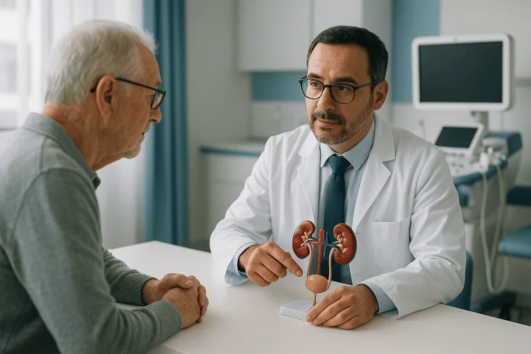
Ureteral Tumors Treatment
Ureteral tumors are usually managed with precise staging and individualized treatment, often combining endoscopic, robotic or open surgery with oncology care when needed.

Quick answer
Ureteral tumors are growths in the narrow tubes that carry urine from the kidneys to the bladder; most are urothelial carcinomas. Diagnosis relies on urine tests, CT urography and ureteroscopy with biopsy. Treatment ranges from kidney-sparing endoscopic removal for small, low-grade tumors to radical nephroureterectomy, removal of the kidney, ureter and a cuff of bladder, sometimes combined with chemotherapy or immunotherapy.
What Are Ureteral Tumors?
Ureteral tumors are abnormal growths that develop in one or both ureters: the narrow, muscular tubes that carry urine from each kidney down to the bladder. Most are urothelial carcinomas, cancers that begin in the specialised lining of the urinary tract, and treatment ranges from removal through a telescope passed along the natural urinary passage to surgery that removes the kidney and the entire ureter together. Which approach is right depends on the tumor’s grade, how deeply it has grown, where it sits in the ureter, and how well your kidneys work.
The ureters are easy to overlook. Each is roughly the width of a drinking straw and moves urine downwards in coordinated waves of muscle contraction. A growth inside this confined channel matters for two reasons. First, it may be malignant. Second, even a small tumor can obstruct drainage from the kidney above it, causing urine to back up and pressure to build. Both problems shape how ureteral tumors are investigated and treated, and both explain why a finding in the ureter is taken seriously even when it looks small on a scan.
Upper tract urothelial carcinoma
Upper tract urothelial carcinoma is the medical name for cancer arising in the lining of the renal pelvis, the urine-collecting chamber inside the kidney, or the ureter. The most common type of ureteral tumor is urothelial carcinoma, also called transitional cell carcinoma. The same lining, the urothelium, covers the renal pelvis, the ureters, the bladder and part of the urethra. Because this entire surface is bathed in the same urine and exposed to the same risk factors, a tumor in the ureter may be related to tumors elsewhere in the urinary tract, particularly the bladder or the renal pelvis. Doctors sometimes call this a field effect. It is the reason surveillance after treatment covers the whole urinary tract, not only the spot where the original tumor was found.
How doctors classify ureteral tumors
Classification is not academic; it decides treatment. Ureteral tumors are described in several ways at once, and each description narrows the options:
- Grade. Low-grade tumors have cells that look closer to normal under the microscope and tend to grow slowly. High-grade tumors look markedly abnormal and are more likely to invade the wall of the ureter and to spread beyond it.
- Depth. Non-invasive tumors are confined to the lining. Invasive tumors have grown into the muscular wall of the ureter or through it, into the surrounding fat or nearby structures.
- Location. Tumors are described as upper, middle or lower ureteral. Location matters because it determines which operations are technically feasible: a tumor near the bladder can sometimes be removed with the lower ureter alone, while a tumor high near the kidney usually cannot.
- Number. A solitary tumor is approached differently from multifocal disease, where several areas of the lining are affected at the same time.
These distinctions strongly influence whether a kidney-sparing approach can be considered or whether more extensive surgery is the safer recommendation. Two patients with “a ureteral tumor” can be given very different, equally correct treatment plans.
Are all ureteral tumors cancer?
No, but the majority found in adults are. Benign growths of the ureter exist, fibroepithelial polyps are one example, and they can produce the same symptoms and the same imaging findings as cancer. Only pathology can settle the question, which is why biopsy and expert microscopic review sit at the centre of the diagnostic pathway. It is also worth knowing that even a benign growth may still need treatment if it blocks the flow of urine from the kidney, because prolonged obstruction can damage kidney function regardless of what the growth is made of.
Facing a Ureteral Tumor Diagnosis
A diagnosis of a ureteral tumor usually arrives unexpectedly. Most people never think about their ureters until a scan, a urine test or an episode of bleeding raises concern. Many patients first learn of a possible tumor after seeing blood in the urine, developing pain in the flank, or undergoing imaging for an entirely different reason. Others arrive with a more complicated history, previous bladder cancer, long-standing kidney problems, or repeated urinary tract investigations that never quite produced an answer.
Because these tumors are relatively uncommon, patients and families tend to have the same set of questions. Is it cancer? Has it spread? Can the kidney be saved? Will treatment mean major surgery? Is chemotherapy needed? How quickly should care begin? These are reasonable questions, and honest answers depend on information that is usually not available on the day of diagnosis: the tumor’s grade, its depth, its exact position, and whether the rest of the urinary tract is clear.
That is why the first phase of care is staging rather than treatment. The goal is to establish where the tumor is, how aggressive it appears, whether it is affecting drainage from the kidney, and whether there is any evidence of spread. Only then can a plan be matched to the actual risk. The alternative, treating on assumption, risks both undertreatment of an aggressive cancer and unnecessary removal of a kidney for a tumor that could have been managed conservatively. At Acibadem, patients with suspected or confirmed ureteral tumors are evaluated through a coordinated pathway that may involve urologic oncology, radiology, pathology, medical oncology, radiation oncology, nephrology, anaesthesiology and supportive care specialists, so that the plan reflects the tumor, the kidneys and the whole person rather than any single test result.
Symptoms and Early Signs of Ureteral Tumors
Symptoms of ureteral tumors are often subtle, intermittent, or absent altogether, which is precisely what makes them easy to dismiss. The pattern below covers what most patients notice first, and what tends to appear later.
What are the early signs of ureter cancer?
The earliest and most common sign is blood in the urine, which may appear pink, red, brown or tea-coloured, or may be invisible and picked up only under a microscope during routine testing. Some people notice a dull ache or pressure in the flank, the side of the back below the ribs, caused by urine backing up behind a partial blockage. Many have no symptoms at all in the early stages, and the tumor is discovered incidentally on an ultrasound, CT or MRI performed for another reason. Later features can include recurrent urinary symptoms, unexplained changes in kidney function, and, in more advanced disease, weight loss and fatigue. The important point is that early signs are usually mild and often come and go, which is why intermittent bleeding should never be interpreted as a problem that has resolved.
Blood in the urine (haematuria)
Blood in the urine deserves proper evaluation in every adult, and particularly in smokers, former smokers, and anyone with a history of urothelial cancer. A ureteral tumor bleeds directly into the stream of urine passing over it, so bleeding can be striking one week and completely absent the next as the tumor surface heals over. Microscopic haematuria, blood detectable only on laboratory testing, carries the same significance as visible bleeding and is investigated the same way. Painless bleeding is, if anything, more suspicious than painful bleeding, because pain usually points towards stones or infection, while a tumor can bleed without causing any discomfort at all.
Flank pain and hydronephrosis
Flank pain develops when a tumor narrows or blocks the ureter and urine can no longer drain freely from the kidney above. The kidney’s collecting system swells with retained urine, a condition called hydronephrosis. The pain is typically a dull, persistent ache rather than the violent, wave-like colic of a passing stone, though the two can overlap. Hydronephrosis found on a scan without an obvious cause is itself a reason for careful evaluation of the ureter, because silent obstruction can gradually erode kidney function even when it causes little or no pain.
Symptoms shared with other conditions
None of these symptoms is specific to cancer. Blood in the urine and flank pain are far more often caused by ureteral stones, and a narrowed ureter with hydronephrosis can equally be the result of benign scarring, see ureteral stricturesfrom previous surgery, stones or inflammation. Infection, injury and certain medications can also discolour urine. This overlap is exactly why the workup is systematic: the task is not to assume cancer, but to rule it in or out with imaging and, where needed, direct inspection of the ureter. A tumor found early behaves very differently from one found after months of attributing symptoms to a presumed stone.
Causes and Risk Factors
Most ureteral tumors arise from long-term exposure of the urinary tract lining to substances excreted in the urine. Tobacco smoking is the most important modifiable risk factor: carcinogens absorbed from smoke are filtered by the kidneys and concentrated in urine, where they sit in contact with the urothelium. The risk persists in former smokers, which is why a smoking history from decades ago still matters when evaluating blood in the urine today.
Other recognised risk factors include a previous urothelial cancer anywhere in the urinary tract, above all bladder cancer, since the same lining is involved, as well as occupational exposure to certain industrial chemicals historically used in dye, rubber, textile and petrochemical work. Long-term exposure to aristolochic acid, a compound found in some traditional herbal preparations, is an established cause of upper tract urothelial carcinoma. A small proportion of cases occur in families with Lynch syndrome, an inherited condition better known for its link to bowel cancer; a strong family history of Lynch-associated cancers is worth mentioning during evaluation. Age matters too: these tumors are predominantly a disease of later adulthood, though they can occur earlier. Having a risk factor does not mean a tumor will develop, and many patients have no identifiable risk factor at all, but the history helps doctors judge how aggressively to investigate ambiguous findings.
How Ureteral Tumors Are Diagnosed
Diagnosis begins with a detailed medical history and physical examination, then proceeds through urine tests, imaging and, in most cases, direct endoscopic inspection of the ureter. Each step narrows the uncertainty left by the one before it. For patients arriving with records from other hospitals, existing scans and pathology are reviewed first, and tests are repeated only when they genuinely add information.
Urine tests and cytology
Urinalysis detects blood and signs of infection. Urine cytology goes a step further: a laboratory specialist examines cells shed into the urine, looking for the abnormal features of cancer. Cytology is most reliable for high-grade tumors, whose cells look distinctly abnormal, and considerably less sensitive for low-grade tumors, whose cells can appear near-normal. A negative cytology result therefore does not exclude a tumor, and a suspicious result does not by itself localise one: it says something abnormal is shedding cells somewhere in the tract, and imaging and endoscopy must find it. In selected cases, urine is collected directly from the upper urinary tract during endoscopy to make the sample more specific.
CT urography and other imaging
CT urography is the imaging cornerstone when it is medically suitable. Contrast material is given intravenously and scans are timed so that the kidneys, ureters and bladder are outlined as the contrast passes through them. A ureteral tumor may show up as a filling defect, a shadow interrupting the column of contrast, or as focal narrowing, wall thickening, obstruction or a visible mass. The study also assesses the opposite kidney and ureter, the lymph nodes and nearby organs, which contributes directly to staging. When CT contrast is not appropriate, for example in some patients with reduced kidney function or contrast allergy, MRI or ultrasound-based protocols are used instead. Imaging of the chest may be added when staging requires it.
Ureteroscopy and biopsy
Ureteroscopy is the definitive way to see the tumor. Under anaesthesia, a thin telescope is passed through the urethra and bladder and up into the ureter, with no external incision. The urologist inspects the bladder first, because coexisting bladder tumors change the plan, and then advances to the suspicious area, examines it directly, and takes biopsy samples with fine instruments. Sampling in the ureter is technically demanding: the channel is narrow and biopsies are necessarily small, which is why biopsy results are always interpreted alongside imaging, cytology and the tumor’s visual appearance rather than in isolation. A temporary internal stent is often placed afterwards to protect drainage while post-procedural swelling settles. In selected patients with small, clearly low-grade tumors, visible disease can be treated during the same session.
Staging: putting the picture together
Staging combines everything, grade from the biopsy, depth and spread as judged on imaging, presence or absence of hydronephrosis, cytology results, and whether disease is solitary or multifocal, into an overall assessment of risk. Broadly, tumors are sorted into lower-risk disease, where kidney-sparing treatment can be discussed, and higher-risk disease, where more radical surgery and possibly systemic therapy are recommended. Features that push a tumor into the higher-risk category include high grade, larger size, invasion on imaging, multifocality, hydronephrosis and suspicious cytology. Getting this sorting right is the single most consequential step in the whole pathway, because it determines whether a kidney is kept or removed.
Who May Need Evaluation for a Ureteral Tumor?
Specialist evaluation is generally appropriate when symptoms, imaging or laboratory results point to an abnormality of the upper urinary tract. The typical triggers are persistent or unexplained blood in the urine, visible or microscopic, hydronephrosis or ureteral obstruction without a clear cause, a suspicious filling defect or mass on imaging, or abnormal urine cytology. A history of bladder cancer raises the stakes for any of these findings, because the shared urothelial lining means upper tract disease is a known pattern after bladder tumors.
Certain situations call for particularly experienced assessment. Patients with a solitary kidney, chronic kidney disease, tumors affecting both sides, or complex anatomy from previous surgery face decisions in which cancer control and kidney preservation pull in opposite directions, and the margin for error is smaller. Tumors that appear technically difficult, high in the ureter, multifocal, or associated with heavy bleeding, also benefit from review by teams that manage upper tract disease regularly. Because the choice of procedure affects both the chance of controlling the cancer and long-term kidney function, the evaluation itself is as consequential as the operation that follows it.
Conditions and Indications Addressed by Ureteral Tumor Treatment
Treatment for ureteral tumors is used to remove or control cancerous and suspicious growths of the ureter, to prevent progression, to preserve kidney function where medically possible, and to reduce the risk of recurrence. The principal indication is upper tract urothelial carcinoma involving the ureter, which may occur alone or alongside tumors in the kidney’s collecting system or in the bladder.
Within that umbrella, the indications vary widely. Some patients are treated for small, low-grade, localised tumors that are candidates for kidney-sparing techniques. Others require treatment for high-grade disease, invasive tumors, multifocal disease, recurrent tumors after previous treatment, or tumors causing obstruction. Treatment is also indicated when a tumor compromises kidney drainage, causes repeated bleeding, or is associated with declining kidney function, problems that need solving regardless of the tumor’s final pathology.
In some cases, surgery proceeds even when biopsy results are limited, because imaging and endoscopic findings together point strongly to cancer. This happens more often with ureteral tumors than with tumors elsewhere, simply because the ureter’s narrow calibre makes generous sampling impossible. Treatment planning also addresses the conditions that travel with the tumor: hydronephrosis, urinary blockage, the infection risk that obstruction creates, impaired kidney function, and suspected tumor involvement near the bladder or the kidney itself.
Finally, care is rarely a single event. A patient may undergo diagnostic ureteroscopy and biopsy first, then definitive surgery, then surveillance or additional oncology treatment. In higher-risk cancers, medical oncology may recommend chemotherapy before surgery, to treat microscopic disease while kidney function is still strong enough for certain drug regimens. After surgery, the final pathology report guides whether further therapy or close monitoring is the right next step.
How Ureteral Tumor Treatment Is Performed
Treatment starts with a complete assessment, not an operation. The team reviews prior imaging, pathology reports, operative notes, laboratory results and cancer history; this step is particularly valuable when records come from several hospitals in different formats. Where needed, imaging is repeated or extended to clarify the tumor’s location, size, extent, and relationship to the kidney, bladder and surrounding structures. Blood tests establish kidney function, blood counts and overall fitness for anaesthesia or systemic therapy.
Preparation also means risk stratification. The team judges whether the tumor is low-risk or high-risk based on grade, size, imaging appearance, obstruction, multifocality and cytology. Complex cases go to multidisciplinary discussion, urologic oncology, radiology, pathology, medical oncology, radiation oncology, nephrology, anaesthesiology, so that the plan aligns with international evidence-based protocols while accounting for kidney function, general health and the patient’s own priorities.
Can a tumor be removed from a ureter?
Yes. A tumor can be removed from a ureter, and there are three main routes: endoscopically through the natural urinary passage, by removing only the affected segment of ureter and reconstructing it, or by removing the kidney and ureter together when the disease demands it. The typical sequence of care looks like this:
- Diagnostic ureteroscopy with biopsy, to see the tumor directly and establish its grade.
- Staging and multidisciplinary review, to sort the tumor into low-risk or high-risk disease.
- Definitive treatment, endoscopic, segmental or radical, matched to that risk, sometimes preceded by chemotherapy in high-risk disease.
- Pathology review of what was removed, which either confirms the plan or changes it.
- Long-term surveillance of the entire urinary tract, with additional therapy if pathology indicates it.
Diagnostic ureteroscopy
Diagnostic ureteroscopy is usually performed under anaesthesia. A small camera passes through the natural urinary tract, so there are no external incisions. The urologist examines the bladder, then advances into the ureter to inspect the suspicious area, take biopsies, and collect urine samples from the upper tract if needed. A temporary ureteral stent is often placed to support drainage and reduce swelling afterwards. Many patients go home the same day or after a short stay, depending on their condition and what was done. Stents can cause urgency, a sensation of incomplete emptying and mild discomfort while in place; this is expected, temporary, and explained in advance along with the plan for removal.
Endoscopic, kidney-sparing treatment
Endoscopic treatment removes or destroys visible tumor tissue through the ureteroscope, using laser energy or fine instruments, with the goal of treating the tumor while keeping the kidney and ureter intact. It is a genuine option, but a selective one. It is considered mainly when the tumor appears low-grade, localised and technically accessible, or when preserving kidney function is especially important, such as in patients with a solitary kidney or chronic kidney disease. The trade-off is stated plainly: because the ureter and kidney remain in place, tumors can recur, and endoscopic treatment therefore commits the patient to strict, long-term endoscopic and imaging follow-up. Repeat procedures are common enough that they should be considered part of the approach rather than a failure of it.
Segmental ureterectomy
Segmental ureterectomy removes only the affected portion of the ureter with a margin of healthy tissue, then restores continuity. Depending on where the tumor sits, the two cut ends may be joined directly, or the remaining ureter may be reimplanted into the bladder, sometimes using anti-reflux techniques related to those applied in correcting vesicoureteral reflux. It is most often considered for tumors of the lower ureter, or for selected localised lesions elsewhere, when adequate cancer margins can be achieved without compromising the reconstruction. The operation may be performed open, laparoscopically or with robotic assistance; robotic platforms help in the confined pelvic space by providing magnified vision and precise instrument movement for the delicate suturing that reconstruction requires.
Radical nephroureterectomy
Radical nephroureterectomy is the standard operation for high-risk, invasive, multifocal or extensive disease. It removes the kidney, the entire ureter, and a small cuff of bladder tissue where the ureter enters the bladder. The bladder cuff is not an optional extra: urothelial tumors have a known tendency to recur at that junction if lining is left behind, so complete removal is part of the cancer operation itself. Depending on tumor characteristics, previous surgery and anatomy, the procedure is performed through minimally invasive or open approaches. In selected patients, regional lymph nodes are also removed, for staging information and, in some situations, as part of treatment. Some teams additionally instil medication into the bladder after surgery with the aim of lowering the chance of later bladder recurrence; whether this is appropriate is an individual decision made by the treating team.
Chemotherapy, immunotherapy and radiotherapy
Systemic therapy treats cancer cells beyond the reach of any instrument. Chemotherapy may be recommended before surgery in high-risk disease, timing matters here, because removing a kidney reduces overall kidney function afterwards, and some chemotherapy regimens require kidneys that are working well. In other patients, chemotherapy or immunotherapy is considered after surgery, based on what the final pathology shows about depth, margins and lymph nodes. Radiation therapy is not a standard primary treatment for ureteral tumors, but it retains a role in selected situations, such as symptom control or specific multidisciplinary plans. All of these decisions belong to the treating team, made with full knowledge of the individual case.
The technology behind the treatment
Modern cross-sectional imaging maps the urinary tract and evaluates lymph nodes and distant organs. High-resolution endoscopic systems allow direct visualisation inside a channel only a few millimetres wide, with targeted biopsy and laser treatment through the same instrument. Pathology laboratories determine tumor type and grade from very small samples. Minimally invasive and robotic-assisted platforms, where appropriate, allow complex operations through small incisions, and enhanced recovery protocols, early mobilisation, structured pain control, early return to eating, support safer recovery after major surgery. The principle is that technology is chosen for what it adds to the individual case, not for its own sake: an open operation remains the right choice for some patients, and a simple ureteroscopy remains the right choice for others.
How long treatment takes
Diagnostic ureteroscopy is comparatively brief and often involves same-day discharge or a single night’s observation. Endoscopic tumor treatment is similar in scale, though the follow-up commitment stretches over years. Segmental ureterectomy and radical nephroureterectomy require a hospital stay, with duration depending on the surgical approach, recovery pace, kidney function, pain control and general health. It helps to plan for the full arc of care: preoperative evaluation, the procedure itself, early recovery, and a follow-up review, including pathology results and any stent or catheter removal, rather than treating the operation date as the finish line.
Why Early Action Matters
Early evaluation matters because ureteral tumors do not stand still. A localised, low-grade lesion has treatment options, including kidney-sparing ones, that an invasive tumor does not. Delay can close doors: a tumor that grows deeper into the ureteral wall, becomes multifocal or causes persistent obstruction narrows the range of safe treatments, and can turn a kidney-preserving plan into a kidney-removing one.
Does ureter cancer spread fast?
It depends almost entirely on grade and depth. Low-grade, non-invasive tumors typically grow slowly and tend to recur locally rather than spread. High-grade tumors behave more aggressively: they invade the ureteral wall earlier, and the ureter’s wall is thin, which means the step from surface disease to invasive disease is shorter here than in the bladder. This anatomical fact is one of the reasons upper tract tumors are treated with respect even when small, and why staging is done promptly rather than on a leisurely schedule. No one can predict an individual tumor’s pace from a scan alone, which is itself an argument for early, complete evaluation rather than watchful delay.
Obstruction is the second reason speed matters. When urine cannot drain from kidney to bladder, pressure builds inside the kidney; over time this impairs kidney function, and it raises the risk of infection. Infection trapped behind an obstruction can become an urgent problem requiring immediate drainage. Even without infection, prolonged blockage makes later treatment more complicated and can eliminate the option of preserving the kidney.
Sequencing is the third reason. In some high-risk tumors, chemotherapy is best given before surgery, while both kidneys still contribute to overall kidney function and certain drug regimens remain feasible. Waiting too long can remove that option. Timely staging and multidisciplinary review make sure decisions happen in the right order, not just eventually.
Finally, early care means early surveillance of the whole tract. Urothelial cancers can appear in more than one location over time, so a patient with a ureteral tumor also needs careful assessment of the bladder and the kidney’s collecting system. Finding related lesions early changes the plan, and prevents disease from being missed while attention is fixed on a single spot.
Benefits of Ureteral Tumor Treatment
The realistic benefits of treatment depend on stage, grade, location, kidney function and the approach selected. Stated plainly, they are these:
| Benefit | What It Means for You |
|---|---|
| Accurate diagnosis and staging | Biopsy, imaging and specialist review define the tumor’s type and extent, so treatment is matched to your actual risk, neither more nor less. |
| Cancer control | Surgery, endoscopic treatment and oncology therapies aim to remove or control the tumor and reduce the risk of progression. |
| Kidney preservation when appropriate | Selected low-risk tumors can be treated with kidney-sparing approaches, maintaining kidney function when it is medically safe to do so. |
| Relief of obstruction and symptoms | Treatment restores urine drainage, reduces bleeding, and addresses pain or kidney swelling caused by the tumor. |
| Personalised long-term surveillance | A structured follow-up plan monitors the bladder, the ureteral area, remaining kidney tissue and the wider urinary tract for recurrence or new tumors. |
Recovery Timeline After Ureteral Tumor Treatment
Recovery varies widely depending on whether treatment was endoscopic, reconstructive, minimally invasive or open. The table below describes the general shape; your own team will translate it into specifics.
| Time Period | What Patients Can Expect |
|---|---|
| Day 1 | After ureteroscopy, most patients are walking soon afterwards and may go home the same day or after observation. After major surgery, patients are monitored in hospital for pain control, urine output, bleeding and early mobility. |
| First week | Mild blood in the urine, urinary urgency, tiredness or stent-related discomfort are common after endoscopic care. After surgery, walking increases gradually, diet advances, and plans for drains, catheter or stent are reviewed. |
| First month | Strength returns progressively. Pathology results arrive and guide the next steps, surveillance, additional imaging, stent removal, or oncology consultation where indicated. |
| Longer term | Follow-up cystoscopy, urine testing, imaging and kidney function checks are scheduled according to risk. Patients treated with kidney-sparing methods need particularly close surveillance. |
A few practical realities are worth spelling out. If a ureteral stent is in place, expect it to make itself felt, urgency, a pulling sensation on movement, occasional pink-tinged urine, and expect a clear plan for when it comes out. After nephroureterectomy, the remaining kidney takes over the work of two; most people with one healthy kidney live entirely normal lives, but kidney function is monitored over time and protected through blood pressure control, hydration as advised, and sensible general health measures. The pace of returning to work, driving and lifting also differs sharply between a ureteroscopy and a major abdominal operation, and the surgical team sets those milestones individually rather than by a fixed calendar.
What Influences Outcomes and a Good Result?
Outcomes for ureteral tumors are driven by a handful of factors, some fixed and some very much within reach of good planning. The most important is stage: tumors confined to the lining or superficial layers have different options and a different outlook from tumors that have invaded deeply or spread beyond the ureter. Grade runs a close second, high-grade tumors behave more aggressively and demand more intensive treatment and closer surveillance.
Location and extent shape the surgery. Lower ureteral tumors are sometimes suitable for segmental removal and reconstruction; upper ureteral tumors are harder to treat conservatively. Size, number of lesions, and the presence of disease in the kidney’s collecting system or bladder all affect the plan. Hydronephrosis, suspicious lymph nodes and abnormal cytology each suggest higher-risk disease and pull the recommendation towards more radical treatment.
Kidney function is the counterweight in every decision. When both kidneys work well, radical surgery is safer from a long-term renal perspective, because the remaining kidney can carry the load. When a patient has chronic kidney disease, a solitary kidney, tumors on both sides, or conditions such as diabetes or hypertension that threaten the kidneys over time, preservation becomes a major priority, and the team must weigh cancer control against the real costs of reduced renal function, including its effect on eligibility for some cancer drugs.
The quality of staging is a factor patients rarely see but always feel. Detailed imaging, expert endoscopic evaluation and experienced pathology review reduce uncertainty, and reduced uncertainty means fewer wrong turns. In high-risk disease, timely involvement of medical oncology matters, because some patients benefit from treatment before surgery while others need therapy afterwards based on final pathology, and the difference is only visible to a team looking at the whole picture.
Surveillance is part of the outcome, not an appendix to it. Urothelial tumors can recur in the bladder, in remaining upper tract tissue, or near the surgical site. Patients treated with kidney-sparing methods need more frequent endoscopic and imaging follow-up, precisely because the ureter and kidney are still there. Patients who undergo nephroureterectomy still need bladder surveillance, since bladder recurrence is a recognised pattern after upper tract urothelial carcinoma. A follow-up schedule that is actually kept detects recurrence early, when treatment options are widest. Occasionally, surveillance also identifies benign narrowing at a surgical join, a stricture, which is treatable in its own right.
Patient factors complete the list. Smoking history, cardiovascular health, kidney function, nutritional status and the ability to tolerate anaesthesia or chemotherapy all influence what can be offered and how recovery goes. Stopping smoking, keeping blood pressure and diabetes under control, staying hydrated as advised, and attending every follow-up appointment are the contributions that belong to the patient, and they are not small ones.
Ureteral Tumor Care at Acibadem
Patients being treated for ureteral tumors need more than a procedure. They need an accurate diagnosis, a credible plan, clear communication, and coordination across specialties, because these tumors sit at the intersection of cancer care and kidney preservation, and no single specialist owns the whole problem. At Acibadem, care for ureteral tumors is led by urologists experienced in urinary tract tumors, with complex cases reviewed through multidisciplinary boards where imaging, pathology, kidney function, surgical options and oncology treatment are weighed together.
The diagnostic pathway is built to define the disease as precisely as possible before anything is decided. Advanced imaging evaluates the kidneys, ureters, bladder, lymph nodes and surrounding organs. Endoscopic evaluation allows direct inspection and biopsy. Pathology assessment separates low-grade from high-grade disease and anchors the choice between conservative and radical treatment. When records arrive from another hospital, they are reviewed carefully, and additional tests are recommended only when they are medically useful, not as a matter of routine.
Surgical planning is individual. Some patients are candidates for endoscopic treatment or kidney-sparing reconstruction; others need radical nephroureterectomy for extensive or higher-risk disease. Minimally invasive and robotic-assisted approaches are used when they suit the tumor and the anatomy; open surgery remains the right answer in selected complex cases where direct access gives the safest or most complete operation. There is no default pathway: the approach follows the medical need. Where systemic treatment is required, medical oncologists determine whether chemotherapy, immunotherapy or other evidence-based therapies belong in the plan and, critically, in what order, since removing a kidney changes what is feasible afterwards. Radiation oncology, nephrology, anaesthesiology and intensive care teams are involved when clinically indicated.
Moving Forward With Clarity
Ureteral tumors demand a plan tailored to the individual: some patients are well served by kidney-sparing endoscopic or reconstructive procedures, others need radical surgery and oncology care, and the difference lies in details, grade, depth, location, kidney function, that only a complete evaluation reveals. Accurate staging is therefore the true first step, ahead of any decision about operating.
Whoever provides your care, a few things consistently make that evaluation faster and better: complete prior imaging rather than written reports alone, original pathology material where available, operative notes from any previous urinary tract procedures, recent kidney function results, and an honest account of smoking history and prior cancers. A well-documented case gets a well-founded plan. And a well-founded plan, matched to the actual tumor rather than to worst-case assumptions, is what allows treatment for a ureteral tumor to control the cancer while preserving as much normal function, and as much normal life, as the disease allows.
Preparation
- Preparation usually includes urine tests, blood tests, CT or MRI imaging, and sometimes ureteroscopy with biopsy to confirm the diagnosis and stage. An anesthesia assessment is performed before surgery. Blood thinners and certain medications may need adjustment under medical guidance.
Aftercare
- After treatment, patients may have a urinary catheter or ureteral stent for a short period. Pain control, hydration, wound care, and gradual return to activity are important. Pathology results guide the need for additional oncology treatment and long-term surveillance.
Frequently Asked Questions
What affects the cost of ureteral tumor treatment?
The main factors are the diagnostic workup, tumor stage and grade, whether kidney sparing treatment is possible, the surgical method, hospital stay, pathology, stent care and any need for chemotherapy, immunotherapy or radiotherapy. Travel and follow up arrangements can also affect the overall budget.
How can I get a personalised quote from Acibadem?
You can request a free consultation and share your medical records, imaging reports, pathology results and current symptoms. The international patient team can coordinate review by the relevant specialists and provide a personalised estimate based on the recommended pathway.
Will the quote include diagnosis and treatment together?
This depends on how much information is already available. Some patients need further imaging, ureteroscopy or biopsy before a final treatment plan can be confirmed. A preliminary estimate may be updated after specialist assessment and staging.
Does robotic or minimally invasive surgery change the cost?
It can. Robotic, laparoscopic, endoscopic and open approaches use different equipment, operating room time, consumables and hospital resources. The safest and most appropriate method is selected by the specialist according to the tumor and patient factors.
Are travel, accommodation and interpreter services part of the medical cost?
They may be handled separately from the medical quote, depending on the package and patient preferences. International patient services can help clarify what is included and assist with language support, scheduling and local logistics.
Is this information a medical or financial recommendation?
No. It is general educational information. A urology and oncology specialist review is needed to define the treatment plan, and a personalised quote is required to understand expected costs for your individual case.
Medically reviewed by the Acıbadem International Medical Board September 1, 2026
See our medical review board →
Update history
- PublishedJune 8, 2026
- Medical review approvedSeptember 1, 2026
- Last content updateSeptember 8, 2026