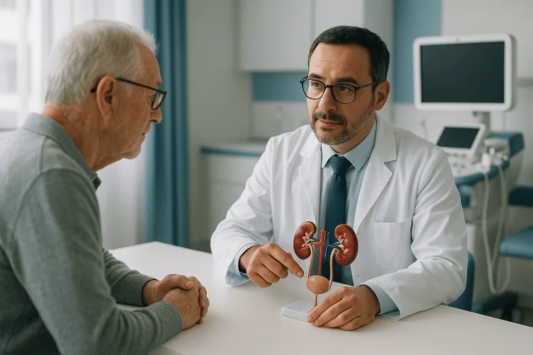
Nephrolithiasis Treatment
Nephrolithiasis, or kidney stones, is treated according to stone size, location and symptoms. Care may include imaging, pain control, medical therapy, shockwave treatment or endoscopic stone removal.
Quick answer
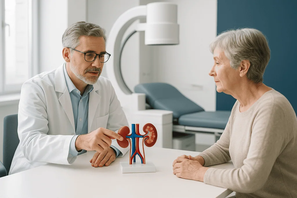
Nephrolithiasis is the medical term for kidney stone disease, hard mineral deposits that form inside the kidney. Treatment ranges from pain control and monitored natural passage for small stones to shockwave lithotripsy, ureteroscopy or percutaneous surgery for stones that are large, blocking urine flow or linked to infection. The right approach depends on stone size, location, composition and the patient's overall health.
Kidney Stone Symptoms and the Decision to Treat
Nephrolithiasis treatment is the medical and procedural care used to manage kidney stones, hard mineral deposits that form inside the kidney and may stay there or move into the urinary tract. It ranges from pain relief and monitored natural passage to shockwave, endoscopic and keyhole procedures that remove stones which cannot pass on their own. It is for anyone with a confirmed stone, and for people with recurrent stones who need a plan to prevent the next one.
Kidney stone symptoms often arrive without warning. You may feel entirely well one day and develop severe flank pain, nausea, vomiting, blood in the urine or a sudden, urgent need to urinate the next. The pain can be intense enough to bring people to emergency departments, and it is frequently the first sign that a stone exists at all. The uncertainty is often as difficult as the pain itself: you want to know whether the stone will pass by itself, whether you need a procedure, and which approach is safest for your situation.
Some stones are small and pass naturally with pain control, hydration guidance and medical therapy. Others become trapped, block the flow of urine, cause infection or damage kidney function. In those cases, timely treatment matters. Modern kidney stone care is highly individualised: the right approach depends on the stone’s size, location and composition, the degree of obstruction, whether infection is present, your kidney function, the severity of your pain and your overall health.
At Acibadem, nephrolithiasis care follows a structured diagnostic pathway, imaging, laboratory evaluation and specialist assessment, to determine whether observation, medication, shockwave treatment or endoscopic stone removal is most appropriate. The goal is not only to relieve pain. Effective treatment aims to clear the current stone safely, protect kidney function, reduce the risk of complications and lower the chance of future stones. Recognising kidney stone symptoms early makes each of those goals easier to reach.
What Is Nephrolithiasis?
Nephrolithiasis is the medical term for kidney stone disease. It refers to solid deposits of minerals and salts that crystallise inside the kidney. A stone may sit quietly in the kidney for months or years, or it may travel into the ureter, the narrow tube that carries urine from the kidney to the bladder, where it typically causes the classic wave-like pain most people associate with stones.
What does nephrolithiasis mean?
Nephrolithiasis literally means “stone condition of the kidney”: it combines the Greek words nephros (kidney) and lithos (stone). You may also see the phrase nephrolithiasis disease used online; it describes exactly the same condition as “kidney stones”: there is no separate illness behind the longer name. Doctors use the technical term in imaging reports and medical records, which is why many patients first encounter it after a scan rather than in conversation.
How do you pronounce nephrolithiasis?
Nephrolithiasis is pronounced NEF-roh-lih-THY-uh-sis, with the main stress on the “THY” syllable. It is a mouthful, which is one reason clinicians often simply say “kidney stone disease” when speaking with patients.
What is nephrolithiasis in the kidney, compared with stones elsewhere?
Nephrolithiasis specifically describes stones located within the kidney itself. When a stone moves into the ureter, doctors call it a ureteral stone (ureterolithiasis), and its behaviour and treatment options change: you can read more about that stage on our ureteral stones page. Stones can also form directly in the bladder, usually for different reasons such as incomplete bladder emptying; these are covered separately under bladder stones. The distinction matters because a stone’s location strongly influences whether it can pass naturally and which procedure suits it best.
Kidney Stone Symptoms: What to Look For
Kidney stone symptoms depend on where the stone is and whether it is moving. A stone resting inside the kidney may cause no symptoms at all and be discovered incidentally on a scan done for another reason. A stone that begins to travel is a different matter. Pain commonly begins in the side or back, below the ribs, and may spread toward the lower abdomen or groin as the stone moves down the ureter. It typically comes in waves, varies in intensity and can be severe enough that no position feels comfortable. Some patients notice urinary changes before severe pain develops; others have both at once.
What are the 7 warning signs of kidney stones?
Seven signs are most commonly reported by people with a symptomatic stone:
- Severe flank pain in the side or back below the ribs, often arriving in waves.
- Pain radiating toward the lower abdomen or groin as the stone descends.
- Blood in the urine, which may look pink, red or brown, or be visible only on testing.
- Burning or pain when urinating, particularly once the stone nears the bladder.
- Frequent or urgent urination, sometimes passing only small amounts.
- Nausea and vomiting, driven by shared nerve pathways between the kidney and the gut.
- Cloudy or foul-smelling urine, fever or chills, which suggest infection may be present.
Fever combined with a suspected obstructing stone is the sign clinicians take most seriously, because it can indicate infection trapped behind a blockage. In that situation, urologists treat the case as an emergency: drainage of the kidney and antibiotics usually take priority over removing the stone itself.
What can be mistaken for a kidney stone?
Several conditions produce pain in the same region and are regularly confused with stones. Urinary tract infections can cause burning, urgency and flank pain without any stone being present. Appendicitis, diverticulitis and other abdominal conditions can mimic a right- or left-sided stone. Gallstones cause upper abdominal pain that some patients initially attribute to a kidney problem. Muscular back strain, shingles affecting the flank, gynaecological conditions such as ovarian cysts, and, rarely, in older adults, vascular problems can all resemble a stone episode. Because kidney stones symptoms overlap with so many other conditions, imaging is essential: a scan confirms whether a stone is actually there, where it sits and whether it is blocking urine flow. This is one reason self-diagnosis based on pain pattern alone is unreliable, and why a formal diagnosis should come before any treatment decision.
What Causes Kidney Stones?
What causes kidney stones is, at its core, urine chemistry: stones form when minerals and salts in the urine become so concentrated that they crystallise into solid deposits. Anything that raises the concentration of stone-forming substances, lowers the concentration of natural inhibitors or reduces urine volume increases the risk.
The likely stone type matters for both treatment and prevention. Common types include:
- Calcium-based stones (calcium oxalate and calcium phosphate), the most frequent type, linked to high urinary calcium, high oxalate, low citrate and low urine volume.
- Uric acid stones, associated with persistently acidic urine, gout, certain diets and some metabolic conditions.
- Struvite stones, which form in association with particular urinary infections and can grow quickly.
- Cystine stones, a less common type caused by an inherited metabolic condition that begins producing stones early in life.
What causes nephrolithiasis in some people and not others?
Nephrolithiasis develops when individual risk factors combine, which is why one person forms stones repeatedly while another never does. Low fluid intake is the most consistent contributor. Dietary patterns high in salt, animal protein or oxalate-rich foods play a role, as do obesity, hot climates, occupations that limit drinking during the day, and family history. Some patients form stones after bariatric surgery or because of digestive disorders that change how the gut absorbs oxalate. Certain medications can promote crystal formation. Underlying metabolic factors, high urinary calcium, low citrate, high oxalate, high uric acid or chronically low urine volume, often explain recurrent stones, and identifying them is the foundation of prevention. A complete treatment approach therefore considers why the stone formed, not only how to remove it.
What Nephrolithiasis Treatment Involves
Nephrolithiasis treatment includes the full range of medical and procedural care used to manage kidney stones. It may begin with diagnosis and symptom control, and progress to stone removal if the stone is unlikely to pass, is causing complications or produces recurrent symptoms.
Conservative management is often suitable for smaller stones that are expected to pass on their own. It typically includes pain relief, anti-nausea medication, medicines that help relax the ureter and ease stone passage, hydration guidance, urine straining to capture the stone for analysis, and follow-up imaging. Interventional treatment is considered when the stone is too large to pass, pain cannot be controlled, urine flow is blocked, infection is present, kidney function is threatened or the patient has a single functioning kidney.
How do you get rid of kidney stones?
There are two honest routes: let the stone pass or have it removed. Many small stones pass naturally over days to weeks with adequate fluids, pain control and, where appropriate, medication that relaxes the ureter, all under medical supervision, with imaging to confirm the stone has genuinely cleared. Stones that will not pass are treated with one of three main procedures: extracorporeal shockwave lithotripsy, which breaks the stone from outside the body; ureteroscopy, which reaches the stone through the natural urinary passage; or percutaneous nephrolithotomy, a keyhole operation for larger or more complex stones. No drink, supplement or home remedy reliably dissolves an established stone, with the partial exception of some uric acid stones, which can sometimes shrink under a doctor-directed programme of urine alkalinisation. Symptom relief is not proof of clearance: a stone can continue to obstruct the kidney even after pain improves, which is why follow-up imaging is part of proper care rather than an optional extra.
Who May Need Kidney Stone Care
Most people first seek care for nephrolithiasis because of sudden pain, but the decision about who needs specialist treatment rests on a structured diagnosis rather than pain alone. Evaluation usually begins with a medical history, physical examination, urine testing and blood tests. Urinalysis can reveal blood, infection markers or crystals. Blood tests assess kidney function, infection signs, calcium levels and other metabolic factors.
Imaging is central. A non-contrast computed tomography (CT) scan is often preferred because it shows the stone’s size, density and location and reveals whether urine flow is obstructed. Ultrasound is used in selected situations, including pregnancy, younger patients and follow-up checks, because it avoids radiation. Plain X-rays help with certain stone types, particularly when monitoring how treatment is progressing.
Specialist kidney stone care is warranted when symptoms are severe, recurrent or complicated: stones that do not pass, repeated emergency visits, persistent blood in the urine, recurrent urinary infections, reduced kidney function or a history of multiple stones. Some patients need particularly careful planning: those with only one kidney, kidney transplant recipients, pregnant patients, people with complex urinary anatomy and patients with established kidney disease. Children who form stones deserve specific attention, because an underlying metabolic or inherited cause is more likely at a young age and untreated recurrent stones can contribute to long-term problems such as paediatric chronic kidney disease.
Where a diagnosis has already been made, a structured second review of imaging, laboratory results and prior treatment can clarify whether immediate intervention is genuinely needed or whether a less invasive approach, or simple observation, is reasonable. Both answers are legitimate outcomes of a good review.
Conditions and Indications Addressed by Nephrolithiasis Treatment
Kidney stone treatment addresses both acute stone episodes and chronic stone disease. The immediate concern is usually pain or obstruction; the longer-term concern is preventing recurrence and preserving kidney health.
Accepted indications for active treatment include symptomatic kidney stones, ureteral stones causing obstruction, stones associated with infection, stones too large to pass naturally, recurrent stone formation, stones in patients with reduced kidney function and stones causing persistent blood in the urine. Treatment may also be advised for certain asymptomatic stones, if they are growing, sit in a high-risk position, are linked to infection or are likely to cause future complications. Not every silent stone needs surgery; some are reasonably monitored with periodic imaging.
Ureteral stones require intervention when they remain lodged, produce uncontrolled pain or block the kidney. Stones that stay inside the kidney may still need treatment if they are large, numerous, associated with repeated infections or positioned where they interfere with urinary drainage. Staghorn stones, branching stones that can fill part or all of the kidney’s collecting system, almost always require active management, because they cause progressive kidney damage and harbour infection.
Nephrolithiasis care also addresses the metabolic side of the condition. Patients who form stones because of high urinary calcium, low citrate, high oxalate, high uric acid or low urine volume benefit from identifying that driver. In adults with repeated stones, prevention becomes a core part of care: stone analysis, 24-hour urine testing, blood tests, dietary changes and targeted medication where appropriate. Removing a stone without asking why it formed leaves the underlying process untouched.
How Nephrolithiasis Treatment Is Performed
Whatever the eventual procedure, the pathway follows a consistent sequence:
- Define the stone. Your urologist reviews symptoms, previous stone history, medical conditions, medications, allergies and prior operations. Imaging establishes stone size, location, hardness, degree of obstruction and the anatomy of the urinary tract.
- Assess the risk. Blood and urine tests identify infection, kidney function changes and metabolic factors. Anaesthesia fitness is evaluated where a procedure is likely.
- Choose the approach. Observation, shockwave treatment, ureteroscopy, percutaneous surgery or urgent drainage, matched to the stone and to you.
- Treat and verify. The chosen treatment is carried out, then follow-up imaging confirms the stone has cleared and the obstruction has resolved.
- Prevent the next stone. Stone analysis and metabolic evaluation feed into a prevention plan.
Observation and medical therapy
If the stone is small and there are no warning signs, the first step may be watchful waiting supported by medical therapy: pain control, medication to reduce ureteral spasm or assist stone passage, anti-nausea treatment, and clear instructions about hydration and urine straining. Follow-up imaging is essential, because symptoms alone do not confirm passage.
Extracorporeal shockwave lithotripsy (ESWL)
Extracorporeal shockwave lithotripsy is a non-incision treatment used for selected kidney stones and upper ureteral stones. Focused shockwaves are directed at the stone from outside the body under imaging guidance, fragmenting it so the pieces can pass naturally in the urine. Sedation or anaesthesia may be needed depending on the situation. Fragments may pass over days or weeks, and follow-up imaging checks whether the stone has cleared sufficiently. ESWL suits stones that are not too large or too dense; harder stones fragment poorly and are often better treated endoscopically.
Ureteroscopy and laser fragmentation
Ureteroscopy is commonly used for stones in the ureter and many stones within the kidney. A thin scope is passed through the natural urinary pathway, urethra, bladder, ureter and, with a flexible instrument, into the kidney itself, so no external incision is required. The surgeon sees the stone directly and either removes it with tiny instruments or breaks it into fragments with laser energy. A temporary ureteral stent is often placed afterwards to keep urine draining while swelling settles; stents are removed later, at a timing decided case by case.
Percutaneous nephrolithotomy (PCNL)
Percutaneous nephrolithotomy is generally reserved for larger, harder or more complex kidney stones, including staghorn stones. Under imaging guidance, the surgeon creates a small tract through the skin of the back directly into the kidney and passes instruments through it to break up and extract the stone. This approach clears a larger stone burden than other methods, requires anaesthesia and usually involves a short hospital stay, with monitoring for bleeding, infection, pain and kidney drainage.
Emergency drainage: stents and nephrostomy tubes
When a stone is blocking urine flow and infection is suspected, definitive stone removal is not the first step, drainage is. This may mean a ureteral stent placed internally or a nephrostomy tube placed through the skin into the kidney, together with antibiotics and supportive care. Where infection is significant, urologists may work alongside colleagues in the infectious diseases department. Once infection and inflammation are controlled, the definitive stone procedure can be planned far more safely.
How long do procedures and hospital stays take?
Duration varies with the treatment. A straightforward ureteroscopy or shockwave session may be completed relatively quickly, while complex stone surgery takes longer. Many patients go home the same day after selected procedures; others stay overnight or for several days after more complex interventions. Technology supports every stage, modern imaging to locate stones and plan the safest route, endoscopic camera systems for direct visualisation, laser systems that fragment stones through very small instruments, and ultrasound, fluoroscopic or CT-based guidance where precision matters. The value of technology lies in choosing the right tool for the right stone, not in using the most complex option for every patient.
Why Acting Early Matters
Not every kidney stone requires urgent intervention, but timely assessment is important. A stone that blocks urine flow raises pressure inside the kidney, and if the obstruction persists, kidney function can be affected. When obstruction and infection occur together, the situation can deteriorate quickly and typically requires emergency drainage.
Delay carries practical costs beyond risk: repeated pain episodes, dehydration from vomiting, worsening infection and, in severe, prolonged cases, loss of kidney function. A stone that could have been treated with a less invasive approach may become more complicated once infection, swelling or prolonged obstruction sets in. Recurrent stones also create ongoing inflammation that contributes to long-term kidney problems.
Early evaluation does not automatically mean surgery. In many cases it produces the opposite: a safe, documented plan for observation, pain control and follow-up. The value is in knowing which stones can be watched and which cannot. Clinicians regard certain features as red flags that change the urgency of a case, fever, chills, uncontrolled pain, persistent vomiting, reduced urine output, pregnancy, a single kidney or known kidney disease. Taking kidney stone symptoms seriously at the first episode, rather than after the third emergency visit, is what keeps treatment simple.
Benefits of Kidney Stone Treatment
The specific benefits depend on the stone and the chosen approach, but the overall aim is consistent: relieve symptoms and protect kidney health.
| Benefit | What It Means for You |
|---|---|
| Pain relief | Treating or removing the stone reduces severe flank pain, urinary discomfort and repeated emergency episodes. |
| Protection of kidney function | Relieving obstruction reduces pressure on the kidney and may prevent avoidable loss of function. |
| Control of infection risk | When stones are associated with infection or blockage, timely drainage and treatment reduce the risk of serious complications. |
| Targeted treatment selection | Imaging and laboratory evaluation match the treatment to the stone’s size, location and composition, no more intervention than the stone requires. |
| Reduced recurrence risk | Stone analysis, metabolic testing and prevention planning help lower the likelihood of future stone episodes. |
Recovery Timeline After Kidney Stone Treatment
Recovery varies with stone size, procedure type, anaesthesia, stent use and overall health, but most patients follow a recognisable pattern.
| Time Period | What Patients Can Expect |
|---|---|
| Day 1 | Monitoring for pain, urination, bleeding, fever and response to treatment. Some patients go home the same day; others remain in hospital for observation. |
| First week | Mild blood in the urine, urinary urgency or flank discomfort may occur. With a stent in place, bladder irritation and frequent urination are common. Medication and hydration guidance matter most here. |
| First month | Gradual return to normal activities. Follow-up usually includes imaging to confirm stone clearance and an appointment for stent removal where one was placed. |
| Longer term | Prevention becomes the focus: stone analysis, urine testing, dietary recommendations and selected medications where appropriate to reduce recurrence risk. |
Expect procedure-specific differences. After shockwave lithotripsy, flank soreness, blood in the urine and passage of fragments are common. After ureteroscopy, urinary burning, urgency and stent-related bladder discomfort typically persist until the stent comes out. After percutaneous nephrolithotomy, recovery includes incision care, drainage tube management in some cases and a slower, staged return to normal activity. Your medical team will give individual instructions covering medications, hydration, activity and follow-up imaging.
Factors That Influence Outcomes
A good result in nephrolithiasis care depends on accurate diagnosis, appropriate treatment selection and disciplined follow-up. Stone size is one of the most important variables: very small stones frequently pass naturally, while larger stones rarely do and usually need a procedure. Location matters too: a stone near the bladder behaves differently from a stone in the kidney or upper ureter.
Stone composition shapes the treatment response. Some stones fragment readily with shockwaves; harder stones respond better to endoscopic laser treatment, and stone density on CT helps predict which is which. Anatomy is another factor: narrowing, abnormal kidney drainage, prior surgery or congenital differences in the urinary tract can change the safest route to the stone.
Infection status is critical. Active infection changes the timing and sequence of care, when a blocked kidney is infected, drainage comes before definitive stone removal, always. Kidney function, pregnancy, obesity, heart or lung disease and anaesthesia risk all influence planning. Blood-thinning medication is a particular consideration before any procedure; whether and how it is adjusted is a decision for the treating doctor, never something to change on your own initiative.
Surgical judgment matters as much as equipment: knowing when to observe, when to intervene, which method to select and when to stage treatment across two sessions for safety. And the procedure itself is only part of the outcome. Follow-up imaging must confirm that obstruction has resolved and that clinically significant fragments are not left behind, because retained fragments can seed the next stone. Finally, patient participation carries real weight, taking prescribed medication correctly, telling the care team promptly about new fever or worsening pain, attending follow-up visits and completing urine testing all support a better long-term result.
How Acibadem Approaches Nephrolithiasis Care
Kidney stone care at Acibadem is organised around one principle: two patients with the same diagnosis may need entirely different treatment. One person needs medication and monitoring. Another needs urgent drainage. A third benefits from planned endoscopic treatment followed by metabolic evaluation. The care plan reflects symptoms, stone features, kidney function, infection risk and practical timing.
Urology specialists assess stone disease using modern diagnostic tools and evidence-based protocols. Imaging, laboratory testing, anaesthesia assessment and infection evaluation are coordinated to decide between conservative management, shockwave treatment, ureteroscopy, percutaneous surgery or temporary drainage. Complex cases draw on related specialties, nephrology, radiology, infectious diseases, anaesthesiology and internal medicine, which is particularly valuable for patients with recurrent stones, kidney disease, infection or complicated anatomy.
Prevention is treated as part of the treatment, not an afterthought. Where appropriate, patients receive recommendations on fluid intake, diet, medication, stone analysis and metabolic testing, and those findings can be shared with the patient’s other physicians to support ongoing care.
Prevention and Long-Term Outlook
Kidney stone disease can be intensely painful, but it is also very treatable when the stone is evaluated correctly and the plan is matched to the patient. The questions that matter most are practical ones: is this stone likely to pass, is it causing obstruction or infection, and which treatment offers the safest path through?
The long-term picture depends less on the procedure and more on what follows it. People who have formed one stone carry a real risk of forming another, and that risk is modifiable. Consistent fluid intake to keep urine dilute, dietary adjustments guided by stone analysis and urine testing, treatment of underlying metabolic factors and, where a doctor prescribes it, targeted medication together form the working toolkit of stone prevention. Combined with follow-up imaging to confirm the current stone has truly cleared, this turns nephrolithiasis from a series of painful emergencies into a managed, largely predictable condition.
Preparation
- Evaluation usually includes urine tests, blood tests and imaging such as ultrasound or CT to define stone size and location. Patients may be asked to stop certain blood-thinning medicines and follow fasting instructions if anesthesia is planned. The urology team reviews infection risk, kidney function and the most suitable treatment option.
Aftercare
- Patients are encouraged to drink fluids as advised and take prescribed pain relief or antibiotics if needed. Temporary urinary discomfort, blood in the urine or stent-related symptoms may occur after endoscopic treatment. Follow-up imaging and stone analysis help prevent recurrence and guide diet or medication changes.
Frequently Asked Questions
What affects the cost of kidney stone treatment?
The main factors are stone size, location, number of stones, symptoms, infection risk, imaging needs, treatment method, anaesthesia, hospital stay, stent use and follow-up requirements. The final quote also depends on the hospital, urologist expertise and whether urgent care is needed.
How can I get a personalised quote for nephrolithiasis treatment in Turkey?
You can request a free consultation and share your recent imaging, laboratory results, symptoms and medical history. A urology team can review your case and provide a personalised plan and quote based on the treatment most suitable for you.
Is shockwave treatment always cheaper than endoscopic stone removal?
Not always. Shockwave treatment may be less complex in selected cases, but the final cost depends on stone features, expected success, need for repeat treatment, anaesthesia, imaging and follow-up. A specialist should decide which option is clinically appropriate.
Can the quote include travel and language support?
For international patients, hospitals may offer coordination such as interpreter support, appointment planning, airport transfer guidance and accommodation assistance. What is included should be confirmed in the written estimate before travel.
Why might emergency kidney stone care cost more than planned treatment?
Emergency care may involve urgent imaging, pain control, infection management, drainage procedures, admission and closer monitoring. These needs can change the treatment plan and affect the final cost.
Medically reviewed by the Acıbadem International Medical Board August 31, 2026
See our medical review board →
Update history
- PublishedJune 8, 2026
- Medical review approvedAugust 31, 2026
- Last content updateSeptember 8, 2026
References1
- Kidney Stones medlineplus.gov
Trusted care for international patients
From the Acibadem Blog
How CT, Ultrasound and Urine Tests Shape a Kidney Stone Treatment Plan
A non-contrast CT scan is the most accurate single test for finding kidney stones and measuring their size and position, but it is not…