

Aortic Aneurysm Treatment
Aortic aneurysm care focuses on monitoring or repairing a weakened, bulging section of the aorta to prevent rupture. Treatment may include endovascular stent grafting or open surgical repair.
Quick answer
An aortic aneurysm is a weakened, enlarged section of the aorta, the body's main artery. Treatment aims to prevent the weakened wall from rupturing or tearing. Small, stable aneurysms are usually monitored with regular imaging and blood pressure control. Larger or faster-growing aneurysms may be repaired, either with a stent graft placed through the blood vessels or with open surgery that replaces the weakened segment.
What Is an Aortic Aneurysm?
An aortic aneurysm is a weakened, enlarged section of the aorta, the main artery that carries blood from your heart to the rest of your body. As the artery wall stretches, it becomes thinner and less able to withstand the pressure of each heartbeat. Treatment exists for one purpose: to stop that weakened section from rupturing or tearing. An aneurysm is not a tumour and not a blockage. It is a structural weakness, and how it is managed depends on where it sits, how large it is, what shape it has and how quickly it is changing.
Learning that you have an aneurysm can be unsettling, partly because most aortic aneurysms cause few or no symptoms until they become dangerous. Patients tend to ask the same questions: how serious is it, does it need surgery now, can it be monitored safely, and if repair is needed, should it be done with a stent graft or open surgery. This page answers those questions as directly as the evidence allows. Some aneurysms can be followed closely with imaging and blood pressure control for years. Others should be repaired at a planned time to reduce the risk of rupture or dissection, both of which are life-threatening emergencies.
The aorta: why location matters
The aorta rises from the heart, curves over in an arch that gives off the arteries to the brain and arms, then descends through the chest and abdomen before dividing into the arteries to the legs. An aneurysm can develop in any of these segments, and each segment has different neighbouring structures, different branch vessels and different technical considerations for repair. A bulge just above the heart raises questions about the aortic valve. A bulge in the abdomen raises questions about the arteries to the kidneys and legs. An aortic aneurysm is one of several conditions affecting this vessel; the wider group, including dissection, narrowing and inflammation of the aorta, is covered on our aortic diseases page.
What is the main cause of an aneurysm?
The main cause of an aneurysm is gradual degeneration of the artery wall, most often driven by a combination of atherosclerosis, long-standing high blood pressure and smoking. Over years, these factors break down elastin and collagen, the proteins that give the aortic wall its strength and recoil. Once the wall begins to stretch, the physics work against it: a wider vessel experiences greater wall tension at the same blood pressure, which encourages further enlargement. This is why aneurysms tend to grow rather than shrink, and why blood pressure control is part of every management plan.
Other causes matter in specific groups. Inherited connective tissue disorders such as Marfan syndrome, Loeys-Dietz syndrome and vascular Ehlers-Danlos syndrome weaken the aortic wall from birth and can produce aneurysms at younger ages. A bicuspid aortic valve is associated with enlargement of the ascending aorta. Less commonly, infection, chest or abdominal injury, or inflammatory disease of the vessel wall is responsible. Family history is relevant even when no named syndrome is present: aneurysms cluster in families, which is why close relatives of affected patients are sometimes offered screening with ultrasound.
What happens if you have an aneurysm?
In most cases, nothing happens for a long time. Aneurysms usually enlarge slowly, and many people live with a small, stable aneurysm for years under structured surveillance. The concern is what can happen as the wall stretches further. Rupture means the wall gives way and blood escapes; this causes sudden internal bleeding and is a hospital emergency. Dissection means the layers of the wall split apart, which can block blood flow to vital organs. Clot can also form inside the aneurysm sac and travel downstream, and a large aneurysm can press on nearby structures such as the airway, the swallowing tube or the spine. Management is therefore an exercise in probability: your medical team weighs the risk of leaving the aneurysm alone against the risk of repairing it, and repeats that assessment every time new imaging arrives.
What are the early signs of an aneurysm?
Usually there are none. Most aortic aneurysms are found incidentally, on an ultrasound, CT scan, MRI, chest X-ray or heart study performed for another reason. When symptoms do occur, they depend on location. An aneurysm in the abdomen may cause a deep, persistent ache in the abdomen, back, flank or pelvis, and some people notice a pulsing sensation near the navel. An aneurysm in the chest may cause chest or upper back pain, hoarseness, a persistent cough, difficulty swallowing, breathlessness or a feeling of pressure. None of these symptoms is specific; each overlaps with far more common conditions. That is precisely why imaging, not symptom-checking, is how an aneurysm is confirmed and measured.
 Dr. Mohamed Al-QadiMDBoard Commentary
An aortic aneurysm should not be managed according to diameter alone; its location, rate of enlargement, symptoms, body size, aortic valve or root involvement, genetic background, surrounding branch vessels and the patient’s heart, lung and kidney function all influence whether surveillance, endovascular repair, open surgery or a hybrid approach is most appropriate. Acıbadem-affiliated research reflects several parts of this decision pathway. A comparative study of 69 patients undergoing aortic root replacement for annuloaortic ectasia evaluated arterial cannulation and cerebral-perfusion strategies during open surgery, illustrating the importance of organ protection in proximal aortic procedures. Acıbadem Kadıköy physicians have also reported endovascular treatment of a rapidly enlarging thoracoabdominal aneurysm after previous type A dissection repair, as well as hybrid reintervention for a late type I endoleak after TEVAR; the latter particularly reinforces why long-term imaging is an integral part of endovascular treatment rather than an optional follow-up measure. In emergency care, a retrospective 53-patient study that included an Acıbadem Hospital cardiovascular-surgery affiliation reported lower early mortality when REBOA was incorporated into hybrid surgery for ruptured abdominal aortic aneurysm, although this observational result should not be interpreted as an Acıbadem-wide success rate or applied to elective cases. Current guidelines support ultrasound surveillance for many smaller abdominal aneurysms and CT angiography, MRI or echocardiography according to aortic location and clinical need. Repair decisions should integrate symptoms, growth, anatomy and hereditary risk, while the choice between EVAR or TEVAR, open reconstruction and complex hybrid repair should be based on secure landing zones, branch-vessel involvement, connective-tissue disease, infection and operative risk. Endovascular treatment may reduce the early physiological burden in anatomically suitable patients, but it creates a continuing obligation to monitor for endoleak, graft migration, loss of seal, branch-vessel complications and changes in aneurysm-sac size.
Types of Aortic Aneurysm
Aneurysms are classified by the segment of the aorta they involve and by the condition of the vessel wall. The classification is not academic. It determines which specialists plan your care, which repair techniques are anatomically possible and how urgent the timeline is.
Abdominal aortic aneurysm (AAA)
An abdominal aortic aneurysm (AAA) develops in the section of the aorta that passes through the abdomen, most often below the arteries that supply the kidneys. It is one of the most common types of aortic aneurysm, particularly in older people with a history of smoking or high blood pressure. Many are picked up on abdominal ultrasound. Treatment may involve surveillance, endovascular stent graft repair or open repair, and the choice depends on the aneurysm’s size and growth rate, the shape of the aortic “neck” above it, whether the iliac arteries to the legs are involved, and your overall health and life expectancy.
Thoracic aortic aneurysm
A thoracic aortic aneurysm sits in the chest. It may affect the ascending aorta just above the heart, the aortic arch, the descending thoracic aorta, or a combination of segments. Repair may require cardiovascular surgery, endovascular thoracic stent grafting or a hybrid plan combining both. When the aortic root or aortic valve is involved, the operation may also address the valve, either by preserving it with a valve-sparing technique or by replacing it. Thoracic aneurysms are more likely than abdominal ones to be linked to genetic conditions, which changes both the thresholds for repair and the plan for long-term follow-up.
Thoracoabdominal aortic aneurysm
A thoracoabdominal aneurysm extends from the chest into the abdomen and can involve the arteries that supply the spinal cord, kidneys, liver and intestines. These are among the most complex aneurysms to treat, because repair must protect every one of those organs while the diseased segment is replaced or excluded. Planning is correspondingly detailed, and treatment may be delivered in stages rather than in a single procedure.
What is a dissecting aortic aneurysm?
A dissecting aortic aneurysm occurs when a tear forms in the inner layer of the aortic wall and blood forces its way between the layers, splitting them apart along the length of the vessel. The modern term for this event is aortic dissection, and it is a distinct emergency rather than a subtype of aneurysm: a dissection can occur in an aorta of normal size, and an aneurysm can exist for years without ever dissecting. The two conditions meet when a dissected aorta continues to enlarge after the initial tear. In that situation, treatment may be needed if the vessel keeps expanding, if blood flow to organs is compromised or if symptoms persist, and the plan depends on whether the ascending aorta, the descending aorta or both are involved. One point of terminology worth clearing up: the word “dissection” here describes the splitting of the vessel wall itself. It has nothing to do with surgical procedures that happen to use the same word, such as neck dissection, which is an operation to remove lymph nodes from the neck.
Aneurysms linked to connective tissue disorders
Connective tissue disorders such as Marfan syndrome, Loeys-Dietz syndrome and vascular Ehlers-Danlos syndrome can lead to aneurysms at younger ages and in less typical locations. These patients are usually managed with different, more cautious size thresholds for repair, closer imaging intervals and lifelong specialist follow-up. Family screening and genetic counselling are often part of the plan, because first-degree relatives may carry the same condition without knowing it. Open surgical repair is frequently preferred over stent grafting in this group, because the fragile vessel wall holds a fabric graft sewn in by hand more reliably than a device anchored by outward pressure.
Inflammatory, mycotic and traumatic aneurysms
Less common aneurysms arise from inflammation of the vessel wall, from infection (historically called mycotic aneurysms) or from injury, typically high-speed deceleration in a road accident. These often need urgent or unusually complex repair, and care commonly involves collaboration across vascular surgery, cardiovascular surgery, infectious diseases, radiology and intensive care. An infected aneurysm, in particular, changes the surgical strategy, because placing a permanent device into an infected field is generally avoided where an alternative exists.
What Is Aortic Aneurysm Treatment?
Aortic aneurysm treatment means preventing rupture and dissection, not necessarily operating. For many patients, especially when the aneurysm is small and stable, the safest plan is active surveillance: scheduled imaging to track size, together with blood pressure management, cholesterol control, smoking cessation and treatment of related cardiovascular risk factors. When the aneurysm reaches a size, growth rate or symptom pattern associated with higher risk, repair is recommended. There are two main categories of repair, endovascular and open surgical, plus hybrid strategies that combine them. The right choice is anatomical and personal at the same time: it depends on where the aneurysm is, what the vessel around it looks like, and what your heart, lungs and kidneys can safely tolerate.
How dangerous is a 4 cm aortic aneurysm?
A 4 cm aortic aneurysm in the abdomen is usually below the size at which repair is recommended, and most vascular teams manage it with surveillance rather than surgery. At that diameter, for most patients, the risk of a planned operation outweighs the near-term risk of rupture, so the sensible strategy is regular imaging to confirm the aneurysm is behaving itself, combined with strict blood pressure control and stopping smoking. That said, the number alone never settles the question. A 4 cm aneurysm that is growing quickly, causing symptoms or found in a small-framed patient is judged differently from a stable one, and a 4 cm measurement in the ascending aorta of someone with a connective tissue disorder carries a different meaning again. Diameter is the headline; location, growth rate, body size, sex and underlying condition are the rest of the story.
Active surveillance: when watching is the right treatment
Surveillance is a deliberate treatment plan, not the absence of one. It involves imaging at intervals set by the aneurysm’s size and location, ultrasound for many abdominal aneurysms, CT or MRI for thoracic disease, alongside medical management of the factors that drive growth. The discipline lies in keeping the appointments. An aneurysm under surveillance that quietly enlarges between missed scans loses the main advantage of being found early: the chance to repair it on a planned schedule rather than in an emergency.
Endovascular aneurysm repair (EVAR)
Endovascular aneurysm repair places a fabric-and-metal stent graft inside the aorta, delivered through the blood vessels rather than through a large incision. Access is usually gained from the femoral arteries in the groin. Once deployed, the stent graft expands to fit the vessel and creates a reinforced channel for blood flow, excluding the aneurysm sac from direct arterial pressure. Endovascular repair is commonly used for suitable abdominal aortic aneurysms and for many descending thoracic aneurysms. Its appeal is obvious: smaller incisions, less physiological stress, typically a shorter hospital stay and a faster early recovery than open surgery. Its condition is equally clear: the anatomy has to fit the device, with healthy landing zones above and below the aneurysm, and the repair requires lifelong imaging follow-up to confirm the graft stays sealed and in position.
Open surgical repair
Open surgical repair replaces the weakened section of the aorta with a durable fabric graft, through an incision in the chest, the abdomen or both, depending on where the aneurysm lies. Blood flow is carefully controlled while the graft is sewn in, and any branch arteries arising from the diseased segment are reattached or reconstructed. Open repair demands more of the patient in the short term, a longer hospital stay and a more gradual recovery, but it remains the preferred option in several situations: complex aneurysms, anatomy unsuitable for stent grafting, connective tissue disorders, infected aneurysms, some younger patients, and cases where long-term durability is the overriding concern.
How is an ascending aortic aneurysm repaired?
An ascending aortic aneurysm is repaired with open surgery in almost all cases, because the segment just above the heart is not well suited to standard stent grafting. Through an incision in the front of the chest, the surgeon replaces the enlarged section with a fabric graft while the heart and circulation are supported. If the aneurysm involves the aortic root, the operation is extended: the aortic valve may be preserved with a valve-sparing root replacement or replaced together with the root, and the coronary arteries are reattached to the new graft. If the arch is involved, the repair may extend further and include strategies to protect the brain during the procedure. These are established cardiovascular operations, planned in detail from CT imaging and echocardiography before anyone reaches the operating theatre.
Hybrid and staged approaches
Some aneurysms need more than one technique. Hybrid procedures combine open surgery and endovascular repair, for example rerouting branch arteries surgically so that a stent graft can then be landed safely across the diseased segment. Staged repairs treat an extensive aneurysm in planned steps, spacing out the physiological load and protecting the spinal cord and organs between stages. These approaches require careful sequencing and are typically reserved for aneurysms involving major branches to the brain, spinal cord, kidneys or abdominal organs.
Who May Need Aortic Aneurysm Care?
Many people arrive at aneurysm care by accident: an ultrasound ordered for gallstones, a CT scan after a fall, a chest X-ray before an unrelated operation. Others are evaluated because of symptoms, a strong family history or a known genetic condition. Care may be needed whenever imaging shows enlargement of the thoracic aorta, the abdominal aorta or both, even if you feel entirely well.
Symptoms, when they occur, track the anatomy. Abdominal aneurysms tend to announce themselves with deep abdominal, back, flank or pelvic pain, or a pulsing sensation in the belly. Thoracic aneurysms lean towards chest or back pain, hoarseness, cough, swallowing difficulty and breathlessness. Rupture and acute dissection behave differently again: they typically cause sudden, severe, often tearing pain in the chest, back or abdomen, sometimes with fainting, weakness or collapse. These events are treated in hospital as emergencies; they are not situations for outpatient appointments or travel planning.
Diagnosis rests on imaging. Ultrasound is the usual tool for detecting and following abdominal aneurysms. CT angiography maps the aneurysm’s size, shape and relationship to branch arteries in the detail needed for treatment planning. MRI angiography suits selected patients, particularly when repeated scans are needed or contrast exposure is a concern. Echocardiography assesses the aortic root, the ascending aorta and the function of the aortic valve.
Referral for specialist evaluation typically happens in one of these situations:
- The aneurysm is approaching a size threshold at which repair is usually discussed.
- Imaging shows the aneurysm growing faster than expected.
- Symptoms attributable to the aneurysm have appeared.
- There is a strong family history or a diagnosed connective tissue disorder.
- The aneurysm involves complex anatomy, such as the arch or visceral branches.
- A previous repair needs structured follow-up.
- Repair has been recommended elsewhere and a second opinion is wanted before deciding.
How Aortic Aneurysm Treatment Is Performed
Initial evaluation and treatment planning
Planning begins with a full review: medical history, current symptoms, previous imaging, medications, allergies, kidney function, heart and lung health and prior operations. If you are travelling for treatment, existing scans and reports are normally reviewed before a plan is proposed, because decisions about the aorta should rest on images, not on written summaries alone. High-quality CT angiography sits at the centre of the process. It lets the team measure the aneurysm precisely, evaluate the branch vessels, assess calcium and clot within the sac, and determine whether the anatomy suits endovascular repair. Depending on the case, MRI, echocardiography, catheter-based angiography, cardiac testing, lung function studies and laboratory work are added. A multidisciplinary discussion, vascular surgeons, cardiovascular surgeons, interventional radiologists, anaesthesiologists, cardiologists and intensive care specialists as required, then settles whether monitoring, endovascular repair, open surgery or a combined strategy is most appropriate.
Preparation before repair
Preparation depends on the planned procedure and your risk profile. Blood pressure, diabetes, cholesterol and blood-thinning medications are reviewed by the treating team, and any adjustments are made under their direction. Smoking cessation is strongly encouraged, because smoking is associated with aneurysm growth and with complications after repair. Patients being considered for open surgery usually undergo cardiac assessment to confirm surgical fitness. Before the procedure, your care team explains fasting instructions, anaesthesia planning, the expected hospital stay, the likelihood of a period in intensive care and the follow-up imaging schedule, so that nothing on the day comes as a surprise.
Endovascular stent graft repair, step by step
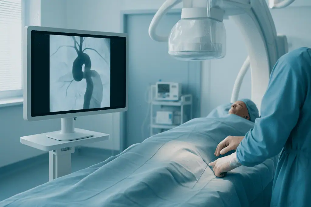
Endovascular repair takes place in a specialised operating room or angiography suite equipped for advanced imaging, under general anaesthesia or regional anaesthesia with sedation. A typical procedure runs like this:
- Small incisions or punctures are made at the groin to access the femoral arteries.
- Guidewires and delivery catheters are advanced through the vessels to the aneurysm under live X-ray guidance.
- The folded stent graft is positioned precisely across the aneurysm, using the measurements taken from your CT scan.
- The graft is deployed and expands to fit the vessel, creating a reinforced pathway for blood and sealing the aneurysm sac away from arterial pressure.
- Completion imaging checks the graft’s position, confirms flow through the aorta and its branches, and looks for any leakage of blood around the graft, known as an endoleak.
- The access sites are closed, and you are moved to a monitored recovery area.
The procedure often takes a few hours, though complex repairs take longer. Many patients spend the first night in a recovery or intensive care setting before moving to a regular room. The hospital stay is typically shorter than after open surgery, but the obligation that comes with a stent graft is permanent: lifelong imaging follow-up to confirm the device remains sealed and the aneurysm sac is stable or shrinking.
Open surgical repair
Open repair is performed under general anaesthesia. The surgeon reaches the aneurysm through an incision in the abdomen, the chest or both, controls blood flow above and below the diseased segment, and replaces it with a fabric graft sewn directly into healthy aorta. Branch arteries arising from the segment are reattached or reconstructed as needed. In thoracic and thoracoabdominal operations, additional strategies protect the brain, spinal cord, kidneys and abdominal organs while the repair is completed. Immediately after surgery you are cared for in an intensive care unit, then transferred to a ward room as your condition stabilises. The trade-off is straightforward: a bigger operation and a slower recovery in exchange for a repair valued for its durability, particularly where stent grafting is unsuitable.
Technology used during aneurysm care
Modern aneurysm care leans on detailed imaging, precise measurement and continuous monitoring. Advanced CT and MRI define the aneurysm; three-dimensional reconstruction helps the team understand complex anatomy and select graft dimensions. During endovascular repair, real-time imaging guides device placement and confirms blood flow. During open surgery, monitoring technology supports anaesthesia management, organ protection and postoperative safety. It is worth saying plainly that technology does not replace judgement. The experience of the treating team, careful patient selection and disciplined follow-up shape the result at least as much as the equipment does.
Recovery after treatment
Recovery depends on the type of repair, your baseline health and whether the aneurysm was treated electively or as an emergency. After endovascular repair, most patients begin walking soon after the procedure and return to many daily activities within several weeks, with heavy lifting and strenuous exercise restricted until the physician confirms healing is on track. After open repair, recovery is more substantial: time in intensive care, then a gradual programme of walking, breathing exercises, nutrition, wound care and medication management on the ward. Fatigue, reduced appetite, incision discomfort and lower stamina are normal in the early weeks. Returning to your previous level of activity can take several months, especially after thoracic or thoracoabdominal surgery, and the pace varies genuinely from person to person.
Why Acting Early Matters
An aortic aneurysm may remain stable for a long time, but the ones that enlarge become more dangerous as the wall stretches. The main risks of delay are rupture, dissection, clot embolisation, compression of nearby structures and, the common thread behind all of them, emergency surgery, which removes every advantage of planning. An elective repair allows time for detailed imaging, medical optimisation, specialist discussion and deliberate selection of the safest technique. A ruptured aneurysm allows none of that. Outcomes are generally better when higher-risk aneurysms are identified and managed before rupture, and the same logic applies to dissection, which can cut off blood flow to the brain, spinal cord, kidneys, intestines or limbs. Acting early does not always mean operating early. In many patients the correct early action is disciplined surveillance with scheduled imaging and aggressive risk-factor control. What matters is that the aneurysm is assessed by specialists who can tell the difference, and keep telling it, scan after scan.
Benefits of Aortic Aneurysm Treatment
The benefits depend on the aneurysm type, the chosen method and your overall health, but the objective is constant: reducing the risk of a life-threatening aortic event.
| Benefit | What It Means for You |
|---|---|
| Reduced risk of rupture | Repair protects the weakened section of the aorta once the aneurysm’s size, growth pattern or symptoms make intervention appropriate. |
| Personalised treatment choice | Some patients are better suited to endovascular repair, others to open surgery or continued monitoring. The plan follows your anatomy and medical condition, not a fixed pathway. |
| Less invasive options for selected patients | Where the anatomy is suitable, endovascular stent grafting allows smaller incisions, a shorter early recovery and less physical stress than open repair. |
| Durable repair for complex anatomy | Open surgery remains the right answer for aneurysms that cannot be safely or effectively treated with a stent graft. |
| Structured long-term follow-up | Ongoing imaging and medical management monitor the repair, detect changes early and address future cardiovascular risk. |
Recovery Timeline After Aortic Aneurysm Repair
Recovery varies widely between individuals and between techniques, but the following timeline reflects what many patients experience after elective repair.
| Time Period | What Patients Can Expect |
|---|---|
| Day 1 | After endovascular repair, close monitoring and often the first gentle movement. After open repair, care in an intensive care setting with attention to breathing, circulation, urine output and pain control. |
| First week | Endovascular patients may be discharged within a shorter timeframe if stable. Open surgery patients usually remain in hospital longer, gradually increasing walking, eating and breathing exercises. |
| First month | Energy improves gradually. Heavy lifting is avoided; medication, wound care and activity instructions are followed. Follow-up visits and imaging are scheduled according to the repair type. |
| Longer term | Cardiovascular risk reduction continues alongside periodic imaging. Endovascular repairs require lifelong surveillance; open repairs also need follow-up, particularly if other aortic segments are enlarged. |
Factors That Influence Outcomes
The single most important factor is whether the aneurysm is repaired electively or in an emergency. Planned treatment allows time to evaluate the heart, lungs, kidneys and vascular anatomy, which removes avoidable risk before it materialises. Everything else on this list works better when there is time to use it.
Location and complexity come next. A small infrarenal abdominal aneurysm with a favourable neck is a very different proposition from a thoracoabdominal aneurysm involving the arteries to major organs. The presence of dissection, connective tissue disease, infection, heavy calcification or previous aortic surgery each adds complexity that the plan must absorb.
Your general health matters more than your age in isolation. Heart disease, lung disease, kidney function, diabetes, frailty, smoking history, blood pressure control and nutritional status all influence both the choice of technique and the pace of recovery. What you do afterwards counts too: adherence to prescribed medication, blood pressure management and staying away from cigarettes all bear on the long-term result.
Anatomy decides whether endovascular repair is even on the table. A stent graft needs secure landing zones of healthy vessel above and below the aneurysm. If the aorta there is too short, too wide, sharply angled or heavily diseased, open surgery or a more complex endovascular strategy is safer, and the more complex strategies bring their own demands for careful assessment and long-term follow-up.
Follow-up is not an afterthought; it is part of the treatment. After endovascular repair, imaging checks for endoleaks, graft movement, changes in the aneurysm sac and branch vessel problems. After open repair, follow-up assesses the graft and watches the rest of the aorta, because aortic disease can involve more than one segment, particularly in patients with genetic conditions or long-standing hypertension. Finally, outcomes depend on coordination. Aneurysm care is a pathway, diagnosis, risk assessment, anaesthesia planning, repair, intensive care, rehabilitation, surveillance, and each handover between specialists is a point where good communication earns its keep.
Can you live a normal life with an aneurysm?
Yes, many people live a broadly normal life with a small aortic aneurysm, provided it is monitored properly. Daily activities, work, walking and moderate exercise are usually unaffected; the practical changes are keeping imaging appointments, controlling blood pressure, taking prescribed medication and not smoking. Some doctors advise avoiding very heavy lifting or intense straining, and that guidance is individual: it depends on the aneurysm’s size and location, so it belongs in a conversation with your own physician rather than a general rule. After a successful repair, most patients also return to a full range of ordinary activities, with periodic imaging as the lasting reminder of the diagnosis.
Aortic Aneurysm Care at Acibadem
At Acibadem, aortic aneurysm care is organised around detailed assessment and individualised decision-making rather than a single standard technique. Cases can be reviewed across the relevant specialties, vascular surgery, cardiovascular surgery, cardiology, radiology, anaesthesiology and intensive care, which matters most when the aneurysm involves the chest, the abdominal branches, a previous repair or coexisting heart disease. Multidisciplinary discussion keeps the recommendation honest: it reflects the full range of appropriate options, not just the one a single department happens to perform.
Diagnostic pathways include advanced vascular imaging and structured preoperative assessment, with the aim of understanding the aneurysm accurately before recommending surveillance, endovascular repair or open surgery. Depending on the type and location of the aneurysm, treatment may involve monitoring, stent graft repair, open surgical repair or a staged approach, planned within the broader framework of aortic diseases care. For international patients, coordination services cover appointment scheduling, medical record review, translation and communication in multiple languages, practical support for decisions that often have to be made quickly but should never be made carelessly. Two patients with aneurysms of similar size can reasonably receive different recommendations because of anatomy, age, kidney function, family history or symptoms; the working principle is to match the treatment to the patient.
Getting a Second Opinion on an Aortic Aneurysm
A second opinion is common in aneurysm care and generally worth the effort, because the central questions, repair now, or watch safely, turn on judgement as well as measurement. A useful second opinion confirms the diagnosis, reviews whether the aneurysm meets accepted criteria for repair, compares the endovascular and open options against your specific anatomy, and identifies any testing still needed before a decision. One practical point makes a large difference: specialists assess an aneurysm far more accurately from the original CT, MRI, ultrasound or echocardiography images than from written reports alone, so the imaging files themselves are the most valuable documents a reviewing team can examine. Whether the eventual answer is surveillance, stent grafting, open repair or a staged plan, the purpose of the exercise is the same: a decision made with full information, at a time chosen by you and your doctors rather than by the aneurysm.
Preparation
- Before treatment, patients usually have imaging tests such as CT angiography to assess aneurysm size, location, and surgical risk. Blood tests, heart evaluation, medication review, and anesthesia assessment are completed. Smoking cessation and blood pressure control are important before the procedure.
Aftercare
- After repair, patients are monitored closely for blood pressure, circulation, kidney function, and wound or access-site healing. Follow-up imaging is needed, especially after endovascular repair, to check the stent graft and aneurysm sac. Patients should follow activity restrictions, take prescribed medications, and attend regular cardiovascular follow-ups.
Frequently Asked Questions
What affects the cost of aortic aneurysm treatment?
The main factors are aneurysm location and complexity, the chosen repair method, stent graft requirements, imaging needs, anesthesia, intensive care, hospital stay, and the patient’s overall health. A personalised quote can only be prepared after a specialist reviews medical records and imaging.
How can I get a personalised quote from Acibadem?
You can request a free consultation by sharing recent imaging reports, medical history, medication details, and any prior treatment records. The international patient team can help coordinate specialist review and provide information on the proposed plan and package inclusions.
Does a package usually include travel and accommodation?
Medical packages typically focus on hospital and treatment-related services, while travel, accommodation, and interpreter support may be arranged or quoted separately depending on patient needs. It is important to confirm inclusions before travel.
Is endovascular repair always less expensive than open surgery?
Not always. Endovascular repair may involve specialized stent grafts and advanced imaging, while open surgery may require longer inpatient and intensive care support. The final cost depends on anatomy, device needs, recovery, and clinical risk.
Why is specialist review needed before confirming cost?
Aortic aneurysm treatment depends heavily on detailed imaging, aneurysm anatomy, rupture risk, and overall fitness for surgery. A vascular or cardiovascular specialist must determine the safest suitable option before an accurate quote can be prepared.
Medically reviewed by the Acıbadem International Medical Board August 30, 2026
See our medical review board →
Update history
- PublishedJune 8, 2026
- Medical review approvedAugust 30, 2026
- Board commentary addedSeptember 8, 2026
- Last content updateSeptember 8, 2026
References8
- Deser SB, Arapi B, Tel Ustunisik C, Bitargil M, Yuksel A. REBOA Improves Outcomes in Hybrid Surgery for Ruptured Abdominal Aortic Aneurysms. Ann Vasc Surg. 2024;100:8-14. doi:10.1016/j.avsg.2023.10.027. PMID: 38122969. (Retrospective cohort study – Included an Acıbadem Hospital cardiovascular-surgery affiliation and 53 consecutive patients with ruptured abdominal aortic aneurysm; directly supports the page’s emergency and hybrid-repair sections, but does not represent an elective or institution-wide success rate.)
- Hokenek AF, Kinoglu B, Gursoy M, Sirin G, Gulcan F. Direct Innominate Artery Cannulation in Surgery for Annuloaortic Ectasia. J Card Surg. 2013;28(5):550-553. doi:10.1111/jocs.12182. PMID: 23931724. (Comparative cohort study – Acıbadem University Aile Hospital and Acıbadem University International Hospital-affiliated evaluation of 69 patients undergoing aortic-root replacement; directly supports the page’s open-surgery, aortic-root and cerebral-protection content.)
- Senay S, Alhan C, Karabulut H, Toraman F, Cagil H. Endovascular treatment of rapidly expanding thoracoabdominal aortic aneurysm after surgical repair of acute type A dissection. Ann Thorac Surg. 2008;85(2):636-638. doi:10.1016/j.athoracsur.2007.08.069. PMID: 18222283. (Case report – Acıbadem Kadıköy Hospital publication describing endovascular management of progressive thoracoabdominal aortic enlargement after previous type A dissection surgery; directly supports the page’s dissection-related aneurysm and complex endovascular-treatment sections.)
- Arıtürk C, Okten M, Dagdelen S, Toraman F, Karabulut H. A hybrid therapy as a third approach for type 1 proximal endoleak of thoracic endovascular aortic replacement: Caroticocarotid bypass and re-redo endovascular therapy. J Saudi Heart Assoc. 2013;25(4):273-276. doi:10.1016/j.jsha.2013.04.005. PMID: 24198453. (Case report – Acıbadem Healthcare Group Kadıköy Hospital publication describing hybrid treatment of a late proximal endoleak after TEVAR; directly supports the page’s statements on hybrid repair, endoleak detection, secondary intervention and long-term imaging surveillance.)
- Wanhainen A, Van Herzeele I, Bastos Goncalves F, Bellmunt Montoya S, Berard X, Boyle JR, et al. Editor’s Choice: European Society for Vascular Surgery (ESVS) 2024 Clinical Practice Guidelines on the Management of Abdominal Aorto-Iliac Artery Aneurysms. Eur J Vasc Endovasc Surg. 2024;67(2):192-331. doi:10.1016/j.ejvs.2023.11.002. PMID: 38307694. (Evidence-based clinical practice guideline – Directly supports abdominal-aortic-aneurysm screening and diagnosis, surveillance of smaller aneurysms, cardiovascular risk reduction, thresholds for repair, open versus endovascular treatment, ruptured AAA management, complex anatomy, endoleaks and long-term follow-up.)
- Wanhainen A, Gombert A, Antoniou GA, Fidalgo Domingos LA, Gouveia E Melo R, Grabenwöger M, et al. European Society for Vascular Surgery (ESVS) 2026 Clinical Practice Guidelines on the Management of Descending Thoracic and Thoraco-Abdominal Aortic Diseases. Eur J Vasc Endovasc Surg. 2026;71(2):172-270. doi:10.1016/j.ejvs.2025.12.050. PMID: 41448425. (Evidence-based clinical practice guideline – Directly supports the page’s sections on descending thoracic and thoracoabdominal aneurysms, chronic dissection, rupture, TEVAR, complex endovascular or open repair, branch-vessel and spinal-cord protection, multidisciplinary planning and surveillance.)
- Mazzolai L, Teixido-Tura G, Lanzi S, Boc V, Bossone E, Brodmann M, et al. 2024 ESC Guidelines for the management of peripheral arterial and aortic diseases. Eur Heart J. 2024;45(36):3538-3700. doi:10.1093/eurheartj/ehae179. PMID: 39210722. (European clinical practice guideline – Directly supports the page’s integrated approach to aortic imaging, cardiovascular risk-factor management, surveillance, intervention selection, acute presentation and long-term care.)
- Isselbacher EM, Preventza O, Hamilton Black J, Augoustides JG, Beck AW, Bolen MA, et al. 2022 ACC/AHA Guideline for the Diagnosis and Management of Aortic Disease: A Report of the American Heart Association/American College of Cardiology Joint Committee on Clinical Practice Guidelines. Circulation. 2022;146(24). doi:10.1161/CIR.0000000000001106. PMID: 36322642. (Evidence-based clinical practice guideline – Directly supports the page’s content on aortic-root and ascending-aortic aneurysms, bicuspid aortic valve, hereditary connective-tissue disorders, genetic counseling and family screening, size and growth criteria, open and endovascular repair and lifelong aortic surveillance.)
Trusted care for international patients
From the Acibadem Blog

When Does an Aortic Aneurysm Move From Monitoring to Repair? Size, Growth and Symptoms
Most aortic aneurysms are monitored with scans and repaired only when the risk of rupture begins to outweigh the risk of surgery. For an…



































