

Valve Surgery
Valve surgery repairs or replaces damaged heart valves to improve blood flow and reduce symptoms such as breathlessness, fatigue, chest discomfort, or heart failure risk.
Quick answer
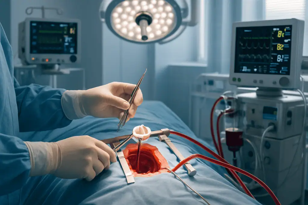
Valve surgery repairs or replaces one or more of the heart's four valves when narrowing or leakage disrupts blood flow. Performed under general anaesthesia, usually with a heart-lung machine, it takes several hours. Surgeons either reconstruct your own valve or implant a mechanical or biological prosthesis. Most patients spend around a week in hospital, with gradual recovery over six to twelve weeks.
What Is Valve Surgery?
Valve surgery is an operation to repair or replace one or more of the heart’s four valves: the aortic, mitral, tricuspid or pulmonary valve. It is recommended when a heart valve has become so narrowed, leaky or damaged that it disrupts blood flow and places strain on the heart muscle. Aortic valve replacement is the most frequently discussed form of this surgery, but the same principles apply across all four valves: restore reliable one-way blood flow before the heart is permanently harmed.
Your heart valves open and close with every heartbeat, directing blood through the heart’s chambers and onward to the lungs or the rest of the body. A healthy valve opens fully to let blood through and closes tightly to stop it flowing backwards. When that mechanism fails, the heart compensates by working harder, for a while. Over time, sustained strain can lead to enlargement of the heart chambers, weakening of the pumping muscle, rhythm disturbances and heart failure. That progression is why heart valve diseases are monitored so carefully, and why surgery is timed to intervene before the damage becomes irreversible.
What problems does valve surgery treat?
Valve surgery treats two core mechanical problems, alone or in combination. Valve stenosis means the valve does not open fully. The opening narrows, often because the leaflets stiffen or calcify, and blood flow is restricted, forcing the heart to generate higher pressure with every beat. Valve regurgitation, also called insufficiency or a leaky valve, means the valve does not close properly, so a portion of the blood leaks backwards and the heart must pump the same blood twice. Some patients have both problems in the same valve, and some have disease in more than one valve. The severity of the narrowing or leakage, your symptoms, the size and pumping strength of your heart, the pressures in your lungs, and which valve is involved all shape the recommendation you receive.
Repair or replacement: how the decision is made
Where the anatomy allows, surgeons prefer to repair your own valve rather than replace it. Repair can involve reshaping leaflet tissue, removing abnormal or excess tissue, reconstructing leaflet edges, repairing or replacing the small cords that anchor the leaflets, and implanting a supportive ring around the valve opening so it closes tightly. Repair matters most in mitral and tricuspid valve disease, where preserving the native valve tends to support more natural heart function and, in selected patients, can reduce the need for long-term anticoagulation. A durable repair, however, needs the right anatomy. A valve that is heavily calcified, scarred by rheumatic disease, destroyed by infection or structurally degenerated may not be repairable in a way that lasts, and an unreliable repair is worse than a good replacement. Your surgical team makes this judgement from detailed imaging before the operation and confirms it by inspecting the valve directly during surgery.
Mechanical and biological valves
Heart valve replacement uses one of two broad types of prosthesis, and the choice shapes your long-term care more than almost any other decision in the process. Mechanical valves are manufactured from durable materials and are designed to last for decades, but they require lifelong blood-thinning medication and regular blood monitoring, because clots can form on the valve without it. Biological valves are made from animal or, less commonly, human tissue. They may not require lifelong anticoagulation for the valve itself, which suits many patients, but tissue valves wear over time and may eventually need a further intervention: a consideration that weighs more heavily the younger you are.
There is no universally correct answer. Your age, lifestyle, occupation, other medical conditions, pregnancy plans, tolerance of anticoagulant medication, access to blood-test monitoring where you live, and personal priorities all belong in the discussion. A frequent traveller, a woman planning pregnancy, an older patient with kidney disease and a young manual worker may each be steered towards different valves for sound reasons. A good surgical team explains the trade-offs plainly and lets the decision be made with you, not for you.
When a Heart Valve Problem Begins to Affect Daily Life
Heart valve disease is unsettling partly because its symptoms arrive gradually. You may notice that climbing stairs takes more effort than it did a year ago, that you tire earlier, or that lying flat at night makes breathing difficult. Some people feel chest pressure, palpitations, dizziness or swelling in the legs and ankles. Others notice nothing at all and are told during a routine examination that a heart murmur needs further evaluation. Both routes into diagnosis are common, and neither should be dismissed: a quiet valve problem can still be a serious one.
Once a valve problem has been confirmed, the decision layers practical questions on top of medical ones. You may be comparing opinions from different doctors, trying to understand whether surgery is truly necessary now or can safely wait, and wondering how long recovery will take. You may also be asking about the safety of open-heart surgery, whether a less invasive approach is possible in your case, how long a repaired or replaced valve will last, and what daily life looks like afterwards. These are legitimate questions, and this page addresses each of them directly.
The central point to hold on to is this: valve surgery is recommended when a diseased valve is significantly disrupting blood flow or straining the heart. Left untreated, severe valve disease can progress to heart failure, rhythm disturbances such as atrial fibrillation, raised pressure in the lung circulation, fainting episodes or, in certain conditions, sudden deterioration. Timely evaluation by an experienced heart team determines which path is right for you, careful monitoring, medication, a catheter-based procedure, valve repair or valve replacement. Not everyone with valve disease needs an operation. Everyone with significant valve disease needs a plan.
Who May Need Valve Surgery?
You may need valve surgery when a heart valve problem is severe, progressive, or causing signs that the heart is under stress. Some patients feel distinctly unwell; others feel relatively normal but have test results showing that the heart is enlarging, weakening or developing pressure changes. This second group matters. In valve disease, symptoms are important, but they are not the only guide: the heart can compensate silently for years, and waiting until symptoms become severe may allow damage to progress past the point where it can be undone.
Symptoms that commonly lead to a valve evaluation include shortness of breath during activity, fatigue, reduced stamina, swelling of the feet or ankles, palpitations, chest discomfort, fainting or near-fainting, difficulty sleeping flat, coughing at night, and a general sense of fluid retention. In advanced disease, patients may have had repeated hospital admissions for heart failure or fluid overload. Some patients also unconsciously scale back their activity to avoid symptoms, which can mask how limited they have actually become: a detail worth being honest about when a doctor asks what you can and cannot do.
How is heart valve disease diagnosed?
Diagnosis begins with a physical examination and medical history; the definitive test is echocardiography. A heart murmur, abnormal pulse, fluid retention or signs of heart strain may prompt further testing, and echocardiographyan ultrasound examination of the heart, then shows valve movement, the degree of leakage or narrowing, chamber sizes, pumping function and pressure estimates. A transthoracic echocardiogram is performed from the chest wall and is non-invasive. When more detailed views are needed, a transoesophageal echocardiogram uses an ultrasound probe passed into the oesophagus under sedation, giving the team a much closer look at the valve structures.
Additional tests are used selectively. An electrocardiogram assesses heart rhythm. Blood tests evaluate heart strain, kidney and liver function, and blood counts. Chest imaging and computed tomography define valve and aortic anatomy, which matters when the aorta itself may need attention. Cardiac magnetic resonance imaging helps in selected cases, particularly for quantifying leakage and assessing heart muscle. Stress testing shows how the heart responds to exercise, which can unmask symptoms a patient has adapted around. Coronary angiography checks for blocked coronary arteries before surgery, if significant coronary artery disease is found, heart bypass surgery can be performed during the same operation when clinically appropriate, sparing you a second procedure.
Patients typically arrive at a surgical recommendation through one of several scenarios: symptoms are interfering with quality of life; imaging shows severe valve disease even without major symptoms; the heart is becoming enlarged or weakened; a valve infection has damaged tissue; a previously implanted valve has deteriorated; or another heart operation is planned and the valve can sensibly be addressed at the same time. In urgent situations, active infection with heart failure, or acute valve failure, surgery may be recommended quickly rather than electively.
Conditions Treated With Valve Surgery
Valve surgery addresses a wide range of diseases affecting one or more valves, and the cause of the problem matters as much as the degree of narrowing or leakage. The same operation is not right for every valve, and the same valve problem is not treated identically in every patient.
Aortic valve stenosis is one of the most common reasons for aortic valve replacement, particularly in older adults. The aortic valve stiffens and narrows, usually through calcification, and the heart must force blood through an ever-smaller opening. Breathlessness, chest discomfort, fainting and heart failure symptoms in a patient with severe aortic stenosis mark a stage that warrants prompt assessment. In younger patients, a bicuspid aortic valve, one born with two leaflets instead of three, can dysfunction earlier in life and is often associated with enlargement of the aorta itself, which changes the surgical plan.
Aortic valve regurgitation occurs when the aortic valve leaks backwards into the heart’s main pumping chamber. Causes include valve degeneration, congenital valve differences, enlargement of the aortic root, infection and inflammatory conditions. Treatment may involve valve repair or replacement and, where the aorta is enlarged, repair or replacement of a section of the aorta in the same operation.
Aorta heart valve replacement surgery: when the aorta is treated too
Aorta heart valve replacement surgery refers to operations that replace the aortic valve and treat part of the aorta: the body’s main artery, at the same time. This combined approach is considered when the aortic root or ascending aorta is enlarged, as can happen with bicuspid valves, connective tissue conditions or an aortic aneurysm. Treating the valve and the vessel together avoids a second major operation later and addresses the whole mechanical problem in one plan. Whether this applies to you depends on precise measurements from CT imaging, which is one reason thorough imaging is insisted upon before any decision. Broader aortic diseases follow their own assessment pathway, which is coordinated with the valve evaluation when both are present.
Mitral valve regurgitation most often develops when the valve leaflets prolapse or their supporting cords stretch or rupture; it can also follow heart enlargement after a heart attack or cardiomyopathy. In suitable patients, mitral valve repair is generally preferred over replacement because it preserves the natural valve and, when the anatomy is favourable, provides durable relief of symptoms. The mechanism and location of the leak determine whether repair is feasible, which is why an experienced imaging review matters before you are told the valve “must” be replaced.
Mitral valve stenosis is commonly related to rheumatic heart disease in many parts of the world. It causes shortness of breath, atrial fibrillation, pulmonary hypertension and fluid retention. Some patients are candidates for balloon-based treatment through a catheter; others require surgical repair or replacement, depending on how scarred and calcified the valve has become.
Tricuspid valve disease frequently accompanies left-sided valve disease, pulmonary hypertension, rhythm disorders, congenital abnormalities or injury from implanted device leads. Surgical repair is often considered when significant tricuspid leakage is present, particularly if another valve operation is being performed anyway, adding the repair at that point avoids a separate future operation on a valve that is technically harder to reach later.
Endocarditisinfection of the heart valves, can destroy valve tissue, cause severe leakage, create abscesses around the valve, or send infected material into the bloodstream. Surgery is considered when the infection cannot be controlled with antibiotics alone, when heart failure develops, or when the risk of embolic complications is high. Timing in endocarditis is a specialist judgement, balanced case by case between infection control and structural repair.
Valve surgery is also used for congenital valve abnormalities, some of which are identified and followed from childhood as part of congenital heart diseasesas well as for degeneration of a previously implanted biological valve, dysfunction of a mechanical valve, valve injury after trauma, and combined disease involving more than one valve. In every case, the clinical question is not simply whether a valve is abnormal. It is whether intervening now will reduce your risk, relieve your symptoms and protect your heart function over the years ahead.
How Heart Valve Surgery Is Performed
Heart valve surgery begins well before the operating theatre, with a preoperative assessment designed to confirm the diagnosis, define the valve anatomy precisely, quantify surgical risk and plan the safest approach. This assessment may include echocardiography, coronary evaluation, blood tests, lung function assessment, an anaesthesia consultation, and imaging of the chest and major blood vessels. Your medications are reviewed carefully by the treating team, particularly blood thinners, diabetes medications, blood pressure treatments and rhythm drugs, and any adjustments before surgery are directed by your doctors, not improvised.
Where you have already been investigated elsewhere, planning often starts with a review of your existing medical records. Recent echocardiograms, angiograms, CT or MRI scans, blood results, physician reports and discharge summaries allow the clinical team to judge whether further tests are needed. Where records are incomplete or imaging quality is limited, tests are repeated rather than guessed around, accurate decisions need accurate pictures.
Before the operation, you receive instructions about fasting, bathing, medication timing and what to bring to hospital. The surgical and anaesthesia teams explain the planned operation, the expected incision, the repair or replacement strategy, the realistic alternatives and the risks. If replacement is expected, the mechanical-versus-biological discussion happens here, in detail, with your circumstances on the table.
What happens during the operation
Most valve operations follow a recognisable sequence under general anaesthesia:
- Step 1: Anaesthesia and monitoring. You are put fully to sleep, and monitoring lines are placed to track heart function, blood pressure, oxygenation and organ perfusion continuously throughout the operation.
- Step 2: Access to the heart. In a traditional operation, the surgeon reaches the heart through an incision in the chest. In selected patients, a smaller incision with specialised instruments and enhanced imaging may be used instead.
- Step 3: The heart-lung machine. A heart-lung machine temporarily takes over the work of circulation and oxygenation, allowing the surgeon to stop and open the heart safely.
- Step 4: Repair or replacement. In a repair, the surgeon reconstructs your own valve, removing excess tissue, closing gaps, rebuilding leaflet edges, replacing or shortening supporting cords, and often implanting a supportive ring. In a replacement, the diseased valve is removed and a mechanical or biological prosthesis is secured in its place. If another problem is present, coronary disease, an enlarged aorta, atrial fibrillation or a second diseased valve: it may be treated under the same anaesthetic when indicated.
- Step 5: Checking the result. Before the chest is closed, high-resolution echocardiography in the operating theatre confirms that the repaired or replaced valve opens freely, closes tightly and functions properly under normal heart pressures. If the result is not right, it is corrected then, not later.
- Step 6: Transfer to intensive care. You are moved to a cardiovascular intensive care unit for close monitoring during the first phase of recovery.
How long does open heart valve replacement surgery take?
Most open heart valve operations take several hours from the start of anaesthesia to transfer to intensive care. The exact duration depends on how many valves are involved, whether the valve is repaired or replaced, whether additional procedures such as coronary bypass or aortic work are performed at the same time, and whether you have had previous heart surgery, which adds scar tissue and time. Family members are typically updated at key stages according to hospital protocols, so a long wait does not, by itself, mean something has gone wrong.
Can a heart valve be replaced without open heart surgery?
Yes, in selected patients, some valves can be treated through a catheter rather than through open surgery. The best-known example is transcatheter aortic valve implantation (TAVI, also called TAVR), in which a new aortic valve is delivered through a blood vessel, usually from the groin, and expanded inside the diseased valve without opening the chest. Catheter-based options also exist for some mitral and tricuspid valve problems, and certain narrowed valves can be treated with balloon procedures. These approaches are genuinely valuable, but they are not universally suitable: anatomy, the mechanism of the valve problem, calcification, your surgical risk profile and your age all determine whether a catheter-based treatment or surgery serves you better in the long run. That judgement belongs to a heart team that offers both and can therefore recommend either honestly.
How can a leaky heart valve be fixed without surgery?
A leaky heart valve cannot be repaired with medication alone, drugs can ease the symptoms and reduce the strain on the heart, but they cannot make a valve close properly. What medication does well is manage fluid retention, blood pressure and rhythm while a valve problem is mild or while the right timing for intervention is being determined. For some patients with mitral regurgitation who are at high surgical risk, catheter-delivered devices that clip the leaflets together can reduce leakage without open surgery. Whether that is appropriate depends on the mechanism of the leak and detailed imaging. Be cautious of any suggestion that a significantly leaking valve can simply be left indefinitely on tablets: the leak itself does not improve, and the heart’s ability to compensate is finite.
Recovery in hospital: intensive care and the cardiac ward
After surgery, you recover first in a cardiovascular intensive care unit. A breathing tube may be in place initially and is removed once you are awake, stable and breathing adequately. Temporary drainage tubes, monitoring lines, pacing wires and a urinary catheter support the early phase and are removed step by step as your condition improves. Pain is managed with medication, and breathing exercises begin early to reduce the risk of lung complications.
As you progress, you move from intensive care to a cardiac ward. Nurses and physiotherapists help with sitting, standing, walking, coughing exercises, wound care and a gradual return to activity. The team monitors your heart rhythm, blood counts, kidney function, lung status, valve function and response to medications. Before discharge, you receive clear instructions covering wound care, medications, activity limits, warning signs, follow-up visits and travel planning.
If a mechanical valve has been implanted, anticoagulation education is an essential part of discharge. You learn how the blood-thinning medication will be monitored, which interactions matter, and how long-term care will be coordinated after you go home. Patients with biological valves may also need temporary or longer-term blood thinners depending on heart rhythm and other conditions: that decision sits with your treating doctors. Thorough discharge documentation allows your own cardiologist to continue your care without guesswork.
Risks, Seriousness and What the Numbers Really Mean
How serious is heart valve replacement surgery?
Heart valve replacement surgery is major surgery: it involves general anaesthesia, opening the chest in most cases, and temporarily stopping the heart while a machine takes over circulation, and it should be described as such, not minimised. At the same time, it is one of the most established and standardised operations in cardiac surgery, performed routinely in experienced centres for decades, with each step of the pathway, imaging, anaesthesia, the operation itself, intensive care and rehabilitation, refined specifically around it. Risks exist, as with any major operation: bleeding, infection, rhythm disturbance, stroke, kidney strain and problems related to the new valve are the ones surgeons discuss most often. What determines how serious the operation is for you is your individual profile: your age, the condition of your heart muscle, your kidneys and lungs, whether the operation is planned or urgent, and whether additional procedures are needed. Established risk-scoring systems allow a surgical team to estimate your individual risk before you decide, ask for that estimate rather than a generic reassurance.
What is the success rate of aortic valve replacement surgery?
There is no single success rate for aortic valve replacement that honestly applies to every patient, because outcomes depend heavily on who is being operated on and when. A planned operation in a patient with strong heart function carries a very different risk profile from an urgent operation in a patient with advanced heart failure, kidney disease or a previous cardiac operation. The same is true of every form of heart valve replacement surgery: headline figures quoted online blend together patients whose situations have little in common. The honest approach is individual: a surgical team reviews your imaging, your test results and your history, and gives you a risk estimate specific to your case, alongside what “success” would mean for you, durable valve function, symptom relief and protection of your heart muscle, not merely getting through the operation.
What is the life expectancy after aortic valve replacement?
Life expectancy after this operation depends on your age, your heart function at the time of surgery, your other medical conditions and the type of valve implanted, which is precisely why timing matters so much. Patients treated before the heart muscle has been permanently damaged generally recover more of their capacity than those treated late, and untreated severe aortic stenosis with symptoms carries a genuinely poor outlook, which is the strongest argument for not deferring evaluation. After a successful operation and recovery, many patients return to work, travel and physical activity, with life shaped far more by their overall health than by the valve. Long-term follow-up with a cardiologist, including periodic echocardiograms, is part of protecting that outcome.
What is the hardest heart valve to replace?
Surgeons generally regard the mitral valve as the most technically demanding of the commonly operated valves, because of its complex anatomy, two asymmetric leaflets, a ring that changes shape with every beat, and a web of supporting cords attached to the heart muscle itself. This complexity is also why mitral repair is such a specialised skill and why repair feasibility varies between centres. The tricuspid valve, historically called the “forgotten valve”, presents its own challenges, partly because patients often come to tricuspid surgery late, with strained right-heart function. The aortic valve, by contrast, is the most standardised to replace, which is one reason aortic procedures, surgical and catheter-based alike, are so well developed. “Hardest”, though, is ultimately about the individual case: a heavily calcified, previously operated or infected valve of any type is harder than a straightforward first-time operation.
Why Acting Early Matters
Severe valve disease can be deceptively quiet. Some patients reduce their activity without noticing and therefore never register how limited they have become. Others attribute breathlessness and fatigue to age, stress, weight or lack of fitness. Meanwhile, a severely narrowed or leaking valve keeps loading the heart, which compensates for a period and then begins to enlarge, weaken or develop rhythm problems. By the time symptoms are undeniable, some of that change may be permanent.
Early specialist evaluation matters because the timing of intervention affects long-term heart function. In several valve conditions, surgery performed before irreversible muscle damage is associated with better recovery of pumping strength and functional capacity. In mitral regurgitation, waiting until the heart is significantly enlarged or weakened may reduce the chance that pumping function returns to normal after repair. In aortic stenosis, symptoms such as chest pain, fainting or breathlessness signal a higher-risk stage of the disease that warrants prompt assessment. In endocarditis, delaying surgery when clear indications exist may allow infection to destroy more tissue or spread to surrounding structures.
Delay carries other costs too: a higher likelihood of atrial fibrillation, pulmonary hypertension, fluid accumulation in the lungs, kidney strain, repeated hospital admissions and reduced physical reserve to tolerate surgery when it eventually becomes unavoidable. None of this means every valve problem requires an immediate operation, many do not. Acting early means obtaining an accurate diagnosis, understanding severity, identifying the right treatment window, and following a structured monitoring plan if surgery is not yet needed. For most patients, that clarity is itself a relief: uncertainty, not surgery, is often the heaviest part of the burden.
Benefits of Valve Surgery
When valve surgery is appropriately timed and carefully planned, it can improve blood flow, relieve symptoms and protect the heart from further strain. The table below summarises what each benefit means in practical terms.
| Benefit | What It Means for You |
|---|---|
| Improved blood flow through the heart | A repaired or replaced valve can reduce obstruction or leakage, allowing the heart to pump more efficiently with every beat. |
| Relief of symptoms | Many patients experience less breathlessness, fatigue, chest discomfort, swelling and exercise limitation after recovery. |
| Protection of heart function | Treating severe valve disease at the right time may help prevent or limit enlargement and weakening of the heart muscle. |
| Reduced risk of valve-related complications | Surgery may lower the likelihood of recurrent heart failure episodes or progressive pressure build-up in the lungs when valve disease is the cause. |
| Opportunity to treat combined heart problems | Where needed, surgeons can address related conditions, coronary artery disease, aortic enlargement, rhythm problems or a second diseased valve, during the same operation. |
| Clearer long-term care plan | After surgery, you receive concrete guidance on follow-up imaging, medications, anticoagulation if required, rehabilitation and a safe return to daily life. |
Recovery Timeline After Valve Surgery
Recovery varies with age, overall health, the type of valve operation, the incision approach and whether additional procedures were performed, but most patients follow a recognisable pattern of gradual, week-by-week progress.
| Time Period | What Patients Can Expect |
|---|---|
| Day 1 | Monitoring in cardiovascular intensive care. Breathing support, drainage tubes, rhythm monitoring, pain control and early breathing exercises are typical. |
| First Week | Walking with assistance begins, followed by transfer to the cardiac ward, increased breathing exercises, and education on medication and wound care. Discharge timing depends on stability and progress. |
| First Month | Energy gradually improves, though fatigue is common and normal. Walking is encouraged; heavy lifting and strenuous activity are restricted. Follow-up visits and blood-thinner monitoring may be required. |
| Six to Twelve Weeks | Many patients return to more normal daily activity, guided by the surgeon’s advice, occupation, wound healing and overall strength. Cardiac rehabilitation supports safe reconditioning. |
| Longer Term | Ongoing cardiology follow-up: periodic echocardiograms, medication adjustments, infection-prevention guidance, and anticoagulation management if a mechanical valve or rhythm condition is present. |
How long does recovery take after aortic valve replacement?
Most patients need roughly six to twelve weeks to return to near-normal daily activity after aortic valve replacement, though the breastbone continues to strengthen for some months and stamina keeps improving well beyond the early period. The pace depends on your age, fitness before surgery, whether the operation was straightforward or combined with other procedures, and how disciplined the early rehabilitation phase is. Walking early and often, doing the breathing exercises, protecting the healing chest from heavy lifting, and attending cardiac rehabilitation where available all move the timeline in your favour. Patients recovering from minimally invasive or catheter-based procedures typically progress faster, which is one of the reasons those approaches are considered where anatomy allows.
A practical question many patients raise is when it is safe to travel, particularly by air. Air travel after cardiac surgery needs to be timed with your surgical team, taking into account wound healing, rhythm stability and any anticoagulation plan; longer journeys also add considerations around prolonged immobility, which your team can plan for before you set a date.
What Influences Outcomes After Valve Surgery?
The result of valve surgery is shaped by a combination of patient factors, valve anatomy, surgical planning, timing and postoperative care. A good result is not defined only by getting through the operation. It includes durable valve function, symptom relief, protection of heart muscle, safe rhythm management, clean wound healing, a confident return to activity, and a follow-up plan you can actually sustain where you live.
Timing is arguably the most important factor. Patients treated before advanced heart muscle damage, severe pulmonary hypertension or repeated heart failure episodes tend to recover more favourably. This is why careful surveillance is essential when severe valve disease exists with few symptoms, in selected cases, surgery is recommended on the basis of heart size, pumping function, valve measurements or exercise testing rather than symptoms alone.
Valve anatomy comes next. Some valves are highly suitable for repair; others are too calcified, infected, scarred or degenerated to repair reliably. In mitral disease, the location and mechanism of the leak determine repair feasibility. In aortic disease, the state of the leaflets, the aortic root and the ascending aorta determines whether repair, replacement or a combined aortic procedure is appropriate. A durable repair requires both technical expertise and the right anatomy, one without the other is not enough.
Overall health shapes both risk and recovery. Diabetes, kidney disease, lung disease, obesity, frailty, previous stroke, liver disease, anaemia and prior chest surgery each add complexity. Coexisting coronary artery disease, atrial fibrillation or reduced pumping function may require additional treatment during the same operation. None of these conditions necessarily rules surgery out, but each one belongs in an honest risk assessment and a tailored perioperative plan.
The type of valve substitute, when replacement is needed, defines long-term care. Mechanical valves are durable but demand careful lifelong anticoagulation and monitoring. Biological valves suit patients who cannot take, or prefer to avoid, long-term anticoagulants, but they can degenerate and may need a future intervention. Younger patients, older patients, women considering pregnancy, athletes, frequent travellers, and patients living far from a laboratory that can monitor blood tests each weigh these trade-offs differently, which is why the discussion should fit your life, not a template.
Rehabilitation and follow-up close the loop. Early mobilisation, breathing exercises, good nutrition, wound care, medication adherence and structured cardiac rehabilitation all support recovery. Patients who understand the warning signs and keep their cardiology appointments are positioned to catch rhythm issues, anticoagulation problems, valve changes or heart-function concerns early, when they are easiest to address.
Living with a repaired or replaced valve
Life after valve surgery has a few standing rules worth knowing early. Any prosthetic valve, and, to a lesser degree, a repaired one, carries a lifelong susceptibility to infective endocarditis, so meticulous dental hygiene matters, and dentists and doctors should always be told about your valve before procedures; in defined situations, your treating team may recommend preventive antibiotics before dental work. Patients with mechanical valves live with anticoagulation as a daily routine: regular blood tests keep the medication in its target range, and diet, alcohol, other medicines and periods of illness can all shift that balance, which is why the monitoring plan agreed at discharge should be practical for the place you actually live. Patients with biological valves attend periodic echocardiograms so that any gradual wear is detected long before it causes symptoms, and if a tissue valve does degenerate years later, options in suitable anatomy now include valve-in-valve catheter procedures rather than automatically a repeat open operation. None of this should dominate life after recovery; for most patients, the valve becomes a background fact rather than a daily preoccupation.
Outcomes also depend on continuity between the operating hospital and the cardiologist who follows you afterwards. Discharge reports, operative notes, the exact valve type and size, medication plans, imaging results and anticoagulation targets should all be documented clearly enough that a cardiologist who has never met you can continue your surveillance without uncertainty. Ask for this documentation explicitly: it is part of the treatment, not an afterthought.
Valve Surgery at Acibadem
Patients facing valve surgery need more than an operation. They need a reliable diagnosis, a carefully reasoned recommendation, experienced surgical and intensive care teams, and a realistic plan for recovery and long-term follow-up. Acibadem’s cardiovascular programmes are structured around those needs, with heart valve care delivered through the cardiovascular surgery departments.
Care is multidisciplinary by design. Cardiologists, cardiac surgeons, cardiovascular imaging specialists, anaesthesiologists, intensive care physicians, electrophysiologists, infectious disease specialists, rehabilitation teams and nursing staff contribute according to the patient’s condition. Complex cases can be reviewed in specialist boards, where imaging, symptoms, surgical risk and alternatives are weighed together: a process that matters particularly for patients who have received differing opinions along the way. Sometimes such a review confirms the original plan; sometimes it refines the timing, identifies missing information, or clarifies whether repair rather than replacement is feasible. Either result is useful, because a decision this significant deserves to rest on complete information.
Evaluation and treatment follow evidence-based protocols. Internationally accepted criteria guide the assessment of valve severity, surgical timing, anticoagulation needs, infection management, heart failure care and follow-up. At the same time, every plan is individualised: a patient with degenerative mitral regurgitation, a patient with rheumatic mitral stenosis, and a patient with a calcified aortic valve plus coronary disease need genuinely different strategies, and treating them identically would serve none of them well.
Diagnostic depth supports those decisions. Echocardiography, transoesophageal imaging, cardiac CT, coronary angiography, rhythm monitoring and laboratory testing are used selectively to build a complete picture before surgery. In the operating theatre, intraoperative echocardiography verifies the quality of a repair or the function of a replacement before the operation is completed. Afterwards, intensive care monitoring supports early recognition of rhythm changes, bleeding, lung issues, kidney stress or blood pressure instability.
Acibadem’s cardiac surgery teams treat the full spectrum of valve disease, from isolated aortic valve operations to complex multi-valve procedures and surgery combined with coronary bypass or aortic work. The technique is chosen according to anatomy and risk rather than preference: some patients are candidates for minimally invasive approaches, while others are safer with a conventional incision that gives wider access for complex repair or combined work. Related cardiothoracic surgery capabilities sit within the same programme, which matters when a valve problem is only part of the picture.
Continuity after discharge is treated as part of the treatment itself. Patients leave with documentation covering the operation performed, the valve type and size if a replacement was used, the medication list, anticoagulation instructions where applicable, wound care guidance, activity restrictions and follow-up recommendations: the material any cardiologist needs to take over care safely.
Making a Considered Decision
Valve surgery is a major decision, and it deserves to be treated as one. It is also a well-established treatment for serious heart valve disease, one that, performed at the right time, can relieve symptoms, restore efficient circulation and protect the heart from progressive strain. The essential ingredients of a good decision are knowable: the exact valve problem, its severity, the realistic options including repair, replacement and catheter-based treatment, the risks specific to your situation, and the shape of the recovery ahead.
If you have been diagnosed with aortic, mitral, tricuspid or pulmonary valve disease, or advised to consider repair or replacement, the most useful things you can do are practical: keep copies of your echocardiograms and reports, understand what your own imaging shows, ask your doctors what would change their recommendation, and make sure any surgical plan comes with an individual risk estimate rather than a general one. Valve disease rewards patients who stay informed and stay in follow-up, before surgery, and for all the years after it.
Preparation
- Before valve surgery, patients usually undergo echocardiography, blood tests, chest imaging, ECG, and sometimes coronary angiography. Blood-thinning medications and certain supplements may need to be adjusted before the operation. Patients are typically asked to fast from midnight and follow individualized instructions from the cardiac surgery and anesthesia teams.
Aftercare
- After surgery, patients are monitored in intensive care before moving to a cardiac ward. Pain control, breathing exercises, wound care, and early walking help support recovery and reduce complications. Follow-up visits, cardiac rehabilitation, and medication management, including anticoagulation if needed, are important after discharge.
Frequently Asked Questions
What affects the cost of valve surgery abroad?
Cost is influenced by the diagnosis, the valve involved, whether repair or replacement is needed, the valve material, surgical approach, hospital stay, intensive care needs, surgeon and anaesthesia fees, and any additional heart procedures. Travel, accommodation, translation, and follow-up arrangements may also affect the total budget.
How can I get a personalised quote for valve surgery in Turkey?
You can request a free consultation by sharing recent cardiology reports, echocardiography results, angiography or cardiac imaging if available, medication details, and a summary of symptoms. The cardiac team reviews the records and provides a personalised plan and quote based on medical suitability.
Does a valve surgery package usually include all hospital costs?
Packages often include core hospital services such as preoperative assessment, the operation, standard inpatient care, and routine support during admission. However, inclusions vary, so patients should confirm what is covered, what may be billed separately, and what happens if extra care is medically required.
Why can the same valve operation have different costs between hospitals?
Differences can come from surgeon experience, hospital accreditation, intensive care resources, the complexity of the valve disease, the type of prosthetic valve or repair materials, diagnostic testing, and the level of international patient services provided.
Is the lowest quote always the best option for valve surgery?
Not necessarily. Valve surgery is complex, so patients should consider the cardiac team, hospital quality standards, intensive care capability, accreditation status, transparency of inclusions, communication support, and follow-up planning as well as cost. This information is general and is not medical or financial advice.
Medically reviewed by the Acıbadem International Medical Board August 30, 2026
See our medical review board →
Update history
- PublishedJune 8, 2026
- Medical review approvedAugust 30, 2026
- Last content updateAugust 30, 2026
References1
- Heart Valve Surgery medlineplus.gov
Trusted care for international patients
From the Acibadem Blog

Blood Thinners After Heart Valve Surgery: Who Needs Them and How They Are Monitored
Not everyone needs blood thinners after heart valve surgery. People who receive a mechanical valve usually take a vitamin K antagonist anticoagulant, such as…

How Long Is Recovery After Heart Valve Surgery? From Intensive Care to Cardiac Rehab
Recovery after heart valve surgery usually unfolds in stages: about a day in intensive care, roughly a week in the hospital, and two to…



































