How Is an MRI of the Prostate Done: Preparation, Procedure and Results
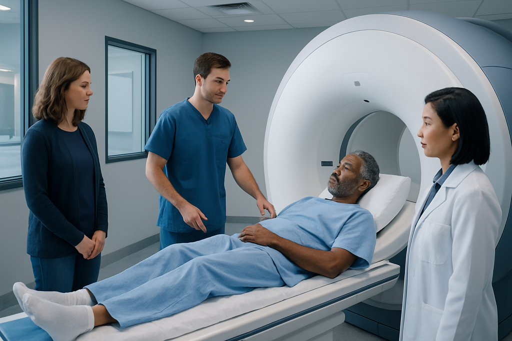
A prostate MRI does not use ionizing radiation and is usually performed as an outpatient examination. Preparation may include completing a safety screening, following local instructions about food or bowel preparation, and removing metal items.
Key Takeaways
- A prostate MRI does not use ionizing radiation and is usually performed as an outpatient examination.
- Preparation may include completing a safety screening, following local instructions about food or bowel preparation, and removing metal items.
- Most prostate MRI examinations take about 30 to 60 minutes, including positioning and image acquisition.
- MRI can identify areas that may need targeted biopsy, but it cannot by itself confirm whether a tumor is cancerous.
- People should tell the imaging team about implants, kidney problems, allergies, claustrophobia, or a prior prostate biopsy.
A prostate MRI is a noninvasive imaging examination that uses a powerful magnet and radio waves to create detailed pictures of the prostate and nearby tissues. It commonly helps doctors assess an elevated PSA result, investigate concerning symptoms, guide biopsy planning, or evaluate known prostate cancer.
Overview: How Is an MRI of the Prostate Done?
How is an MRI of the prostate done? The person lies still on a padded table that moves into an MRI scanner while the machine uses magnetic fields and radio waves to take detailed images of the prostate. The examination is painless, does not use X-rays, and is usually completed without an overnight hospital stay.
Most modern examinations are multiparametric MRI (mpMRI). This combines several image types to show the prostate’s anatomy, water movement within tissues, and blood flow patterns after contrast when contrast is appropriate. Together, these images can help a radiologist identify areas that look suspicious and help the urology team decide whether monitoring, biopsy, or further assessment is needed.
A prostate MRI may be requested because of a raised or rising prostate-specific antigen (PSA) level, an abnormal digital rectal examination, urinary symptoms, a previous negative biopsy, or a diagnosis of prostate cancer. It is also useful for estimating whether a known cancer may extend beyond the prostate and for planning care.
How a Prostate MRI Works and Who May Need One
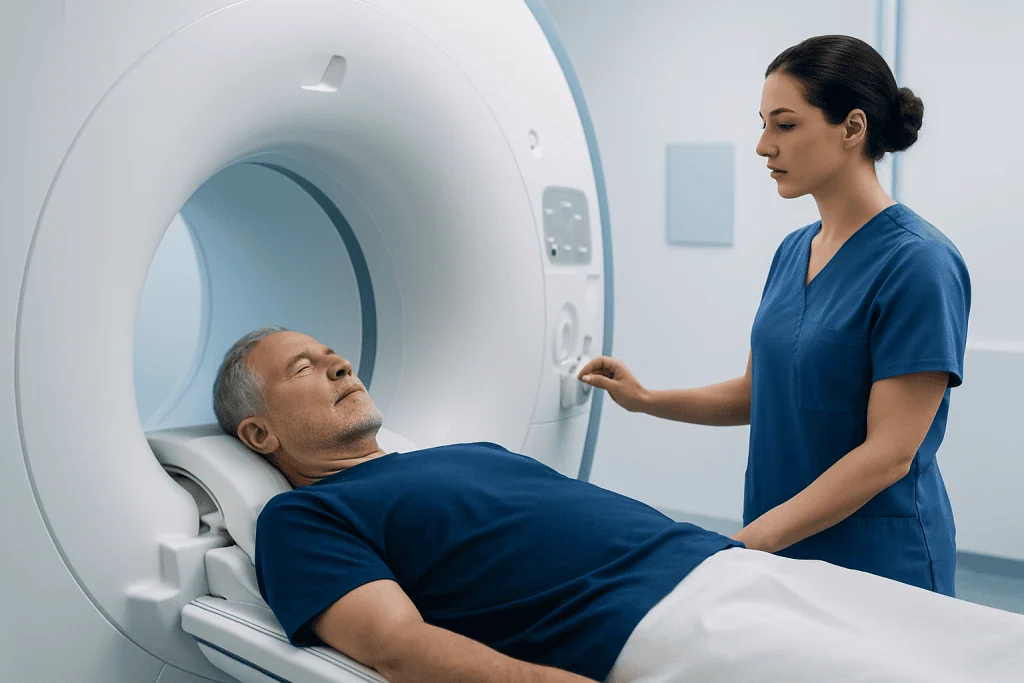
MRI produces images by detecting signals from hydrogen atoms in the body after they are exposed to a strong magnetic field and radiofrequency pulses. Unlike CT scanning, it does not use radiation. A specialized surface coil is usually placed around the pelvis to receive detailed signals from the prostate region.
Doctors may recommend prostate MRI before a first biopsy or after a biopsy that did not explain an ongoing PSA concern. If a suspicious area is seen, the images can be used to plan an MRI-targeted biopsy, in which samples are directed toward the area of concern. MRI can also support active surveillance and treatment planning for people with confirmed prostate cancer.
Not everyone can have an MRI in the same way. Some implanted devices, metal fragments, certain clips, or older pacemakers may make MRI unsuitable or require special safety checks. Many modern implants are MRI-conditional, meaning scanning may be possible under specified conditions. The imaging department should always receive complete information about implants, prior surgery, and any possible metal exposure.
A prostate MRI is one part of a wider urologic assessment. PSA results, examination findings, symptoms, family history, and any earlier biopsy findings are considered together rather than relying on one test alone.
What Do I Need to Do to Prep for a Prostate MRI?

The imaging center provides the most important preparation instructions, as protocols can differ. A person may be asked to avoid eating for several hours before the scan, especially if contrast is planned. Some centers recommend a light diet, a small enema, or other measures to reduce stool and gas in the rectum, because these can affect image quality.
Before the appointment, the person should complete an MRI safety questionnaire and report any implanted medical device, metal fragment injury, surgery, medication patch, hearing aid, dental device, or pregnancy possibility. Kidney disease, prior reactions to MRI contrast, and severe claustrophobia should also be discussed in advance.
On the day, comfortable clothing without metal is helpful, although changing into a gown is common. Jewelry, watches, hair accessories, credit cards, removable dental appliances, and electronic devices must be removed before entering the scanner room. Regular medicines are often continued unless the clinician gives different instructions.
If a biopsy has recently been performed, the MRI may need to be delayed. Post-biopsy bleeding and inflammation can make interpretation more difficult. The referring urologist and radiology team can advise on the appropriate timing for each individual.
Step by Step: What Happens During the Procedure?
After arrival, a radiology professional reviews safety information, explains the examination, and answers questions. The person lies on their back on the scanning table, usually with a padded imaging device placed over the lower abdomen and pelvis. Modern prostate MRI commonly uses this external coil and does not routinely require an internal rectal coil.
The table then moves into the scanner. The body generally goes into the machine far enough for the pelvis to be centered, so the person’s head may be inside the scanner depending on body size and equipment design. An intercom allows communication with the MRI staff throughout the test, and many centers offer headphones or earplugs because the scanner makes loud tapping and knocking sounds.
Remaining as still as possible is important because movement can blur the images. The technologist may give simple instructions through the intercom. For some examinations, gadolinium-based contrast material is injected through a small intravenous line partway through the scan to assess tissue blood flow. Contrast is not needed for every protocol.
After imaging is complete, the table moves out and the intravenous line is removed if one was used. There are no lingering magnetic effects in the body. The person can usually dress and leave shortly afterward.
How Long Does a MRI of the Prostate Take?
The actual imaging portion of a prostate MRI commonly takes about 30 to 45 minutes. Allowing time for registration, safety screening, changing clothes, positioning, and possible intravenous contrast, the full visit may take approximately one to one and a half hours.
Scan time can vary according to the equipment, the imaging protocol, whether contrast is used, and whether additional images are needed. The radiology team may repeat a sequence if movement affects image clarity. This is normal and helps ensure the examination provides useful information for the referring clinician.
Afterward, most people can return to usual daily activities immediately. If a sedating medicine has been arranged for claustrophobia or anxiety, a responsible adult may need to accompany the person home, and driving or making important decisions should be avoided until the medication has worn off.
Can You Tell if a Prostate Tumor Is Cancerous From an MRI?
MRI can show areas that have features associated with clinically significant prostate cancer, but it cannot confirm cancer on its own. Radiologists commonly use the Prostate Imaging Reporting and Data System (PI-RADS) to describe how likely a visible area is to represent clinically significant cancer. A higher category may lead a clinician to recommend biopsy, depending on the full clinical picture.
Some noncancerous conditions can look suspicious on MRI. Inflammation, infection, benign prostatic enlargement, scarring, and changes after a biopsy may affect the appearance of prostate tissue. Conversely, a normal or low-suspicion MRI reduces the likelihood of significant cancer but does not eliminate it completely.
If tissue confirmation is needed, a urologist may recommend a targeted biopsy using MRI images alongside ultrasound guidance. Pathology examination of biopsy tissue remains the standard way to diagnose prostate cancer and determine its grade. For information about the condition and its evaluation, see prostate cancer.
Results are normally sent to the clinician who ordered the scan. That clinician explains what the findings mean in the context of PSA values, examination findings, personal health, and preferences before recommending next steps.
Do You Have to Go All the Way In for a Prostate MRI?
For a prostate MRI, the pelvis must be positioned at the center of the scanner, so a substantial part of the body goes into the MRI tunnel. In many scanners, the head may also be inside the machine for at least part of the examination. The experience can feel enclosed, but the person is monitored continuously and can communicate with staff at any time.
The scanner is open at both ends and is well lit and ventilated. Ear protection, music where available, a call button, and clear communication can make the test more manageable. People who feel anxious in enclosed spaces should tell the referring doctor or imaging department before the appointment; relaxation strategies, a wider-bore scanner, or prescribed sedation may be options.
There is generally no need for an instrument to be inserted into the rectum for contemporary prostate MRI. An endorectal coil was used more often in the past, but many centers now obtain high-quality images with external pelvic coils. The exact approach depends on the MRI system and local protocol.
Benefits, Risks, Recovery and When to Seek Medical Care
The key benefit of prostate MRI is detailed, noninvasive information that can help avoid unnecessary biopsy in some people and improve the accuracy of biopsy when tissue sampling is needed. It may also help clinicians assess the location and possible extent of known cancer, supporting individualized treatment discussions.
MRI itself is generally very safe when screening procedures are followed. The main practical challenges are noise, the need to lie still, and discomfort for people with claustrophobia. Gadolinium contrast reactions are uncommon, but the team should know about previous contrast reactions and significant kidney disease. The strong magnet can be hazardous around certain metal objects or implants, which is why safety screening is essential.
Recovery is usually immediate. No special aftercare is required after a scan without sedation, and people can eat, drink, work, and exercise as usual unless given different instructions. Drinking fluids after contrast may be advised as part of normal hydration, but specific restrictions are not typically necessary.
When to seek medical care: A prostate MRI is not emergency care. However, prompt medical assessment is sensible for fever with urinary symptoms, inability to pass urine, visible blood in urine that is heavy or persistent, severe pelvic pain, or rapidly worsening symptoms. A person should contact the imaging center or a clinician urgently after contrast if they develop symptoms of a serious allergic reaction, such as trouble breathing or swelling of the face or throat.
Acibadem International’s multidisciplinary urology, radiology, and oncology specialists at JCI-accredited hospitals support international patients undergoing prostate assessment and care planning, including prostate cancer treatment.
Frequently asked questions
Is a prostate MRI painful?
A prostate MRI is generally painless. The person may find it uncomfortable to lie still on the table and may notice loud scanner noises, but the scan itself does not cause pain. An intravenous line for contrast may cause a brief pinch.
Can I eat before a prostate MRI?
Instructions vary between imaging centers. Some people may be asked not to eat for several hours before the examination, while others may be allowed a light meal. The appointment instructions from the radiology department should be followed closely.
Do I need contrast for a prostate MRI?
Not every prostate MRI requires contrast material. Many multiparametric MRI protocols include gadolinium-based contrast to assess blood flow, but radiologists may use a non-contrast approach in selected situations. The decision depends on the clinical question and local protocol.
What happens if my prostate MRI shows a suspicious area?
A suspicious finding does not automatically mean cancer. The urologist will review the MRI report alongside PSA results, examination findings, and medical history. A targeted biopsy, monitoring plan, or further testing may be recommended depending on the overall level of concern.
Can I have a prostate MRI with a pacemaker or joint replacement?
Many people with joint replacements and some modern pacemakers can undergo MRI, but each implant must be checked carefully. The person should provide device details to the imaging team well before the appointment. Never enter an MRI room without completing the required safety screening.
When will I get prostate MRI results?
A radiologist needs time to review the images and prepare a report, so results are not always available immediately after the scan. Timing varies by center and clinical urgency. The clinician who ordered the MRI will explain the findings and any recommended next steps.
References
- American College of Radiology
- European Association of Urology
- National Cancer Institute
- Radiological Society of North America
- Prostate Imaging Reporting and Data System (PI-RADS)
This article is for general information only and is not a substitute for professional medical advice. Please consult a qualified doctor about your individual situation.
Oncology care in Turkey — second opinion and treatment plan
JCI-accredited · board-certified surgeons · reply within 24h

More from the Health Library


Related Specialists

Assoc. Prof. Dr. Ali Harman
Interventional Radiology
Dr. Zeynep Ece Demirbaş
Internal Medicine
Dr. Seyfettin Aslan
Ear Nose & Throat
Yaren Kasal
Physical Medicine & Rehabilitation