Actinic Keratosis Pictures: Symptoms, Causes, and Treatment Options

Actinic keratosis often appears as rough, scaly patches on sun-exposed areas such as the face, scalp, ears, hands, and forearms. Pictures can help people recognize common features, but a medical exam is needed to confirm the diagnosis.
Key Takeaways
- Actinic keratosis often appears as rough, scaly patches on sun-exposed areas such as the face, scalp, ears, hands, and forearms.
- Pictures can help people recognize common features, but a medical exam is needed to confirm the diagnosis.
- Long-term exposure to ultraviolet light from the sun or tanning beds is the main cause.
- Treatment may include freezing, prescription creams, light-based therapy, or minor procedures depending on the number and location of lesions.
- Daily sun protection and regular skin checks can help prevent new lesions and detect changes early.
Actinic keratosis pictures typically show small, rough, scaly patches on sun-exposed skin, often pink, red, tan, or skin-colored. These spots are caused by long-term ultraviolet damage and should be checked by a clinician because some can progress to skin cancer over time.
Overview: What Actinic Keratosis Pictures Usually Show
Actinic keratosis pictures usually show dry, rough, scaly areas on skin that has had years of sun exposure. The spots may look pink, red, tan, gray, or close to the natural skin tone, and they often feel easier to notice than to see at first. Common locations include the face, lips, ears, bald scalp, forearms, and backs of the hands.
These lesions are considered a sign of chronic ultraviolet damage. They are not the same as a simple rash or temporary irritation. Although many actinic keratoses remain stable, some can develop into a type of skin cancer called squamous cell carcinoma, which is why assessment by a dermatologist is important.
Photos found online can be helpful for general recognition, but appearance varies widely by skin tone, body site, and how advanced the lesion is. Some spots are flat and barely visible, while others are thicker, crusted, or tender. A clinical examination is the best way to tell actinic keratosis apart from other sun-related skin changes or from conditions such as skin cancer.
How It Can Look and Feel on the Skin
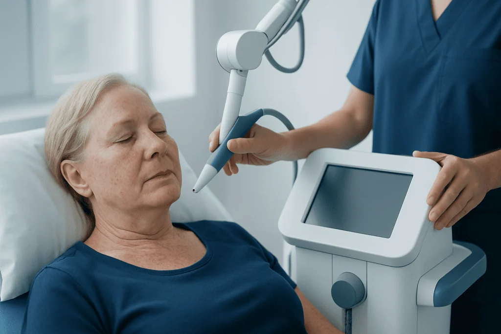
Many people search for pictures because actinic keratosis does not always stand out clearly. In real life, it may present as a patch with a sandpaper-like texture, a persistent scab-like spot, or a small area of scaling that returns after seeming to heal. The borders may be distinct or blend gradually into surrounding sun-damaged skin.
The feel of the lesion is often an important clue. A person may notice roughness when washing the face, applying moisturizer, or running fingers over the scalp or hands. Some lesions itch, sting, burn, or become sensitive, especially after sun exposure, while others cause no symptoms at all.
Actinic keratosis can also appear in special forms. On the lip, it may cause chronic dryness, scaling, or a pale or rough area. On the scalp, particularly in people with thinning hair, several lesions may develop close together. In some cases, multiple spots occur across a broader area of sun-damaged skin, sometimes described as a “field” of change rather than a single isolated lesion.
- Rough or scaly patch
- Flat or slightly raised spot
- Pink, red, tan, white, or skin-colored area
- Crusting or flaking
- Mild tenderness, itching, or burning
- Most often on sun-exposed skin
Causes and Risk Factors
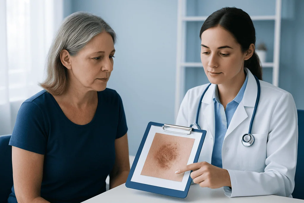
The main cause of actinic keratosis is cumulative ultraviolet exposure over many years. This includes sunlight and artificial UV sources such as tanning beds. UV radiation damages skin cells gradually, so lesions often appear later in adulthood after repeated exposure rather than after one single sunburn.
Several factors increase risk. Fair skin, light eyes, and light hair are associated with greater UV sensitivity, but actinic keratosis can affect people of any skin tone. A history of frequent outdoor work or recreation, living in sunny climates, past severe sunburns, and increasing age can all contribute.
People with weakened immune systems, including some transplant recipients or those taking immunosuppressive medicines, may be more likely to develop multiple lesions. A personal history of actinic keratosis or previous skin cancers also raises the chance of future lesions. These factors make regular skin checks especially important and may overlap with broader risks discussed on pages about sun-related skin cancer.
How Doctors Diagnose Actinic Keratosis
Diagnosis usually starts with a skin examination. A dermatologist looks at the spot closely, asks how long it has been present, whether it has changed, and whether there are symptoms such as bleeding, pain, or rapid growth. The texture and location can be as informative as the color or shape.
In many cases, an experienced clinician can recognize actinic keratosis during the exam. A dermatoscope, a handheld device that magnifies the skin, may help assess surface patterns and blood vessels. This can improve accuracy when distinguishing actinic keratosis from eczema, seborrheic keratosis, superficial skin cancers, or inflamed sun spots.
If a lesion is thick, very tender, ulcerated, bleeding, growing quickly, or not responding as expected to treatment, a biopsy may be recommended. A biopsy involves removing a small sample, or sometimes the whole lesion, and checking it in a laboratory. This helps confirm the diagnosis and rule out early squamous cell carcinoma or other conditions.
Treatment Options and What to Expect
Treatment depends on how many lesions are present, where they are located, how thick they are, and whether there is concern for early skin cancer. A single visible spot may be treated differently from a broad area of sun-damaged skin with several barely visible lesions. The aim is to remove abnormal cells, reduce the chance of progression, and improve the health of the surrounding skin.
Common office-based treatments include cryotherapy, which freezes the lesion with liquid nitrogen, and minor surgical removal or scraping for selected thicker spots. In some cases, doctors may recommend light-based treatments such as photodynamic therapy for multiple lesions in one area. Prescription field treatments may include medicated creams or gels that target damaged cells over a wider surface.
When there is concern that a lesion may already have become cancerous, additional evaluation or a more definitive procedure may be needed. Depending on the findings, management may overlap with broader dermatology care or, in confirmed cancer cases, treatment pathways used for skin cancer treatment. At Acibadem International, multidisciplinary specialists in JCI-accredited hospitals evaluate and treat actinic keratosis and related skin conditions for international patients.
- Cryotherapy for individual lesions
- Prescription topical treatments for field damage
- Photodynamic therapy for selected widespread lesions
- Curettage or minor excision for thicker or suspicious spots
- Follow-up skin checks to monitor for recurrence or new lesions
Prevention and Self-Care After Diagnosis
Because actinic keratosis is linked to cumulative UV damage, prevention focuses on limiting further exposure. Daily broad-spectrum sunscreen, protective clothing, wide-brimmed hats, and seeking shade during peak sunlight hours are practical steps. Tanning beds should be avoided, as they add to the same type of skin injury caused by the sun.
Self-care also includes learning the pattern of one’s own skin. After diagnosis, doctors often advise regular self-checks for rough new spots or changes in existing lesions. Taking note of areas that repeatedly crust, bleed, or fail to heal can help bring useful information to follow-up appointments.
Moisturizers may soften scaling, but they do not treat the underlying damaged cells. Home remedies should not replace medical treatment. If a clinician has prescribed a cream or arranged a procedure, following instructions carefully and attending review visits can improve outcomes and help catch any early changes promptly.
When to Seek Medical Care
A medical review is advisable for any persistent rough or scaly spot on sun-exposed skin, especially if it has been present for weeks or months. Even if pictures online look similar, visual comparison alone cannot confirm the diagnosis. A professional exam is the safest way to know whether the lesion is actinic keratosis or something else.
Prompt assessment is especially important if a spot becomes thicker, painful, rapidly enlarges, bleeds, ulcerates, or does not heal. These changes do not always mean cancer, but they do need evaluation. People with many lesions, a previous history of skin cancer, or a weakened immune system may need more regular dermatology follow-up.
Anyone unsure about a changing skin lesion should arrange a consultation with a qualified doctor or dermatologist. Early diagnosis usually makes treatment simpler and supports better long-term skin health.
Frequently asked questions
What do actinic keratosis pictures usually look like?
They often show small rough patches or scaly spots on sun-exposed skin. The color can vary from pink and red to tan, white, gray, or skin-colored, and the texture is often more noticeable than the appearance.
Can actinic keratosis go away on its own?
Some lesions may seem to fade temporarily, but many persist or return because the underlying sun damage remains. Since it is not possible to predict which lesions may progress, medical assessment is recommended.
Is actinic keratosis cancer?
Actinic keratosis is generally considered a precancerous skin lesion rather than an established skin cancer. However, some lesions can develop into squamous cell carcinoma over time, which is why diagnosis and follow-up matter.
How is actinic keratosis different from a normal age spot?
Age spots are usually flat, smooth, and evenly pigmented, while actinic keratosis tends to feel rough, scaly, or crusted. Because sun-related skin changes can overlap in appearance, a doctor may need to examine the area to tell them apart.
What is the best treatment for actinic keratosis?
There is no single best treatment for every case. Doctors choose treatment based on the number of lesions, their thickness, where they are located, and whether a broader area of sun damage needs treatment.
Should a person worry if an actinic keratosis becomes sore or bleeds?
A sore, thickened, bleeding, or non-healing spot should be checked promptly. These changes do not always indicate cancer, but they can signal the need for closer evaluation or a biopsy.
References
- American Academy of Dermatology
- National Cancer Institute
- Skin Cancer Foundation
- British Association of Dermatologists
- Mayo Clinic
This article is for general information only and is not a substitute for professional medical advice. Please consult a qualified doctor about your individual situation.
Hair transplant in Turkey — costs, graft counts & a free assessment
JCI-accredited · board-certified surgeons · reply within 24h
Add us as a Preferred Source to see more of our trusted health content across Google Search, AI Overviews and Discover.

More from the Health Library



Related Specialists

Dr. Okan Acıcbe
Aesthetic Plastic & Reconstructive Surgery
Dr. Gamze Uçar
Pulmonology
Assoc. Prof. Dr. Gülfem Başol
Gynecology & Obstetrics
Dr. Siyavuş Muhammedrezaï
Neurosurgery