Actinic Keratosis
Actinic Keratosis is a sun-related precancerous skin change. Learn symptoms, causes, diagnosis, treatment and when to see a dermatologist.
Quick answer
Actinic keratosis is a rough, sun-damaged skin lesion that can develop into skin cancer, so it is evaluated and treated based on its size, location, and risk features. At Acibadem in Turkey, dermatology specialists assess suspicious areas with clinical examination and use appropriate options such as cryotherapy, topical medicines, curettage, laser treatment, or surgical removal when needed.
What is actinic keratosis?
Actinic keratosis is a rough, scaly patch that develops on skin that has been exposed to the sun for many years. The name comes from “actinic,” meaning caused by light rays, and “keratosis,” meaning a thickened, scaly growth of the outer skin layer. Doctors sometimes call these patches solar keratoses. In medical coding systems, the condition is listed under ICD-10 code L57.0.
Understanding what is actinic keratosis begins with understanding what it is not. An actinic keratosis is not skin cancer. It is considered a precancerous lesion, which means it is an area of abnormal skin cells that has the potential, in some cases, to develop into a type of skin cancer called squamous cell carcinoma. Squamous cell carcinoma is a cancer that starts in the flat cells of the upper skin layers. Most individual actinic keratoses never become cancer, and some even fade on their own. However, because it is not possible to predict which patches will progress, doctors generally recommend evaluating and, in many cases, treating them.
Actinic keratosis is one of the most common conditions seen in skin clinics. It mainly affects adults over the age of 40, and it becomes more common with each decade of life. People with fair skin, light-colored eyes, and blond or red hair are affected most often, because their skin has less natural protection against ultraviolet (UV) light. The condition is especially common in people who have worked or spent leisure time outdoors for many years, and in those who live in sunny climates or at high altitudes. Men are affected somewhat more often than women, partly because of occupational sun exposure and hair loss on the scalp.
Symptoms of actinic keratosis
Actinic keratosis symptoms are usually seen and felt rather than experienced as illness. Many people notice a rough spot with their fingertips before they can clearly see it. The patches appear on sun-exposed areas of the body and often develop slowly over months or years.
Common actinic keratosis symptoms include:
- A rough, dry, or scaly patch of skin, often less than an inch across, that feels like sandpaper when touched.
- A flat or slightly raised bump on the top layer of skin.
- Color changespatches may be pink, red, brown, skin-colored, or a mixture of these.
- A hard, wart-like surface in some thicker lesions.
- Itching, burning, stinging, or tenderness in the affected area.
- Crusting or occasional bleeding when the patch is scratched or rubbed.
- Location on sun-exposed skinthe face, ears, lips, scalp (especially bald areas), neck, forearms, and the backs of the hands.
Symptoms can vary depending on the thickness and type of the lesion. Early or thin actinic keratoses may be nearly invisible and detectable only by touch. Thicker, more established lesions are raised, scaly, and easier to see. A hypertrophic actinic keratosis is a thickened form with a heavy, crusted scale. In some cases, a lesion produces so much hardened keratin (the protein that makes up the outer skin) that a small horn-like projection forms, called a cutaneous horn. When actinic damage affects the lips, most often the lower lip, the condition is called actinic cheilitis, which causes persistent dryness, cracking, scaling, and blurring of the border between the lip and the surrounding skin.
Actinic keratoses often appear in groups rather than as a single spot, because the surrounding skin has received the same sun exposure. Doctors sometimes describe this as “field damage,” meaning a whole area of skin carries sun-related changes even where no visible patch has formed yet.
Warning signs that a patch may be changing into something more serious include rapid growth, thickening, hardening, persistent pain or tenderness, ulceration (an open sore), or bleeding without an obvious cause. These changes do not prove that cancer has developed, but they should always be checked by a doctor.
Causes and risk factors
The main cause of actinic keratosis is long-term, cumulative exposure to ultraviolet (UV) radiation. UV radiation is the invisible part of sunlight that damages the genetic material (DNA) inside skin cells. Over years of exposure, this damage builds up in the flat cells of the outermost skin layer, called keratinocytes. When damaged cells begin to grow abnormally, they form the rough, scaly patches recognized as actinic keratoses. Tanning beds and sunlamps emit the same type of radiation and contribute to the same damage.
Because actinic keratosis causes are directly linked to lifetime sun exposure, the risk rises steadily with age. Sun damage received in childhood and early adulthood contributes to lesions that appear decades later.
Factors that increase the risk of developing actinic keratosis include:
- Fair skin that burns easily and tans poorly, along with light-colored eyes and blond or red hair.
- Older age, most often over 40, with risk continuing to rise afterward.
- A history of frequent or intense sun exposure or sunburns, including outdoor occupations and outdoor sports.
- Living in a sunny climate, at a low latitude, or at high altitude, where UV radiation is stronger.
- Use of tanning beds or sunlamps.
- A weakened immune system, for example due to organ transplantation, certain cancers, or medications that suppress immunity. Immunosuppressed people tend to develop more lesions, and their lesions carry a higher risk of progressing.
- Certain rare genetic conditions, such as xeroderma pigmentosum or albinism, that reduce the skin’s ability to repair or protect against UV damage.
- A personal history of actinic keratoses or skin cancer, which signals that the skin has already sustained significant sun damage.
Actinic keratosis is not contagious. It cannot be passed from person to person, and it is not caused by poor hygiene or diet.
Diagnosis of actinic keratosis
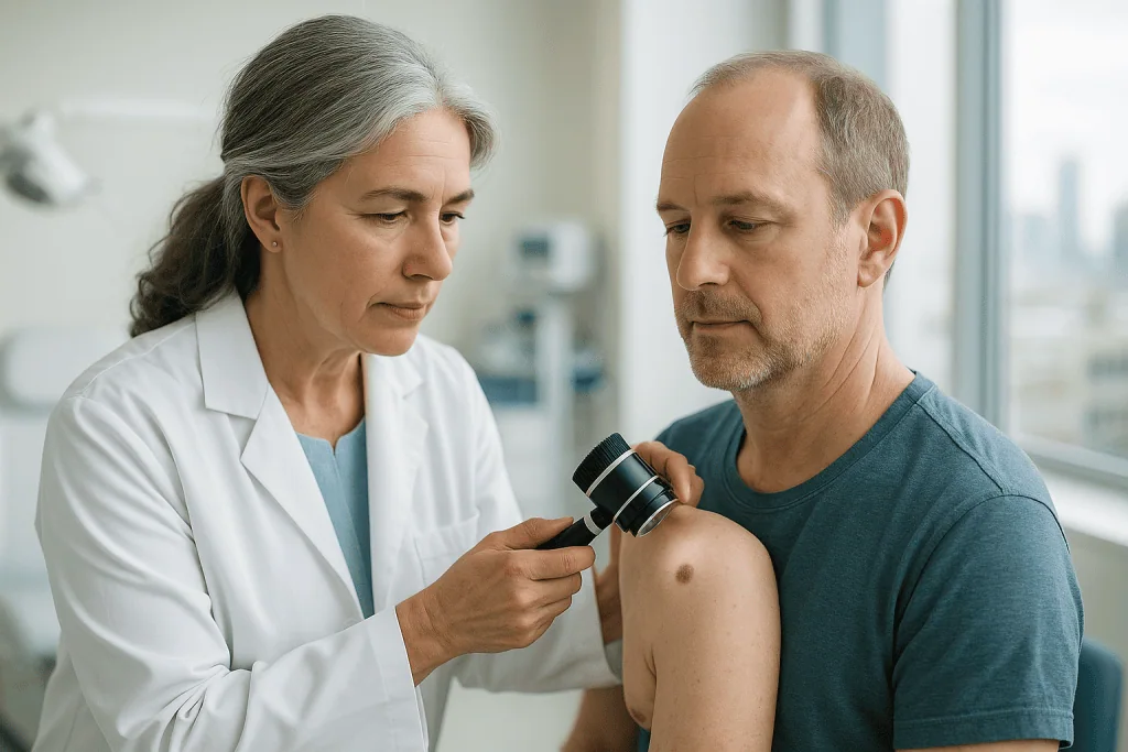
Actinic keratosis diagnosis is usually made through a careful clinical examination of the skin. A doctor, most often a dermatologist, a physician specializing in skin conditions, inspects the lesion, feels its texture, and asks about its history: how long it has been present, whether it has changed, and how much sun exposure the person has had over their lifetime. The characteristic sandpaper-like feel on sun-exposed skin is often enough for an experienced clinician to make the diagnosis.
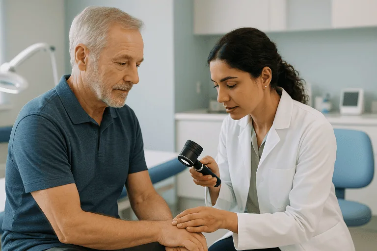
To examine a lesion more closely, the doctor may use a dermatoscope. This is a handheld magnifying instrument with a light that lets the clinician see structures and patterns in the skin that are not visible to the naked eye. Dermoscopy, the technique of using this instrument, helps distinguish actinic keratosis from other conditions such as seborrheic keratosis (a harmless, warty growth), psoriasis, eczema, or early skin cancer.
In some situations, the doctor may recommend a skin biopsy. A biopsy is a minor procedure, done with local numbing medicine, in which a small sample of the lesion is removed and examined under a microscope by a pathologist. A biopsy is often considered when:
- The lesion is unusually thick, large, or painful.
- It bleeds, ulcerates, or grows quickly.
- It does not respond to treatment or returns after treatment.
- The doctor cannot confidently rule out squamous cell carcinoma or another skin cancer.
Blood tests and imaging scans are generally not needed for diagnosing actinic keratosis itself, because the condition affects only the skin surface. As part of the visit, the doctor will usually examine the rest of the sun-exposed skin as well, since additional lesions or other sun-related skin changes are frequently found. In hospital settings such as Acibadem, this condition is evaluated and managed within the dermatology department.
Treatment options for actinic keratosis
Actinic keratosis treatment aims to remove or destroy abnormal cells before they can progress, relieve symptoms such as roughness and irritation, and reduce the overall burden of sun-damaged skin. The best approach depends on how many lesions are present, where they are located, how thick they are, the person’s overall health, and personal preference. Treatment for this condition is typically planned and carried out by specialists in dermatology.
Watchful waiting
Because some actinic keratoses fade on their own and most never turn into cancer, a doctor may in selected cases suggest monitoring a lesion rather than treating it immediately. This usually involves strict sun protection and regular skin checks so that any change can be caught early. Watchful waiting is generally reserved for thin, stable lesions in people who can attend follow-up visits reliably. Any lesion that thickens, becomes tender, or bleeds should be re-evaluated promptly.
Procedures that treat individual lesions
- Cryotherapy (freezing): The doctor applies liquid nitrogen, an extremely cold liquid, to the lesion with a spray or cotton applicator. The freezing destroys the abnormal cells, and the treated spot blisters, crusts, and peels off over the following days to weeks. Cryotherapy is one of the most commonly used treatments for a small number of visible lesions. It may leave a lighter-colored spot on the skin.
- Curettage: The doctor scrapes away the lesion with a small, spoon-shaped instrument called a curette, sometimes followed by electrosurgery, in which a mild electrical current seals the area and destroys remaining abnormal cells. Local numbing medicine is used. This approach also allows the removed tissue to be sent for microscopic examination when needed.
Treatments for larger areas of sun-damaged skin
When many lesions are present, or when the surrounding skin shows widespread sun damage, doctors often prefer “field treatments” that address a whole area rather than one spot at a time.
- Prescription creams and gels: Several topical medications are used, including fluorouracil (a cream that destroys abnormal cells), imiquimod (a cream that stimulates the immune system to attack damaged cells), diclofenac gel (an anti-inflammatory medication), and tirbanibulin (an ointment that interferes with abnormal cell division). These treatments typically cause temporary redness, crusting, and soreness of the treated skin while they work; this reaction is expected and settles after the course finishes. Your doctor will explain how long to apply the medication and what reaction to expect.
- Photodynamic therapy (PDT): A light-sensitizing solution is applied to the skin and allowed to absorb into the abnormal cells. The area is then exposed to a special light, which activates the solution and destroys the damaged cells. The treated skin is usually red and sensitive for several days afterward, and it must be protected from sunlight during the recovery period.
- Chemical peels and laser resurfacing: In selected cases, a doctor may use a medical-grade chemical peel or a laser to remove the damaged outer layer of skin so that healthier skin can regrow. These are performed in a clinical setting and are chosen based on the individual situation.
Surgery
Surgical excision, cutting out a lesion under local anesthesia and closing the skin with stitches, is not usually needed for a straightforward actinic keratosis. It may be recommended when a lesion is suspicious for skin cancer, when a biopsy shows early cancerous change, or when other treatments have failed. Excision provides a complete tissue sample for laboratory examination.
Whatever treatment is chosen, follow-up matters. New lesions can appear on other sun-damaged areas, and treated lesions occasionally return, so periodic skin examinations are generally advised.
Living with actinic keratosis and outlook
For most people, the outlook with actinic keratosis is good, especially when lesions are found and managed early. The condition itself is not cancer, and treatment of individual lesions is usually straightforward. That said, having actinic keratoses is a sign that the skin has significant sun damage, which means an increased lifetime risk of developing further lesions and, in some people, skin cancer. This is why ongoing skin care and monitoring are important, rather than treating the condition as a one-time problem.
Daily habits that help manage the condition and reduce new damage include:
- Using broad-spectrum sunscreen (protecting against both UVA and UVB rays) on exposed skin every day, and reapplying it during prolonged time outdoors.
- Wearing protective clothing, including wide-brimmed hats, long sleeves, and sunglasses.
- Avoiding peak sun hours, generally late morning through mid-afternoon, and seeking shade when outdoors.
- Avoiding tanning beds entirely.
- Checking your own skin regularly for new rough patches or changes in existing spots, including hard-to-see areas such as the scalp and ears, with help from a mirror or another person if needed.
- Attending scheduled skin checks with your doctor, especially if you have had many lesions, a weakened immune system, or previous skin cancer.
It is honest to say that no treatment can guarantee that new actinic keratoses will not form, because the underlying sun damage in the surrounding skin remains. However, consistent sun protection, prompt treatment of new lesions, and regular monitoring together give most people a very manageable long-term course.
Frequently asked questions
What is actinic keratosis in simple terms?
Actinic keratosis is a rough, scaly patch of skin caused by years of sun exposure. It develops on sun-exposed areas such as the face, scalp, ears, and hands, most often in adults over 40 with fair skin. It is considered a precancerous condition, meaning it is not cancer but has the potential, in some cases, to develop into squamous cell carcinoma over time if left unchecked.
Can actinic keratosis heal or go away on its own?
Some actinic keratoses fade on their own, particularly when the skin is protected from further sun exposure. However, lesions that disappear can return, and it is not possible to predict which patches will resolve and which will persist or progress. For this reason, doctors generally recommend having lesions evaluated rather than assuming they will clear by themselves, and many are treated to remove the risk of progression.
How serious is actinic keratosis? Is it cancer?
Actinic keratosis is not cancer, and most individual lesions never become cancer. It is taken seriously because a small proportion of lesions can progress to squamous cell carcinoma, a treatable but genuine form of skin cancer, and because there is no reliable way to tell in advance which lesions will progress. Having multiple lesions or a weakened immune system increases the overall level of concern, which is why medical evaluation and follow-up are recommended.
What does actinic keratosis look and feel like?
A typical lesion is a small, rough, dry, or scaly patch that may be pink, red, brown, or skin-colored, sometimes slightly raised or crusted. Many people describe a sandpaper-like texture that they feel before they can clearly see the spot. Some lesions itch, sting, or feel tender. They appear on skin that gets the most sun: the face, ears, lips, bald scalp, neck, forearms, and backs of the hands.
How is actinic keratosis diagnosed?
Actinic keratosis diagnosis is usually made by a doctor examining the skin, often with a dermatoscope, a lighted magnifying instrument that reveals fine skin structures. In most cases no further testing is needed. If a lesion is thick, painful, bleeding, growing quickly, or otherwise suspicious, the doctor may perform a skin biopsy, removing a small sample under local numbing medicine for examination under a microscope, to rule out skin cancer.
What is recovery like after actinic keratosis treatment?
Recovery depends on the treatment used. After cryotherapy, the treated spot typically blisters, crusts, and heals over days to weeks. Prescription creams often cause temporary redness, peeling, and soreness during the treatment course, which settles afterward. Photodynamic therapy usually causes several days of redness and sun sensitivity. Most treatments are done in an outpatient setting without downtime from normal activities, though your doctor will give specific aftercare instructions, including strict sun protection of the healing skin.
Can actinic keratosis come back after treatment?
Yes, it can. Treatment removes or destroys the visible lesion, but the surrounding skin still carries sun damage, so treated spots occasionally recur and new lesions can appear elsewhere. This does not mean the treatment failed; it reflects the underlying field of sun-damaged skin. Regular skin checks and daily sun protection help catch and manage new or returning lesions early.
When to see a doctor
Any new, persistent, or changing skin patch on sun-exposed skin deserves a professional evaluation, especially if you are over 40, have fair skin, or have a history of significant sun exposure. Early assessment allows simple treatment and helps rule out skin cancer.
Seek medical attention promptly if you notice any of these red-flag warning signs:
- A lesion that grows rapidly or becomes noticeably thicker or harder.
- Bleeding, oozing, or an open sore on a scaly patch that does not heal within a few weeks.
- Persistent pain or tenderness in a rough or crusted spot.
- A hard, horn-like growth projecting from the skin.
- A lesion that returns or worsens after previous treatment.
- A persistent rough, cracked, or scaly area on the lip, particularly the lower lip.
- Any new or changing spot if you have a weakened immune system or a previous skin cancer, since your risk of progression is higher.
None of these signs proves that cancer has developed, but each one warrants a timely examination by a doctor so that the lesion can be properly assessed and, if needed, treated.
Medically reviewed by the Acıbadem International Medical Board September 2, 2026
See our medical review board →
Update history
- PublishedJune 8, 2026
- Medical review approvedSeptember 2, 2026
- Last content updateSeptember 2, 2026