

Pleurx Catheter: An Evidence-Based Patient Guide

A PleurX catheter provides planned, intermittent drainage of recurrent fluid around the lungs or in the abdomen. The procedure is usually performed using local anesthetic and imaging guidance, with sedation when appropriate.
Key Takeaways
- A PleurX catheter provides planned, intermittent drainage of recurrent fluid around the lungs or in the abdomen.
- The procedure is usually performed using local anesthetic and imaging guidance, with sedation when appropriate.
- Patients and caregivers receive training on sterile drainage, dressing changes, and symptoms that need prompt medical advice.
- Potential benefits include symptom relief and fewer repeat fluid-drainage procedures; infection, blockage, leakage, and pain are possible risks.
- The drainage schedule, volume limits, and management of blood-thinning medicines must be individualized by the treating team.
A PleurX catheter is a soft, tunneled tube placed to drain recurrent fluid from the chest or abdomen, often allowing drainage at home rather than repeated hospital procedures. It is commonly used for pleural effusion or ascites when fluid returns and causes symptoms such as breathlessness, chest pressure, abdominal swelling, or discomfort.
Overview: what is a PleurX catheter?
A PleurX catheter is an indwelling drainage catheter: a thin, flexible tube placed under the skin to remove fluid that repeatedly collects in the body. It is most often used for fluid around the lungs, called a pleural effusion, and may also be used for fluid in the abdomen, called ascites. A valve at the outer end stays closed between drainages and connects to a dedicated drainage bottle when fluid needs to be removed.
For pleural fluid, the catheter can reduce symptoms caused by pressure on the lung, including shortness of breath, cough, chest heaviness, and reduced ability to be active. For abdominal fluid, it may help ease tightness, discomfort, early fullness, and breathlessness associated with pressure from ascites. It does not treat the underlying cause of fluid buildup, but it can be an important part of symptom-focused care.
A PleurX catheter is one type of tunneled pleural or abdominal catheter. The term is sometimes used broadly, but the care team will explain the specific device being used and provide device-specific instructions. Written instructions are useful, but a general pleurx catheter patient information pdf or pleurx catheter patient education pdf should never replace the individual plan provided by the clinical team.
How the catheter works and who may benefit
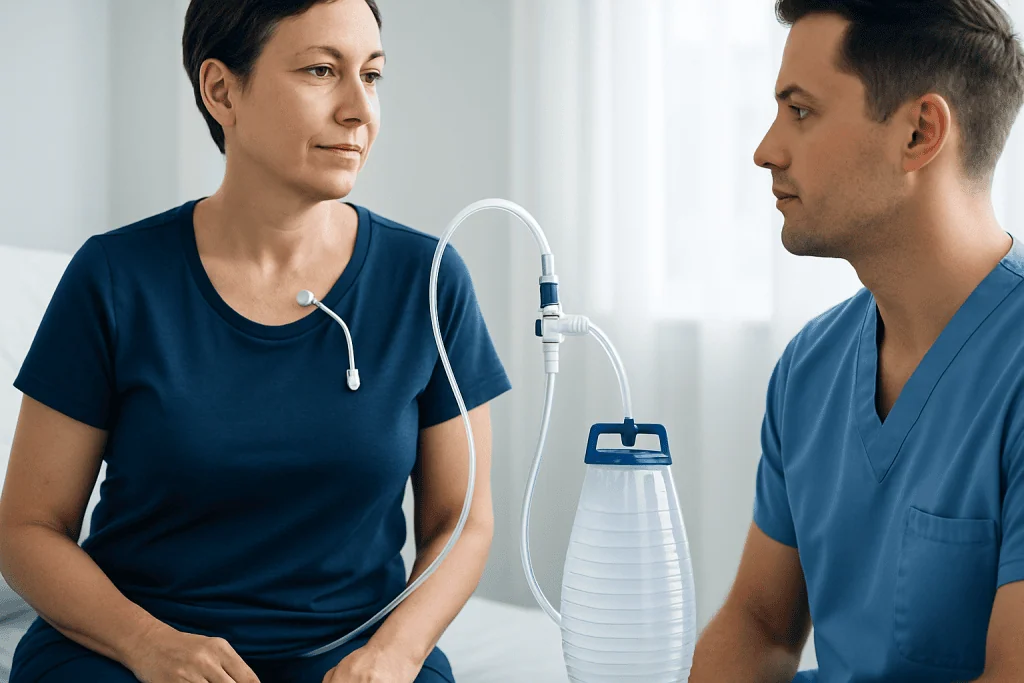
The catheter has a cuff that sits beneath the skin. Over time, tissue grows around this cuff, helping secure the catheter and creating a barrier that can reduce infection risk. The external valve allows controlled drainage into a vacuum bottle. Depending on the reason for placement and the amount of fluid produced, drainage may be needed daily, every few days, or less often.
Clinicians may consider a pleural catheter for a recurrent pleural effusion that returns after thoracentesis, particularly when repeated needle drainage would be burdensome or when the lung does not fully re-expand after fluid removal. Malignant pleural effusion is a common reason for placement, although recurrent non-cancer causes may also be considered in selected circumstances. Readers can learn more about pleural effusion and the conditions that can lead to it.
For recurrent ascites, an indwelling abdominal catheter may be considered when fluid causes substantial symptoms and repeated drainage is no longer the most suitable approach. Candidacy depends on the underlying illness, expected drainage needs, skin condition, infection risk, ability to manage the device at home, and personal care goals. A pulmonologist, oncologist, interventional radiologist, surgeon, or palliative-care clinician may be involved in the decision.
A catheter is not appropriate for everyone. Active infection at the proposed insertion site, uncontrolled bleeding risk, certain complex fluid collections, or lack of safe home support may change the plan. The team weighs these factors alongside the likely benefits of symptom relief and reduced procedures.
Before placement: assessment and anticoagulation planning

Before placement, the team reviews symptoms, medical history, medications, allergies, prior chest or abdominal procedures, and imaging results. Ultrasound, chest X-ray, CT, or other imaging helps confirm the location and amount of fluid and guides safe placement. Blood tests may be requested when clinically indicated, especially if there is concern about bleeding, infection, kidney function, or nutritional status.
Pleurx catheter anticoagulation needs careful, personalized planning. Blood-thinning medicines, including anticoagulants and antiplatelet medicines, can increase bleeding risk during insertion. Patients should tell the care team about every prescription medicine, over-the-counter pain reliever, supplement, and herbal product. They should not stop, restart, or alter blood-thinning treatment independently; the prescriber and procedural team will give specific instructions based on the medicine and the reason it is prescribed.
The team also explains eating and drinking instructions if sedation is planned, how the person will travel home, and whether a family member or caregiver should attend teaching. It is helpful to ask who will arrange drainage supplies, whom to contact after hours, and how frequently follow-up will occur.
What happens during the procedure
PleurX catheter placement is commonly performed in a procedure room or radiology suite using ultrasound or other imaging guidance. The person is positioned so the clinician can access the fluid safely. The skin is cleaned and covered with sterile drapes, and local anesthetic numbs the insertion and tunneling areas. Some patients also receive medication to help them relax, depending on their health and the planned setting.
The clinician makes a small incision, inserts the catheter into the pleural or abdominal space, and tunnels part of it beneath the skin before bringing it out through a second small opening. The catheter is secured, connected briefly if fluid needs to be removed, and covered with a sterile dressing. The full process often takes less than an hour, although preparation and recovery add time.
After placement, imaging may be used for a chest catheter to confirm position and check for complications. The care team monitors comfort, breathing, blood pressure, and the dressing. Most people go home the same day, but some remain under observation or are already in hospital for other medical care.
Placement is part of a broader plan for managing recurrent fluid. When a patient needs an evaluation for recurring chest fluid and potential catheter-based drainage, pleural effusion treatment may involve diagnostic testing, treatment of the underlying cause, drainage procedures, and individualized symptom support.
Recovery timeline and daily drainage care
It is common to have mild soreness, bruising, or pulling discomfort around the catheter site for several days. The care team may recommend suitable pain relief and will explain when bathing, showering, and usual activities can resume. Heavy lifting, vigorous upper-body activity, and swimming are generally avoided until the insertion area has healed and the clinician says it is safe.
Before discharge, a nurse usually provides hands-on pleurx catheter patient education. This includes hand hygiene, preparing a clean work area, checking the site, connecting the drainage bottle, watching the flow, closing the valve, and applying a new dressing. A caregiver can often learn the process as well. Follow the prescribed drainage schedule and the volume limit set by the team; removing fluid too quickly or in a larger amount than advised can cause pain, coughing, dizziness, or low blood pressure.
Drainage often produces less fluid over time, especially with pleural catheters when the pleural layers adhere together naturally. The team reviews drainage records and symptoms to decide whether the schedule can change. In some cases, a pleural catheter can later be removed if drainage remains low and imaging and symptoms support removal.
Healthcare organizations may use a formal pleurx catheter policy and procedure for staff training, sterile technique, documentation, and escalation of concerns. At home, patients should use only the supplies and instructions provided by their care team rather than relying on a generic online protocol.
Benefits, limitations, and possible risks
A major potential benefit of a PleurX catheter is that fluid can be drained at home on a planned schedule. This may reduce repeated visits for needle drainage and help people manage symptoms more consistently. For some individuals, it can support comfort, mobility, and time spent in their usual surroundings while treatment for the underlying condition continues.
However, the catheter requires regular care and does not eliminate fluid formation in every case. Drainage may slow, stop, or remain ongoing depending on the cause of the effusion or ascites. A catheter can also become a practical and emotional adjustment for patients and caregivers, so teaching, written plans, and access to a clinical contact are important.
Possible complications include pain, bleeding, skin irritation, leakage around the tube, blockage, catheter displacement, infection of the skin or deeper fluid space, and—in chest procedures—air around the lung. Rarely, fluid drainage can cause significant symptoms or require urgent assessment. The care team explains the individual risk profile before consent and monitors for complications at follow-up.
Contact the team if drainage becomes unexpectedly difficult, the amount changes markedly, the fluid becomes cloudy or foul-smelling, or there is increasing pain. Do not force a connection, attempt to repair the catheter, or use non-approved equipment. The treating team can advise whether a blockage, dressing issue, or infection needs assessment.
When to seek medical care
Patients should contact their care team promptly for fever, chills, spreading redness, warmth, swelling, pus-like drainage, increasing tenderness, or a persistent rash around the catheter. These symptoms can indicate infection or skin irritation and are best assessed early. New leakage, a loose dressing, damage to the catheter, or inability to drain as instructed also warrants a call.
Urgent medical assessment is needed for severe or worsening shortness of breath, chest pain, fainting, confusion, uncontrolled bleeding, blue or gray lips, or a sudden major deterioration in general condition. These symptoms may not always be caused by the catheter, but they require timely evaluation.
Follow-up appointments help the team review symptoms, drainage amounts, dressing technique, and whether the catheter is still needed. Acibadem International’s multidisciplinary specialists and JCI-accredited hospitals can assess recurrent fluid buildup and provide coordinated catheter-based care for international patients.
Frequently asked questions
Is a PleurX catheter permanent?
A PleurX catheter may remain in place for weeks to months, depending on why it was inserted and how much fluid continues to form. It can sometimes be removed when drainage stays low, symptoms are controlled, and the treating team confirms that removal is appropriate. Some people need it for longer-term symptom management.
How often is a PleurX catheter drained?
The schedule is individualized and may begin with daily or every-other-day drainage, then change according to symptoms and fluid output. The clinician sets a maximum amount to drain at one time. Patients should follow the supplied plan rather than changing the schedule on their own.
Can a person shower with a PleurX catheter?
Showering may be possible once the insertion site has healed and the care team approves it. The dressing generally needs protection from water, and soaking in a bath, hot tub, or swimming pool is usually avoided because it can increase infection risk. Device-specific instructions should be followed closely.
What should patients know about PleurX catheter anticoagulation?
People taking blood thinners should tell the procedural team well before catheter placement and at every follow-up visit. The team may give instructions about temporarily holding or adjusting a medicine, but this decision depends on the individual medicine and reason it is needed. Blood thinners should never be stopped without medical advice.
Does draining a pleural catheter hurt?
Some people experience brief pressure, coughing, or discomfort as fluid drains, particularly if drainage is rapid or the lung is re-expanding. Significant pain, dizziness, worsening breathlessness, or persistent coughing should prompt stopping the drainage and contacting the care team for advice. The team may adjust the drainage amount or frequency.
What information should be recorded after each drainage?
Patients or caregivers are often asked to record the date, amount and appearance of fluid, symptoms before and after drainage, and any concerns with the site or equipment. This record helps the clinical team identify changes and decide whether the drainage plan should be adjusted. It should be brought to follow-up appointments or shared as instructed.
References
- American Thoracic Society
- British Thoracic Society
- National Cancer Institute
- Society of Interventional Radiology
- Memorial Sloan Kettering Cancer Center
This article is for general information only and is not a substitute for professional medical advice. Please consult a qualified doctor about your individual situation.
Explore treatments in Turkey — costs, top hospitals & a free quote
JCI-accredited · board-certified surgeons · reply within 24h

More from the Health Library


Related Specialists

Dr. Ecem Yurdadoğan
Physical Medicine & Rehabilitation
Prof. Dr. Murat Sungur
Internal Medicine
Dr. Kumsal Kurucu
Acibadem Life Clinical Service
Atilla Murat Ünver
Physical Medicine & Rehabilitation