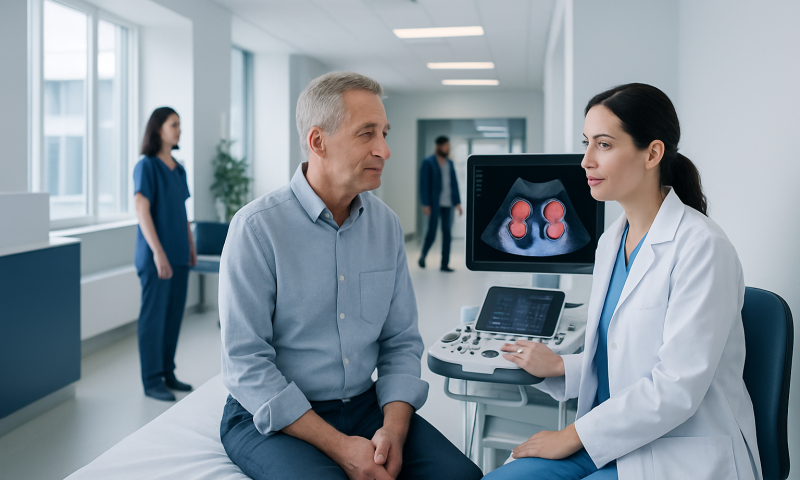
MRI Induction Units: Preparation, Procedure and Results

An MRI induction unit is a pre-scan preparation area, not the scanner itself. Patients should disclose all implants, previous surgery, pregnancy, allergies and possible metal exposure before MRI.
Key Takeaways
- An MRI induction unit is a pre-scan preparation area, not the scanner itself.
- Patients should disclose all implants, previous surgery, pregnancy, allergies and possible metal exposure before MRI.
- Most MRI scans do not require fasting or fluid restriction, but instructions can differ for contrast studies or specific examinations.
- MRI does not use ionizing radiation, but its strong magnet requires strict metal-safety procedures.
- The imaging team can assist if anxiety, pain, claustrophobia or a need to urinate occurs during the scan.
MRI induction units are dedicated areas near the scanning room where patients are prepared safely for magnetic resonance imaging (MRI). They support careful screening, comfort measures and clear communication before a scan, helping the imaging team obtain accurate images while protecting patients from MRI-related hazards.
Overview: What Are MRI Induction Units?
MRI induction units are clinical preparation areas used before a magnetic resonance imaging (MRI) scan. Often called an MRI induction room, this space allows staff to complete safety screening, confirm the examination, explain what will happen and prepare the patient before entry into the scanner environment. It is an important part of MRI procedure preparation because the MRI system uses a powerful, continuously active magnet.
In the induction area, a radiology nurse, radiographer or MRI technologist may review medical information, check for metal-containing devices or objects, and ask patients to change into MRI-safe clothing. If contrast material or sedation is planned, additional checks may be completed. The exact process depends on the body area being scanned and the reason for the examination.
MRI creates detailed pictures of organs, soft tissues, joints, blood vessels and the nervous system. Unlike X-rays and CT scans, it does not use ionizing radiation. However, safe imaging depends on careful planning, particularly for people with implants, prior metal injuries or certain health conditions.
How MRI Induction Works and Who May Need Extra Preparation
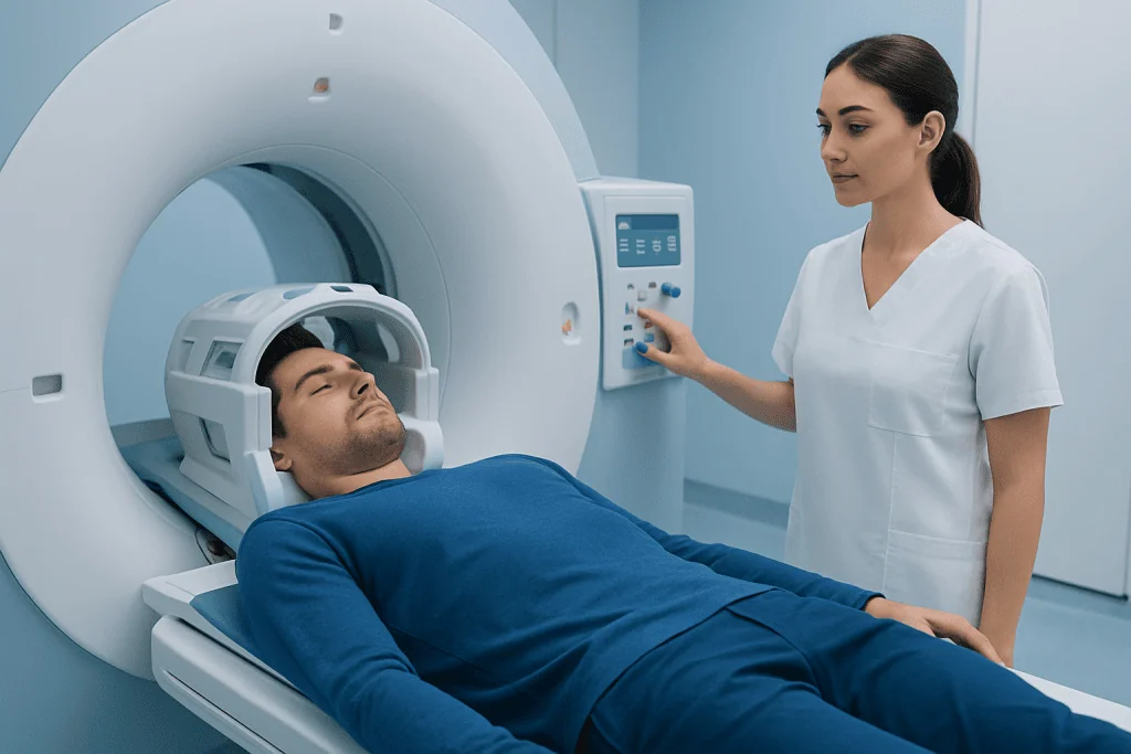
During MRI induction, the team verifies the identity of the patient and the requested scan, then uses a structured safety questionnaire. This identifies factors that may affect whether MRI is suitable, whether special scan settings are needed or whether another imaging test would be safer. The team also discusses any symptoms that could make lying still difficult, such as pain, breathing problems or anxiety in enclosed spaces.
Many modern implants are MRI-conditional, meaning they can be scanned only under specific conditions. Examples include some pacemakers, cochlear implants, neurostimulators, vascular clips, joint replacements and medication pumps. A device card, implant records or the name of the implanting hospital can help the imaging team confirm compatibility. Patients should never assume that an implant is safe for MRI without professional verification.
Extra MRI preparation instructions may be needed for people who are pregnant, have reduced kidney function, have previously reacted to MRI contrast, are receiving dialysis, or may need sedation. Children and people who cannot remain still may require tailored support, and in some situations sedation or anesthesia may be considered after an individual assessment.
- Tell the team about any implanted device, surgery, retained metal fragment or occupational metal exposure.
- Report allergies, kidney disease, pregnancy or a prior reaction to contrast material.
- Explain if claustrophobia, pain, hearing difficulties or mobility limitations could affect the scan.
MRI Pre-Procedure Instructions: What to Do Before the Scan

Patients should follow the instructions provided by the imaging center, as requirements vary by scan type. For many routine MRI examinations, people can eat, drink and take their usual prescribed medicines. Some studies, especially abdominal, pelvic or contrast-enhanced scans, may involve fasting or other specific directions. If instructions are unclear, patients should contact the radiology department before attending rather than changing medication or fasting unnecessarily.
Before entering the MRI induction room, patients are usually asked to remove all metal and electronic items. This includes jewelry, watches, hearing aids, hairpins, removable dental appliances, belts, coins, keys, bank cards and phones. Makeup, skin patches and some clothing may contain metallic particles or backing, so changing into a gown may be advised. The team will provide a secure place for belongings where available.
Patients should bring their referral, relevant previous scans or reports if requested, a list of medicines and details of any implant. They should arrive early enough for screening and consent processes, especially when contrast or sedation is planned. Comfortable clothing without metal fasteners can simplify the process, although the imaging team may still ask the patient to change.
Is there anything I shouldn’t do before an MRI scan?
Patients should not enter the MRI area with metal objects, electronic devices or medical patches unless the staff have confirmed they are safe. They should not withhold regular medicines, eat or drink differently, or use a sedative on their own unless specifically instructed by a clinician. It is also important not to minimize or omit information about implants, metal injuries, pregnancy, kidney problems or prior contrast reactions, as this information guides safe care.
Step by Step: What Happens During an MRI Scan?
After MRI induction and final safety checks, the patient is taken to the scanner room. The technologist helps position them on a padded table, and a specialized receiver coil may be placed around the part of the body being examined. Cushions, straps or supports may be used to improve comfort and reduce movement, as staying still is essential for clear images.
The table moves slowly into the scanner, which is open at both ends. During image acquisition, the machine makes loud tapping, thumping or knocking sounds. Earplugs or headphones are provided to protect hearing, and some centers offer music. The technologist monitors the patient from a nearby control room and communicates through an intercom. A call button or alarm device is usually available so the patient can alert staff at any time.
Some scans use gadolinium-based contrast material, usually given through a small intravenous cannula. Contrast can help show inflammation, tumors, blood vessels or certain organ changes more clearly. The clinician considers the expected benefit, kidney health, medical history and any previous contrast reaction before deciding whether it is appropriate.
The scan duration varies by body region and number of image sequences. Many examinations take between 20 and 60 minutes, though complex studies can last longer. The patient should expect periods of noise and stillness, but the scan itself is not painful.
What happens if I drink water before an MRI?
For most MRI scans, drinking water beforehand is safe and does not affect image quality. In fact, maintaining normal hydration may be helpful unless the imaging team has given different instructions. Certain abdominal, pelvic, bladder or sedation-related examinations may have specific requirements about fluids or a full bladder, so patients should follow their individual appointment guidance.
Pineapple Juice, Bladder Comfort and Other Practical Questions
Why drink pineapple juice before MRI scan?
Pineapple juice is not a routine requirement for MRI. In selected centers and for certain abdominal or pelvic MRI protocols, pineapple juice may be used as an oral contrast agent because its natural manganese content can change the appearance of fluid in parts of the digestive tract on MRI images. This is a protocol-specific instruction, not a general MRI recommendation. Patients should drink it only when their radiology department has advised it and should mention diabetes, dietary restrictions, food allergy or swallowing difficulties beforehand.
What if I have to pee during an MRI?
Patients should use the toilet before the scan whenever possible, particularly if it may be lengthy. If the need to urinate becomes uncomfortable during imaging, they can use the call button or speak to the technologist. The scan can often be paused safely so staff can help; it is better to communicate early than to struggle to remain still and compromise comfort or image quality.
For a small number of pelvic or urinary tract scans, the team may intentionally ask a patient to arrive with a comfortably full bladder. This is different from simply drinking extra fluids before every MRI. The patient should ask the imaging department if they are unsure whether bladder filling is part of their particular MRI procedure preparation.
Benefits, Risks and Recovery Timeline
MRI can provide high-quality images without ionizing radiation and is especially useful for assessing the brain, spine, joints, muscles, many internal organs and soft tissues. It can help clinicians investigate symptoms, monitor known conditions, plan treatment and evaluate response to care. The induction process helps make the examination as safe, comfortable and informative as possible.
For most people, there is no recovery period after a standard MRI. They can leave shortly after the scan and return to normal activities, including eating, drinking and driving, unless they have received sedation. If sedation or anesthesia was used, the patient may need observation, a responsible adult to accompany them home and temporary restrictions on driving or making important decisions, according to the care team’s advice.
MRI-related risks are uncommon when screening is thorough, but the strong magnet can cause serious harm if unsafe metal objects or incompatible implants enter the scanner room. Contrast material may occasionally cause side effects or an allergic-type reaction, and rare complications require prompt clinical attention. People with significant kidney impairment need individualized assessment before some contrast-enhanced studies.
Results are interpreted by a radiologist, who prepares a report for the referring clinician. Timing varies with scan complexity and clinical urgency. The referring doctor explains what the findings mean in the context of symptoms, examination results and any other tests; an MRI report alone does not always establish a diagnosis.
When to Seek Medical Care
Patients should contact the imaging department before their appointment if they discover an implant, possible metal fragment, pregnancy, kidney disease, a previous reaction to contrast material or a need for sedation that was not discussed. They should also call if they cannot follow fasting, medication or transport instructions. Early communication allows the team to adapt the plan or arrange a safer alternative if needed.
During or after an MRI, patients should tell staff immediately about breathing difficulty, swelling, widespread rash, severe dizziness, chest discomfort, intense burning or new severe pain. These symptoms are unusual, but prompt assessment is appropriate. After leaving, urgent medical care is appropriate for severe or rapidly worsening symptoms, especially after contrast or sedation.
For people arranging imaging from abroad, Acibadem International’s multidisciplinary specialists and JCI-accredited hospitals can coordinate MRI assessment and treatment planning for international patients. The most suitable imaging approach should always be chosen with a qualified clinician based on the individual’s medical history and current concerns.
Frequently asked questions
What is an MRI induction unit?
An MRI induction unit is a preparation area used before a patient enters the MRI scanner room. Staff use it to complete safety screening, confirm the planned examination, explain the scan and prepare any needed equipment, contrast access or support measures.
Do MRI induction units use anesthesia?
Most MRI scans do not require anesthesia. However, sedation or anesthesia may be considered for some children, people with severe claustrophobia, or patients who cannot stay still because of pain, disability or another medical reason. This requires an individual clinical assessment and additional safety planning.
Can I wear makeup or deodorant for an MRI?
Some cosmetics, hair products and skin products can contain metallic particles that may affect image quality or, rarely, cause warmth or irritation. The imaging team may ask patients to remove makeup or change into a gown. Deodorant is usually not a concern, but local instructions should be followed.
Can I have an MRI if I have a pacemaker?
Some pacemakers and other cardiac devices are MRI-conditional, meaning they can be scanned under defined conditions. The imaging team must confirm the exact device model and coordinate with relevant specialists before the scan. Patients should bring their device card or implant information whenever possible.
How long does it take to get MRI results?
A radiologist reviews the images and sends a report to the clinician who requested the scan. The timing depends on the type of MRI, the complexity of the findings and whether the examination is urgent. The referring clinician can explain when and how results will be shared.
Is MRI contrast safe?
Gadolinium-based MRI contrast is widely used when it is expected to improve diagnostic information, and most people tolerate it well. The team checks for factors such as prior reactions and kidney function before use. Patients should report any concerning symptoms during or after administration to staff promptly.
References
- American College of Radiology
- Radiological Society of North America
- U.S. Food and Drug Administration
- National Health Service
- World Health Organization
This article is for general information only and is not a substitute for professional medical advice. Please consult a qualified doctor about your individual situation.
Explore treatments in Turkey — costs, top hospitals & a free quote
JCI-accredited · board-certified surgeons · reply within 24h