Temporal Artery Biopsy: Procedure, Recovery and Results

A temporal artery biopsy is commonly used when giant cell arteritis is suspected. The procedure is usually performed with local anaesthetic and often takes less than an hour.
Key Takeaways
- A temporal artery biopsy is commonly used when giant cell arteritis is suspected.
- The procedure is usually performed with local anaesthetic and often takes less than an hour.
- Treatment for suspected giant cell arteritis may begin before biopsy results are available to protect vision.
- Mild tenderness, bruising and temporary numbness around the incision can occur during recovery.
- A normal biopsy result does not always fully exclude giant cell arteritis, so results are interpreted alongside symptoms, tests and imaging.
A temporal artery biopsy is an outpatient procedure used mainly to help confirm or assess suspected giant cell arteritis, an inflammatory condition affecting blood vessels. It involves removing a small section of an artery near the temple for laboratory examination and usually has a short recovery period.
Overview: what is a temporal artery biopsy?
A temporal artery biopsy is a minor surgical procedure in which a clinician removes a small piece of the temporal artery, a blood vessel located just beneath the skin at the side of the head. A laboratory specialist examines the sample under a microscope for inflammation and other changes that can support a diagnosis of giant cell arteritis (GCA), also called temporal arteritis.
Giant cell arteritis is an inflammatory disease of medium and large arteries that occurs most often in older adults. It can affect arteries supplying the head, scalp, jaw and eyes. Because untreated inflammation may sometimes threaten eyesight, clinicians may start treatment promptly when suspicion is high, rather than waiting for biopsy results.
The biopsy is one part of a wider assessment. Symptoms, physical examination findings, blood tests for inflammation and, in some settings, ultrasound or other vascular imaging all help the medical team decide whether GCA is likely and how treatment should proceed.
Why it may be recommended and who is a candidate
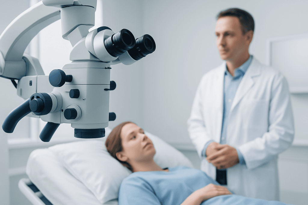
A doctor may recommend a temporal artery biopsy when a person has symptoms or test results that raise concern for giant cell arteritis. Typical symptoms may include a new persistent headache, scalp tenderness, pain or tiredness in the jaw while chewing, unexplained fever, fatigue, weight loss, or changes in vision. Some people also have polymyalgia rheumatica, which can cause aching and stiffness around the shoulders, neck or hips.
Vision symptoms deserve urgent assessment. These may include blurred vision, double vision, a shadow or curtain-like area in the visual field, or brief episodes of vision loss. Such symptoms do not always mean GCA is present, but they require prompt medical evaluation because early treatment is important when GCA is suspected.
The clinician will review a person’s medical history, medicines and bleeding risk before arranging the procedure. People taking anticoagulants, antiplatelet medicines, diabetes medicines or supplements that may affect bleeding should not stop them independently; the prescribing clinician or procedural team can advise on an individual plan.
A biopsy may be taken from the side where symptoms are more noticeable. In selected circumstances, such as when clinical suspicion remains strong after an initial negative result, the team may discuss sampling the artery on the other side or using additional imaging.
How a temporal artery biopsy works

In giant cell arteritis, inflammation can affect the artery wall in patches rather than continuously. This is sometimes called a skip pattern. For this reason, the surgeon usually removes a short segment of artery rather than a very small fragment, allowing the laboratory to examine several levels of the tissue.
The sample is preserved and reviewed by a pathologist. Findings that may support GCA include inflammatory cells within the artery wall, damage to the inner elastic layer and, in some cases, giant cells. The name “giant cell arteritis” comes from these cells, although they are not present in every positive biopsy.
Biopsy findings are interpreted carefully in context. A positive result strongly supports the diagnosis, but a negative result may occur if inflammation is patchy, if a different artery is involved, or if the clinical condition is not GCA. The treating clinician combines the pathology report with the full clinical picture.
What happens during the procedure
A temporal artery biopsy is generally performed as an outpatient procedure. Before it begins, the clinician identifies the artery by feeling its pulse and may use ultrasound to help locate it. The skin near the temple is cleaned, and local anaesthetic is injected to numb the area. A person remains awake, but should not feel pain after the area is numb.
A small incision is made in the skin, and the surgeon carefully isolates and removes a short section of the artery. The incision is then closed with stitches, which may dissolve or need removal at a follow-up visit depending on the type used. A dressing may be applied over the area.
The procedure commonly takes less than an hour, although preparation and post-procedure observation add time to the appointment. Most people return home the same day. If sedation is used or a person has received medicines that can affect alertness, they will need someone else to drive them home.
The sample is sent to the pathology laboratory. The medical team will explain how and when results will be communicated, as turnaround times can vary depending on laboratory processing and whether further tissue review is needed.
Recovery timeline and aftercare
After the biopsy, it is common to have mild soreness, tightness, bruising or swelling around the incision for several days. Some people notice temporary numbness or altered sensation in the scalp near the wound. These effects often improve as the area heals, although sensation can occasionally take longer to return.
The care team will provide wound-care instructions. In general, the incision should be kept clean and dry as advised, and rubbing, scratching or placing pressure on the site should be avoided. Normal light activities can often be resumed soon after the procedure, but strenuous exercise, heavy lifting and swimming may need to wait until the wound is healing well.
Simple pain relief may be appropriate for some people, but medication choices should be confirmed with the clinician, particularly for those using blood-thinning medicines or undergoing treatment for suspected GCA. Stitches that require removal are commonly removed at a follow-up appointment after the incision has healed sufficiently.
Recovery from the procedure itself is usually straightforward, but recovery from the underlying illness depends on the diagnosis and treatment plan. If GCA is confirmed or remains strongly suspected, follow-up commonly involves coordinated care with specialists experienced in inflammatory and vascular conditions.
Benefits, limitations and possible risks
The main benefit of temporal artery biopsy is that it can provide direct evidence of artery inflammation. A confirmed diagnosis can help guide treatment decisions, monitoring and discussions about expected follow-up. It can also help clinicians consider other explanations when the tissue does not show features of GCA.
The biopsy has limitations. Because inflammation may be patchy, a normal result cannot by itself rule out giant cell arteritis. Starting corticosteroid treatment before biopsy may be necessary when vision is at risk, and clinicians can often still obtain useful biopsy information after treatment has begun. The timing should be decided by the treating team and should never delay urgent care.
Complications are uncommon but can include bleeding, infection, wound separation, scarring, persistent numbness, temporary weakness of a nearby forehead muscle or injury to surrounding small nerves. The team takes steps to reduce these risks and will review any individual factors that may increase them.
Contact the procedural team if there is increasing redness, warmth, swelling, drainage, fever, persistent bleeding, worsening pain or a wound that opens. These signs do not always indicate a serious problem, but they should be assessed promptly.
When to seek medical care
Urgent medical care is needed for new or sudden visual changes, temporary loss of vision, double vision, severe new headache, or jaw pain when chewing accompanied by other symptoms of possible giant cell arteritis. These symptoms require same-day assessment, especially in adults over age 50, because clinicians may need to begin treatment quickly to help protect vision.
A non-urgent medical appointment is appropriate for a persistent new headache, scalp tenderness, unexplained fatigue, fever, weight loss, shoulder or hip stiffness, or jaw discomfort with chewing. These symptoms have many possible causes, and a qualified clinician can determine whether blood tests, imaging or biopsy are appropriate.
People recovering from a biopsy should also seek advice if they have concerns about their incision or recovery. Acibadem International’s multidisciplinary specialists and JCI-accredited hospitals support international patients who need assessment and treatment for suspected inflammatory vascular conditions.
Frequently asked questions
Is a temporal artery biopsy painful?
The area is numbed with local anaesthetic, so the person may feel pressure or movement but should not feel sharp pain during the biopsy. Mild soreness or tenderness afterward is common and usually improves over several days.
How long does a temporal artery biopsy take?
The procedure itself often takes less than an hour. Preparation, consent, local anaesthetic and recovery observation mean the full visit may take longer.
How long does it take to get temporal artery biopsy results?
Results may take several days to around one or two weeks, depending on laboratory processes and whether additional tissue sections are reviewed. The care team can provide the expected timeframe for the individual hospital or laboratory.
Can steroid treatment start before a temporal artery biopsy?
Yes. When giant cell arteritis is strongly suspected, particularly if there are visual symptoms, clinicians may start corticosteroid treatment immediately. Protecting vision is the priority, and the biopsy can often still be useful after treatment has started.
Does a negative temporal artery biopsy rule out giant cell arteritis?
No. Inflammation can occur in patches, so the sampled section may not contain affected tissue. Doctors interpret a negative result together with symptoms, examination findings, blood tests and imaging.
Will hair be shaved for a temporal artery biopsy?
Usually, only a small area of hair may need to be trimmed if necessary, and some procedures can be performed with little or no shaving. The surgical team can explain what to expect based on the planned incision location.
References
- American College of Rheumatology
- European Alliance of Associations for Rheumatology
- National Institute for Health and Care Excellence
- Mayo Clinic
- National Institute of Arthritis and Musculoskeletal and Skin Diseases
This article is for general information only and is not a substitute for professional medical advice. Please consult a qualified doctor about your individual situation.
Oncology care in Turkey — second opinion and treatment plan
JCI-accredited · board-certified surgeons · reply within 24h