Vestibular Schwannoma MRI: Preparation, Procedure and Results

MRI is the preferred imaging test for diagnosing and monitoring vestibular schwannoma. A dedicated MRI of the internal auditory canals can identify small tumors near the hearing and balance nerve.
Key Takeaways
- MRI is the preferred imaging test for diagnosing and monitoring vestibular schwannoma.
- A dedicated MRI of the internal auditory canals can identify small tumors near the hearing and balance nerve.
- Contrast-enhanced MRI is commonly used, but selected non-contrast MRI protocols may be appropriate for some people.
- Most vestibular schwannomas grow slowly, and some do not grow during follow-up.
- MRI results help specialists decide between observation, radiation treatment, or surgery.
A vestibular schwannoma MRI is a detailed scan used to detect and monitor a usually noncancerous tumor on the balance and hearing nerve. The examination is painless, does not use radiation, and often includes contrast dye to show very small tumors more clearly.
Vestibular Schwannoma MRI: An Answer-First Guide
A vestibular schwannoma MRI is the main imaging examination used to find, measure, and follow a vestibular schwannoma, also called an acoustic neuroma. It produces highly detailed images of the brain, inner ear, and internal auditory canals, where the hearing and balance nerve travels.
The scan is usually requested when a person has one-sided hearing loss, persistent ringing in one ear, imbalance, or other symptoms that may involve the vestibular nerve. MRI does not use ionizing radiation and is generally well tolerated. Depending on the clinical question, the examination may be performed with and without a gadolinium-based contrast agent.
A vestibular schwannoma is typically a benign, slow-growing tumor arising from Schwann cells that cover the vestibulocochlear nerve. MRI findings allow the care team to confirm the diagnosis, assess the tumor’s size and position, and plan monitoring or treatment when needed. For broader information about the condition, see vestibular schwannoma.
How a Vestibular Schwannoma MRI Works
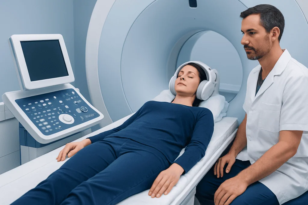
MRI uses a powerful magnetic field and radiofrequency waves to create cross-sectional images of body tissues. Unlike CT scanning or standard X-rays, it does not expose the patient to radiation. During a vestibular schwannoma MRI, the radiology team obtains thin, high-resolution images through the internal auditory canals, cerebellopontine angles, brainstem, and nearby structures.
These images can show whether a mass is present on the vestibular nerve and whether it extends beyond the narrow bony canal into the area between the brain and inner ear. MRI also helps distinguish a vestibular schwannoma from other possible causes of similar symptoms, including inflammation, vascular changes, or less common tumors.
Contrast is often useful because vestibular schwannomas commonly enhance clearly after gadolinium is administered into a vein. This can improve confidence in detecting very small lesions and defining their edges. Modern high-resolution non-contrast sequences can also provide useful information in selected situations, particularly for surveillance imaging.
Who May Need This MRI and How to Prepare

A clinician may recommend MRI for unilateral or asymmetric sensorineural hearing loss, ringing mainly in one ear, unexplained balance difficulty, facial numbness, or a prior finding that needs follow-up. It may also be used after treatment to check for residual or recurrent tumor growth. Hearing tests are commonly reviewed alongside MRI findings.
Before the appointment, the patient should tell the imaging team about any implanted device, prior surgery, metal fragments, medication patch, kidney disease, pregnancy, or history of a reaction to MRI contrast. Many modern implants are MRI-compatible, but the team must confirm the device type and scan conditions in advance.
Most people can eat, drink, and take usual medicines before an MRI unless they are given different instructions. If contrast is planned, the care team may ask about kidney function or arrange a blood test in selected patients. People with claustrophobia should mention this when booking, as practical support, a wider-bore scanner, or clinician-prescribed medication may help.
All jewelry, hearing aids, removable dental items, bank cards, and metal-containing accessories must be removed before entering the scan room. The radiology staff will provide a safety checklist and explain each step before the examination begins.
What Happens During the Procedure and Afterward
On arrival, the patient changes into appropriate clothing and completes MRI safety screening. If contrast is being used, a small intravenous line is placed, usually in the arm or hand. The patient then lies on a padded table, with the head positioned in a special coil designed to capture detailed images of the brain and inner ear.
The table moves slowly into the MRI scanner. The machine makes repeated loud tapping or knocking sounds while images are acquired, so earplugs or headphones are provided. It is important to lie still, but the person can communicate with the radiographer throughout the scan using an intercom and alert device.
A dedicated vestibular schwannoma MRI often takes about 30 to 60 minutes, although timing varies with the protocol and whether contrast is included. The scan itself is painless. Some people notice warmth around the injection site or a cool sensation when contrast is administered; these effects are usually brief.
There is no physical recovery period after MRI. Most people return to normal activities immediately. If sedating medication was used for anxiety, they should arrange for a responsible adult to accompany them home and avoid driving or making important decisions until the medication has worn off.
Is it Safe to Have a Vestibular Schwannoma MRI Without Contrast?
Yes, a vestibular schwannoma MRI without contrast can be safe and informative. High-resolution non-contrast MRI sequences can often identify or monitor many vestibular schwannomas, especially when a known tumor is being followed over time. This approach may be considered when contrast is unsuitable or when the specialist believes it will adequately answer the clinical question.
However, contrast-enhanced MRI remains the standard approach in many diagnostic settings because it may provide clearer detection and characterization of very small tumors or uncertain findings. The best protocol depends on symptoms, previous scan results, kidney function, pregnancy considerations, and the reason for imaging.
Gadolinium-based contrast agents are generally considered safe for most people when used appropriately. Rare allergic-like reactions can occur, and special caution is needed in people with severe kidney impairment. A radiologist and referring clinician can explain whether contrast is recommended in an individual case.
Understanding Results, Growth, and Treatment Decisions
The MRI report usually describes whether a vestibular schwannoma is present, its dimensions, the side involved, and its relationship to the internal auditory canal, brainstem, hearing nerve, facial nerve, and nearby fluid spaces. Results are interpreted alongside symptoms, hearing assessment, age, general health, and comparisons with previous scans when available.
How serious is a vestibular schwannoma? Vestibular schwannomas are usually benign and do not spread to distant parts of the body. Even so, a growing tumor can affect hearing, balance, facial sensation, or nearby brain structures because of its location. The seriousness therefore depends on its size, growth pattern, symptoms, and effect on surrounding nerves and tissues.
How quickly does a vestibular schwannoma grow? Growth is variable. Many tumors grow slowly over years, while some remain stable on serial MRI scans. Less commonly, a tumor may enlarge more quickly. Repeat MRI at intervals recommended by the treating team is the most reliable way to understand an individual tumor’s behavior.
For small, stable tumors with manageable symptoms, active surveillance with repeat MRI and hearing testing may be appropriate. If treatment is needed, options can include microsurgery or focused radiation such as Gamma Knife radiosurgery. Decisions should be individualized through discussion with otology, neurosurgery, radiation oncology, and radiology specialists.
Can a Brain MRI Detect Vestibular Schwannoma?
Yes, a brain MRI can detect a vestibular schwannoma when it includes suitable high-resolution images of the internal auditory canals and cerebellopontine angles. A routine brain MRI may identify a larger tumor, but a dedicated internal auditory canal protocol is often preferred when symptoms specifically suggest a problem affecting the hearing and balance nerve.
The dedicated protocol uses thinner image slices and sequences optimized for the small structures of the inner ear and nearby nerves. Contrast-enhanced images may be added to improve detection of tiny lesions. If a previous routine brain MRI was normal but symptoms remain concerning, a clinician may recommend a targeted MRI examination.
MRI is also valuable after a diagnosis has been made because it provides a consistent method for measuring the tumor over time. The same or comparable imaging protocol may be used during surveillance so that small changes can be assessed more reliably.
When to Seek Medical Care
Medical assessment is appropriate for new or persistent hearing loss in one ear, ringing that is mainly on one side, gradually worsening balance, recurrent dizziness, or altered facial sensation. These symptoms are common and can have many causes, but they should not be self-diagnosed. A primary care clinician, ear specialist, or neurologist can determine whether hearing testing and MRI are needed.
Urgent medical care is important for sudden hearing loss, sudden severe vertigo with new weakness, facial drooping, trouble speaking, severe headache, or difficulty walking. These symptoms can occur with conditions other than vestibular schwannoma and may require prompt evaluation.
People with a confirmed vestibular schwannoma should attend recommended follow-up appointments even if symptoms are stable, since MRI monitoring guides safe decision-making. Acibadem International’s multidisciplinary specialists and JCI-accredited hospitals assess and treat vestibular schwannoma for international patients, including coordinated imaging and treatment planning.
Frequently asked questions
What does a vestibular schwannoma look like on MRI?
A vestibular schwannoma usually appears as a well-defined mass along the vestibulocochlear nerve, often beginning in the internal auditory canal. With gadolinium contrast, it commonly becomes brighter than surrounding tissue. The radiologist also assesses its size and whether it is affecting nearby nerves or the brainstem.
Do all vestibular schwannoma MRIs need contrast?
No. Non-contrast high-resolution MRI can be suitable for some surveillance scans and for people who should avoid contrast. For an initial diagnosis or an unclear finding, contrast is often helpful because it can improve visualization of small tumors.
How long does an MRI for acoustic neuroma take?
A dedicated MRI commonly takes around 30 to 60 minutes. The exact duration depends on the scanner, imaging protocol, need for contrast, and whether additional brain imaging is required.
Can MRI tell whether a vestibular schwannoma is cancerous?
MRI can provide strong evidence that a lesion has the typical appearance of a vestibular schwannoma, which is usually benign. Imaging alone does not always establish every tissue detail with absolute certainty, but biopsy is not routinely needed when MRI features are characteristic.
How often is MRI repeated for vestibular schwannoma?
Follow-up timing is individualized. It may depend on tumor size, symptoms, age, prior growth, and whether the person has had surgery or radiation. The treating specialist will recommend an imaging schedule designed to monitor for meaningful change.
What if MRI shows a small vestibular schwannoma?
A small tumor does not automatically require immediate treatment. Many people are managed with observation and scheduled MRI scans, particularly when the tumor is stable and symptoms are limited. Treatment may be discussed if there is documented growth, worsening symptoms, or concern about effects on nearby structures.
References
- National Institute on Deafness and Other Communication Disorders
- National Cancer Institute
- American College of Radiology
- Congress of Neurological Surgeons
- Radiological Society of North America
This article is for general information only and is not a substitute for professional medical advice. Please consult a qualified doctor about your individual situation.
Oncology care in Turkey — second opinion and treatment plan
JCI-accredited · board-certified surgeons · reply within 24h