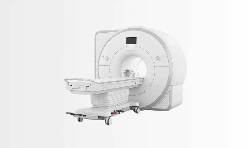
Gamma Knife
Gamma Knife is a precise form of stereotactic radiosurgery that treats certain brain tumors and other brain conditions without an incision by delivering highly focused radiation beams.
Quick answer
Gamma Knife is a form of stereotactic radiosurgery that treats brain tumours, brain metastases, vascular abnormalities and some functional disorders without an incision. Around 200 fine radiation beams converge on a precisely mapped target, delivering a high dose to the lesion while limiting exposure to healthy brain tissue. Most patients are treated in a single outpatient session and go home the same day.
What Is Gamma Knife?
Gamma Knife is a form of stereotactic radiosurgery: a non-surgical treatment that focuses many fine beams of radiation on a precisely defined target inside the head. It is used for brain tumours, brain metastases, certain blood-vessel abnormalities and selected functional disorders such as trigeminal neuralgia. Despite the name, there is no knife, no incision and no physical entry into the brain.
Hearing that you may need treatment for a condition inside the skull is unsettling. Many people immediately picture open brain surgery: a long hospital stay, a visible scar, the fear of losing speech, movement or memory. Those concerns are understandable: the brain controls almost everything you do, so any treatment decision in this area carries weight. Gamma Knife exists precisely because, for carefully selected conditions, focused radiation can do the work that would otherwise require opening the skull.
For some patients, gamma knife treatment is recommended instead of open surgery. For others, it comes after surgery to treat residual disease, alongside systemic cancer therapy, or as one part of a longer-term plan for a benign or functional neurological disorder. Which of these applies to you depends on the diagnosis, the size and position of the lesion, your symptoms, your general health and what you want treatment to achieve.
What most patients actually want to know is simple: will this control the problem, protect quality of life and cause as little disruption as possible? In appropriate cases, that is exactly what this treatment is designed to do. It is a well-established approach in modern neuro-oncology and neurosurgical care, particularly for lesions that are hard to reach surgically or that sit close to structures where accuracy is everything.
What is gamma knife surgery?
Gamma knife surgery is the everyday name for the same procedure: the word “surgery” refers to the surgical level of precision, not to cutting. The formal term is stereotactic radiosurgery, often shortened to SRS. “Stereotactic” means the target is located using a fixed three-dimensional coordinate system, so the treatment team knows its position to within a fraction of a millimetre. “Radiosurgery” means a high dose of radiation is delivered to that target in a single session or, in some cases, a small number of sessions, rather than spread over many weeks. Nothing is removed from the head. The effect happens at the cellular level and develops over the weeks and months that follow.
How does a gamma knife work?
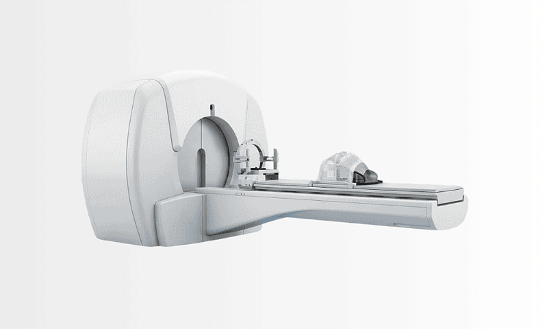
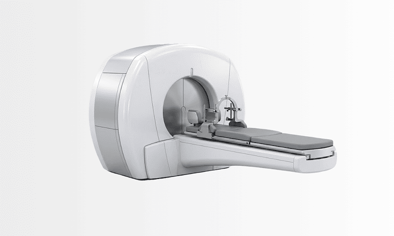
A Gamma Knife works by aiming roughly two hundred separate beams of gamma radiation, generated by cobalt-60 sources, at one carefully mapped point. Each individual beam is weak as it passes through healthy tissue on its way in. Where all the beams cross, at the lesion, the combined dose is high enough to damage the DNA of the target cells so they lose the ability to divide and grow. This convergence principle is what makes the treatment so precise: the surrounding brain receives only a small fraction of the dose concentrated at the target. The machine can also treat irregularly shaped lesions by combining many small overlapping focal points, effectively “painting” the dose to match the shape of the abnormality while steering it away from structures such as the optic nerves or brainstem.
What is gamma knife radiotherapy?
Gamma knife radiotherapy is a phrase people often use interchangeably with radiosurgery, but there is a meaningful difference worth understanding. Conventional radiation therapy spreads a moderate dose over many daily sessions across several weeks, and typically treats a broader field. Radiosurgery concentrates a high dose on a small, sharply defined target in one session or a handful of sessions. Both approaches have their place. Larger lesions, or lesions wrapped around sensitive structures, are sometimes better served by fractionated treatment; small, well-defined targets are often ideal for single-session radiosurgery. Your treatment team chooses between them based on the biology and geometry of what is being treated, not on preference for one machine over another.
What does gamma knife radiation do to a tumour?
Gamma knife radiation damages the genetic material of the cells inside the target so that they can no longer multiply. It does not remove the lesion the way a surgeon physically removes tissue, and it does not vaporise anything on the day of treatment. Instead, the biological effect unfolds gradually. Some tumours simply stop growing, which for many conditions is the goal. Some shrink slowly over months. Vascular malformations close off progressively as the treated vessel walls thicken. In functional disorders such as trigeminal neuralgia, symptom relief typically builds over weeks rather than arriving immediately. For patients and families, this is the single most important expectation to set: Gamma Knife is not instant removal. It is a targeted strategy for controlling disease over time while preserving as much neurological function as possible.
Who May Need Gamma Knife Treatment?
Gamma Knife may be considered whenever imaging shows a condition in the brain or adjacent structures that focused radiation can treat effectively. In oncology, the most frequent scenario is brain metastasescancer that has spread to the brain from elsewhere in the body. It is also used for selected primary brain tumours, non-cancerous tumours and several non-tumour conditions, depending on the diagnosis.
Some patients arrive at this point because of symptoms; others learn about a lesion incidentally, on a scan performed for an unrelated reason. Symptoms, when present, depend heavily on size and location. They may include headaches, seizures, weakness or numbness on one side, changes in speech or vision, balance problems, facial pain, hearing changes, or difficulty with concentration and memory. Small lesions frequently cause no symptoms at all.
Diagnosis usually starts with a neurological evaluation and detailed imaging, most often contrast-enhanced magnetic resonance imaging, sometimes supplemented by computed tomography. For cancer patients, the team also reviews the primary cancer type, previous treatments, the state of disease elsewhere in the body and overall fitness. If surgery has already taken place, pathology results help determine whether radiosurgery is appropriate for residual disease or for the surgical cavity itself.
Gamma Knife is commonly discussed in situations such as these:
- One or more small brain metastases need focused local treatment.
- A tumour sits in a part of the brain where open surgery would carry higher risk.
- A benign tumour is growing or causing symptoms, and its location makes a non-invasive option attractive.
- Disease remains after surgery and needs additional local control.
- A previously treated tumour has recurred and a new strategy is needed.
- Age, other health conditions or personal priorities make a patient a poor candidate for open surgery.
Not every brain lesion can or should be treated this way. Larger tumours, lesions causing significant pressure on the brain, or conditions that require immediate surgical decompression are usually better managed with neurosurgery, other forms of radiation, or a combined approach. That is why the decision is rarely made by one doctor alone. In experienced centres, radiation oncologists, neurosurgeons, neuroradiologists and medical oncologists review the case together before recommending a path.
Conditions Gamma Knife Radiosurgery Can Treat
Gamma knife radiosurgery is best known in cancer care for treating brain metastases, which are among the most common reasons patients are referred for stereotactic radiosurgery. Because the dose is so tightly focused, metastatic lesions can often be treated while sparing healthy brain tissue to a far greater degree than broader-field radiation allows. For suitable patients, this supports local disease control with less impact on cognitive function than more extensive radiation exposure may carry.
It is also used for selected primary and benign intracranial tumours, particularly when the lesion is small, well-defined, or positioned near delicate structures. Typical examples include certain meningiomas, vestibular schwannomas (acoustic neuromas), pituitary adenomas, and some tumours that have recurred or remained after surgery. Suitability depends on imaging characteristics, tumour behaviour, previous treatment and, critically, the lesion’s relationship to structures such as the optic nerves, the cochlea and the brainstem.
Beyond tumours, Gamma Knife treats some vascular abnormalities, most notably arteriovenous malformations, where the radiation causes the abnormal vessels to close off gradually over time. It is also an established option for certain functional neurological disorders, above all trigeminal neuralgia in selected patients whose facial pain has not responded adequately to other approaches. These are not cancer diagnoses, but they sit firmly within the recognised range of conditions this technology was designed for.
Whatever the diagnosis, the central question is always the same: can focused radiosurgery deliver a meaningful dose to the target while respecting the tolerance of the surrounding brain? Where the answer is yes, Gamma Knife is a strong candidate. Where it is no, the team will recommend microsurgery, conventional radiotherapy, fractionated stereotactic radiotherapy, systemic treatment, watchful observation, or a combination of these instead.
Gamma Knife vs CyberKnife: What Is the Difference?
CyberKnife is the other widely known radiosurgery platform, and patients researching one almost always encounter the other. Both deliver highly focused radiation without an incision, and for many intracranial conditions either can be appropriate. The differences lie in engineering and workflow rather than in the underlying principle. You may also see the name written as cyber knife; it refers to the same system.
Gamma Knife uses fixed cobalt-60 sources arranged around the head and treats only the brain, head and upper cervical region: it is a dedicated intracranial instrument, and that specialisation is the source of its precision. CyberKnife mounts a compact linear accelerator on a robotic arm that moves around the patient, tracking position in real time. Cyberknife radiation can therefore be delivered to targets anywhere in the body, spine, lung, prostate and beyond, and is often split over a small number of sessions, which suits somewhat larger lesions.
| Feature | Gamma Knife | CyberKnife |
|---|---|---|
| Radiation source | Fixed cobalt-60 sources arranged around the head | Linear accelerator on a moving robotic arm |
| Body areas treated | Brain, head and upper cervical spine only | Anywhere in the body, including the brain |
| Immobilisation | Rigid head frame or fitted mask | Fitted mask with real-time image tracking |
| Typical sessions | Usually a single session; occasionally staged | One to a small number of sessions |
Neither platform is universally “better”. For a small lesion deep in the brain, the dedicated design of Gamma Knife is often ideal. For a target outside the head, or one better treated over several sessions, the robotic system may be preferred. What matters is that the choice is made by a team experienced with both, based on your anatomy and diagnosis.
How Gamma Knife Is Performed
Treatment is carefully planned and highly individualised. The details differ between patients and conditions, but the pathway almost always includes evaluation, immobilisation and imaging, treatment planning, delivery and follow-up.
Before the Procedure
The process begins with a specialist consultation. Your doctors review your symptoms, medical history, current medicines, any previous cancer treatment and all available imaging. If more detail is needed, further scans are arranged, contrast-enhanced brain MRI usually plays the central role, because it provides the anatomical resolution needed to define the target accurately.
You will receive clear instructions about eating, drinking and medicines beforehand. Tell the team about everything you take, particularly blood thinners, insulin or anti-seizure medicines, so your treating doctor can advise whether anything needs to be adjusted around the procedure. If anxiety or claustrophobia is a concern, raise it in advance; there are practical ways the team can help you prepare.
Immobilisation and Imaging on Treatment Day
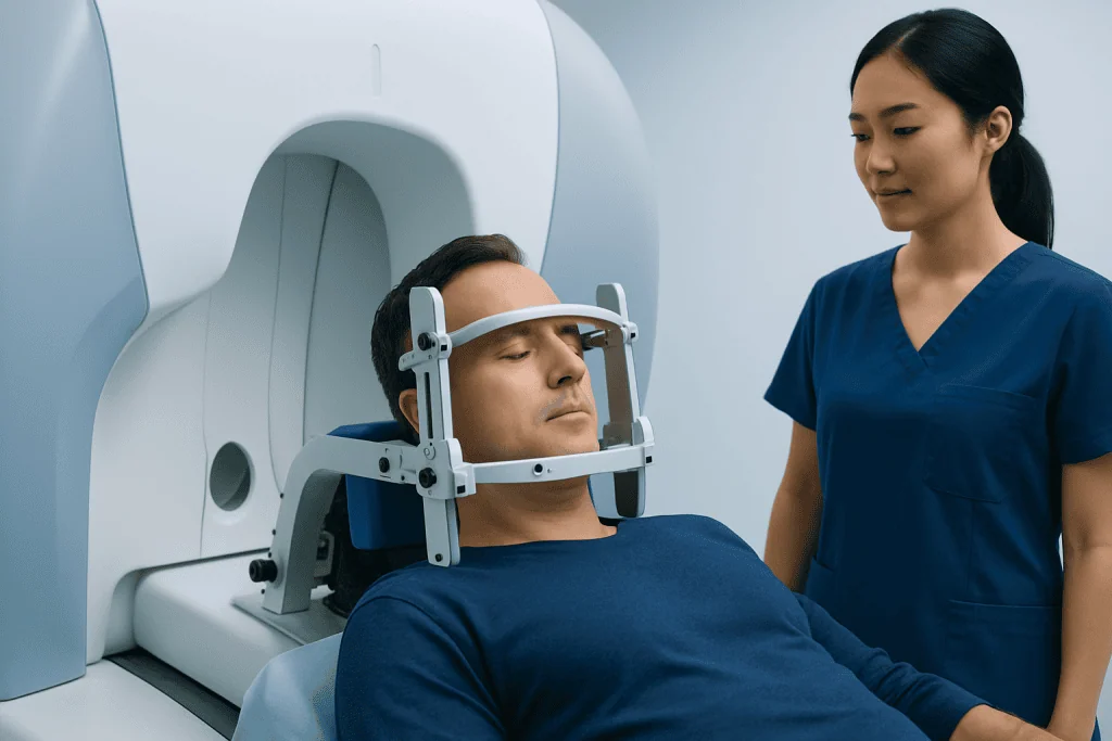
Precision depends on the head staying exactly where the plan expects it to be. Depending on the clinical protocol, this is achieved with either a lightweight rigid frame, fixed to the head under local anaesthetic, or a custom-fitted thermoplastic mask. The frame provides absolute fixation and has been the traditional standard; mask-based systems, used on newer platforms, allow frameless and sometimes multi-session treatment. Your team will explain which approach suits your case and exactly what it involves.
Once immobilisation is in place, imaging is performed so the target can be located in three dimensions. This may include MRI, CT and, for vascular malformations, angiographic imaging. The images are loaded into specialised planning software, and the lesion and every nearby critical structure are outlined with great care.
Treatment Planning
This stage is invisible to the patient but central to the quality of the result. A radiation oncologist, a neurosurgeon and a medical physicist work together on the plan: they define the exact shape of the target, decide the dose, and arrange the beam geometry so the prescribed dose covers the lesion while falling away sharply at its edges. Specialists in radiation oncology model precisely how the dose will distribute within the brain: a step that matters most when a lesion sits near areas responsible for vision, hearing, balance, speech or movement. Some lesions are treated in a single session; for others, the team may recommend a staged or fractionated approach better suited to the lesion’s size or position.
Are You Awake for a Gamma Knife?
Yes, most adults are awake throughout Gamma Knife treatment. General anaesthesia is usually unnecessary because nothing is cut and nothing is felt during beam delivery. You lie on the treatment couch, the machine positions you according to the plan, and the team monitors you continuously by camera and intercom; you can speak to them at any point. Mild sedation can be arranged for patients who feel anxious or find it difficult to lie still, and young children are typically treated under anaesthesia so they remain motionless. Many patients listen to music or simply rest during the session.
Is Gamma Knife Radiation Painful?
You cannot feel the radiation itself: there is no heat, pressure or sensation as the beams are delivered. Where discomfort occurs, it usually relates to the immobilisation rather than the treatment: the pin sites of a rigid head frame can feel tight or tender despite local anaesthetic, and some people find the frame or mask claustrophobic. After frame removal, mild soreness or a headache at the pin sites is common and short-lived. If you are worried about any of this, say so beforehand, teams deal with these concerns every day and can adjust their approach.
How Long Does the Procedure Take?
Duration depends on the number of targets, their size, the complexity of the plan and the prescribed dose. Beam delivery for a single small lesion may be relatively brief; treating multiple metastases or a complex vascular malformation takes longer. Counting immobilisation, imaging, planning and delivery together, expect the day to run from a few hours to most of a working day. Almost all patients are treated as outpatients and go home the same day.
The Technology Behind the Precision
Several layers of technology work together in every treatment. High-resolution imaging defines the target. Stereotactic localisation provides the coordinate system that makes accuracy measured in fractions of a millimetre possible. Planning software lets the clinical team sculpt the dose around the lesion. Quality-assurance systems verify, before and during treatment, that delivery matches the plan exactly. For patients, the practical benefit of all this is not the machinery itself: it is the ability to treat lesions only a few millimetres across, or lying against critical neurological structures, without an incision and usually without general anaesthesia.
After Treatment and Early Recovery
You are observed for a period after the session. If a frame was used, it is removed and the pin sites are dressed. Mild headache, scalp tenderness, fatigue or nausea can occur in the first hours or days and usually pass quickly. If swelling around the treated area is a concern, your doctor may prescribe a short course of steroids or other medication.
Most patients return home the same day and resume light activities within a day or two, depending on how they feel. Follow-up imaging is scheduled from the outset, because the effect of radiosurgery is judged over time rather than on the day. The first scan does not always tell the whole story: some lesions swell temporarily before stabilising or shrinking, so the team interprets each image in the context of timing, symptoms and the expected response pattern for your specific condition.
Why Acting Early Matters
With brain tumours and other intracranial lesions, timing shapes both the available options and the likely course. A small, well-defined lesion is generally easier to treat with focused radiosurgery than a larger one causing mass effect or widespread symptoms. Earlier treatment can also reduce the risk of further neurological decline, seizures or pressure-related complications developing while a lesion grows.
In oncology specifically, delay can allow metastases to enlarge or new lesions to appear. As lesions grow, they may pass beyond the range suited to single-session radiosurgery and instead require open surgery, broader-field radiation or several modalities combined. Tumours near sensitive structures become progressively harder to treat safely the more they expand.
None of this means every lesion needs immediate intervention. In some situations, a small, stable, symptom-free benign tumour, for example, careful observation with scheduled imaging is entirely appropriate. The important point is that a specialist should decide whether waiting is safe, rather than the decision being made by default. Prompt evaluation preserves options: it lets the team weigh Gamma Knife against surgery, fractionated radiotherapy and medical treatment while all of them are still realistic.
Potential Benefits of Gamma Knife Treatment
For appropriately selected patients, radiosurgery offers several meaningful advantages over more invasive alternatives:
| Benefit | What It Means for You |
|---|---|
| Non-invasive treatment | No surgical incision into the brain, which may spare you many of the burdens associated with open surgery. |
| High precision | Radiation is concentrated on the target while exposure to nearby healthy brain tissue is kept as low as possible. |
| Outpatient care for many patients | Most people need no extended hospital stay and return home the same day. |
| Useful for difficult-to-reach lesions | Tumours or abnormalities near delicate structures can be treated without physically entering the brain. |
| Often shorter recovery than open surgery | Many patients resume normal daily activities relatively quickly, depending on their condition and overall health. |
| Fits into a broader cancer plan | Radiosurgery can be combined with surgery, systemic therapy or other radiation strategies as part of coordinated care. |
Set against these advantages are honest limits. Gamma Knife cannot relieve pressure from a large mass the way surgery can, it provides no tissue for pathology, and its effect takes time. Weighing those trade-offs for your particular case is exactly what the multidisciplinary evaluation is for.
Typical Recovery Timeline
Recovery varies with the diagnosis, the area treated, your symptoms beforehand and whether radiosurgery is one element of a larger plan. The timeline below is a general guide, not a schedule:
| Time Period | What Patients Can Expect |
|---|---|
| Day 1 | Observation after treatment, then discharge in most cases. Mild headache, fatigue or scalp discomfort may occur temporarily. |
| First week | Most patients return to light routine activities. Some feel tired or take prescribed medication for swelling or symptom control. |
| First month | Symptoms may begin to stabilise, but imaging changes are usually gradual. Follow-up appointments assess recovery and any treatment-related effects. |
| Longer term | Serial MRI scans evaluate the response over time. Depending on the condition, the lesion may stop growing, shrink gradually, or prompt further treatment planning. |
What Influences Outcomes and a Good Result
Patients understandably ask how successful Gamma Knife is. The honest answer is that it depends on what is being treated and on what success means in that situation. For one patient, success is local control of a brain metastasis. For another, it is preserved hearing, relief from facial pain, or simply avoiding open surgery. Radiosurgery is an effective tool for appropriately selected lesions, but outcomes genuinely vary with diagnosis and individual circumstances, and no responsible clinician will promise a particular result before evaluating your case.
Several factors consistently shape results:
- The type of lesion: benign tumours, metastases, vascular abnormalities and functional disorders behave differently and respond on different timelines.
- Size and number of targets: smaller, well-defined lesions are generally better suited to radiosurgery than larger lesions causing significant pressure.
- Location: lesions near the optic apparatus, brainstem, cranial nerves or motor pathways demand especially careful planning.
- Previous treatments: prior surgery, prior radiation and current systemic cancer therapies all affect planning and expected response.
- Overall health and neurological status: your baseline condition influences recovery, tolerance of treatment and the broader strategy.
- Expert planning and follow-up: good outcomes rest on patient selection, imaging review, dose planning and long-term monitoring, not on the machine alone.
It also matters to know that some side effects appear late rather than immediately. Swelling, radiation-related changes on imaging, or delayed effects on nearby structures can emerge months after treatment and may need medication or simply observation. This is one of the strongest arguments for structured follow-up with an experienced team: the aim is never just to deliver radiation, but to integrate it into a considered, evidence-based plan before, during and after treatment.
What is life expectancy after gamma knife surgery?
Life expectancy after Gamma Knife is determined mainly by the underlying condition, not by the procedure itself. For benign diagnoses, a vestibular schwannoma, a meningioma, trigeminal neuralgia, the treatment addresses a condition that was not typically life-limiting, so the relevant questions are about symptom control and lesion stability rather than survival. For patients with brain metastases, prognosis depends chiefly on the primary cancer, how well it is controlled elsewhere in the body, and overall health; radiosurgery contributes by controlling disease within the brain. Because the range across diagnoses is so wide, only your own oncology team, looking at your complete picture, can discuss prognosis meaningfully.
How long does it take for a tumor to shrink after a Gamma Knife?
A tumor typically responds over months, not days or weeks. The radiation stops target cells dividing at the time of treatment, but visible change on MRI develops gradually as those cells die off and the body clears them. Some lesions shrink steadily over several months to a few years; others stop growing without ever becoming much smaller, which, for many conditions, still counts as a successful result. Some lesions swell slightly before they settle, which is why an early scan can look worse before it looks better. Vascular malformations generally take the longest, closing off progressively over an extended period. Your follow-up imaging schedule is designed around the expected pattern for your specific diagnosis.
How Gamma Knife Care Is Organised at Acibadem
At Acibadem, Gamma Knife is approached as part of a broader neuroscience and oncology pathway rather than as a stand-alone procedure. Patients are evaluated by physicians across the relevant specialties, radiation oncology, neurosurgery, neuroradiology and medical oncology, with neurology, endocrinology or ENT input when the condition calls for it. Complex cases can be reviewed in multidisciplinary boards, so surgery, radiosurgery, conventional radiotherapy and systemic treatment are weighed together rather than in isolation.
That coordinated review matters most for exactly the patients most often referred for radiosurgery: those with brain metastases, recurrent tumours or lesions near critical structures. It grounds the recommendation in the full clinical picture, imaging, pathology, symptoms, prior treatment and your own priorities.
Just as important, plans are individual. Not every patient referred for Gamma Knife is best served by Gamma Knife. Some are advised to have surgery first; others benefit more from fractionated radiation, systemic therapy, surveillance or a combined approach. A careful centre says so plainly when another option is more appropriate, and for many patients, that balance is the most reassuring feature of an experienced team.
Deciding Whether Gamma Knife Is Right for You
If you have been told that a brain tumour, a brain metastasis or another intracranial condition may need treatment, it is reasonable to want clarity before choosing a path. Gamma Knife can be a highly effective option for selected patients, precise, non-invasive and often compatible with getting back to daily life quickly. The decisive question is never whether the technology is impressive, but whether it is the right tool for your diagnosis, anatomy, symptoms and overall plan.
Second opinions are common and entirely normal in radiosurgery, especially where surgery and radiation are both realistic options. Questions worth putting to any treating team include:
- Why is radiosurgery preferred over surgery, fractionated radiotherapy or observation in my case, and what would change that recommendation?
- Will a frame or a mask be used, and will treatment be single-session or staged?
- Which structures near my lesion are at risk, and how does the plan protect them?
- What response should I expect on follow-up imaging, and on what timescale?
- Which side effects are common early, and which can appear late?
- How does this treatment fit with the rest of my cancer care, if relevant?
Clear answers to those questions, from a team that reviews imaging carefully and explains its reasoning, turn an intimidating diagnosis into a decision you can actually weigh. That, more than any single machine, is what a good result is built on.
Our Specialists Explain
 Gamma Knife Treatment for Brain Tumors
Preparation
- Before Gamma Knife, patients usually have imaging such as MRI to map the treatment area accurately. A head frame or mask may be used for precision, and your doctor will review medicines, medical history, and fasting instructions if sedation is planned.
Aftercare
- Most patients go home the same day and can return to light activities within a day or two. Mild headache, nausea, or temporary scalp discomfort can occur, and follow-up imaging is needed to monitor the treatment response over time.
Frequently Asked Questions
What is Gamma Knife and how does it work for brain conditions?
Gamma Knife is a non-surgical form of radiosurgery used to treat certain brain tumors, vascular malformations, and some functional disorders. It delivers many precise beams of radiation to a defined target in the brain while limiting exposure to nearby healthy tissue. Despite the name, there is no knife or incision. Treatment is usually planned with high-resolution imaging, and Acibadem specialists create a personalized approach based on the size, type, and location of the condition.
Which conditions can be treated with Gamma Knife?
Gamma Knife is commonly used for brain metastases, meningiomas, acoustic neuromas, pituitary adenomas, arteriovenous malformations, and selected cases of trigeminal neuralgia. It may be recommended as a primary treatment or after surgery, depending on the diagnosis. Not every brain condition is suitable for radiosurgery, especially if the lesion is very large or causing urgent pressure symptoms. At Acibadem, neurosurgery, radiation oncology, and neuroradiology teams assess each case to decide the best option.
Is Gamma Knife a surgery, and will I need general anesthesia?
Gamma Knife is not open surgery. There are no incisions, no scalp shaving in most cases, and general anesthesia is usually not needed for adults. Most patients are awake during the procedure and may receive local anesthesia, mild sedation, or supportive medication to help them stay comfortable. Children or selected adults may need different anesthesia planning. Your care team at Acibadem explains each step in advance and tailors the plan to your medical needs and comfort.
How long does Gamma Knife treatment take, and do I need to stay in hospital?
Gamma Knife is often completed in a single day, although the exact schedule depends on imaging, treatment planning, and the complexity of the target. Some sessions are short, while others take longer if there are multiple lesions or detailed planning requirements. Many patients go home the same day after a period of observation. In certain situations, an overnight stay may be advised. Acibadem specialists provide a personalized timeline before treatment begins.
Is Gamma Knife painful, and what should I expect during the procedure?
Most patients tolerate Gamma Knife well. The treatment itself is painless because radiation cannot be felt. You may notice some pressure or mild discomfort during frame placement if a head frame is used, although local anesthesia helps reduce this. Some centers may use a mask-based setup for selected cases. During treatment, you lie still while the machine delivers radiation. The Acibadem team monitors you closely and explains what to expect before, during, and after the session.
What are the possible side effects and risks of Gamma Knife?
Side effects vary depending on the area treated, the size of the lesion, and previous treatments. Common short-term effects can include headache, fatigue, nausea, or temporary swelling around the treated area. Some patients may need medication such as steroids to manage swelling. Less common risks depend on the condition being treated and the nearby brain structures. Your doctors at Acibadem review potential benefits and risks in detail and provide a personalized follow-up plan.
How soon will I see results after Gamma Knife treatment?
Gamma Knife does not usually produce instant results. The treated lesion often changes gradually over weeks to months, and the timing depends on the diagnosis. For example, some tumors may stop growing and slowly shrink, while vascular malformations can take much longer to respond. Symptom relief may happen earlier in some conditions, but not always. Follow-up MRI scans are important to track progress. Acibadem specialists schedule imaging and consultations based on your individual treatment goal.
Can Gamma Knife treat multiple brain metastases or recurrent brain tumors?
Yes, Gamma Knife may be used for multiple brain metastases and for some recurrent tumors, depending on factors such as the number of lesions, their total volume, prior radiation, and your overall health. It is often chosen because of its precision and its ability to target specific areas while sparing more healthy brain tissue. However, some patients may need surgery, whole-brain radiotherapy, or systemic treatment instead. Acibadem specialists evaluate all options in a multidisciplinary setting.
How do I know if I am a good candidate for Gamma Knife in Turkey?
Suitability depends on the diagnosis, lesion size, exact location, symptoms, previous treatments, and general medical condition. Patients usually need MRI and sometimes additional imaging or pathology reports for evaluation. International patients can often begin with a remote review of their medical records and scans before traveling. At Acibadem, specialists provide a personalized assessment and treatment recommendation, helping you understand whether Gamma Knife is appropriate or whether another approach may be safer and more effective.
Why do international patients choose Acibadem Hospitals in Turkey for Gamma Knife?
International patients often look for experienced multidisciplinary teams, advanced imaging, modern treatment technology, and coordinated care in one center. Acibadem brings together specialists in neurosurgery, radiation oncology, neuroradiology, and oncology to plan treatment carefully and follow patients after therapy. Support for international visitors may include help with medical documentation, travel planning, and language assistance. Before treatment, Acibadem specialists review your records in detail and create a personalized plan based on your diagnosis and needs.
Medically reviewed by the Acıbadem International Medical Board September 1, 2026
See our medical review board →
Update history
- PublishedJuly 19, 2026
- Medical review approvedSeptember 1, 2026
- Last content updateSeptember 1, 2026
Compare options, costs and recovery
From the Acibadem Blog
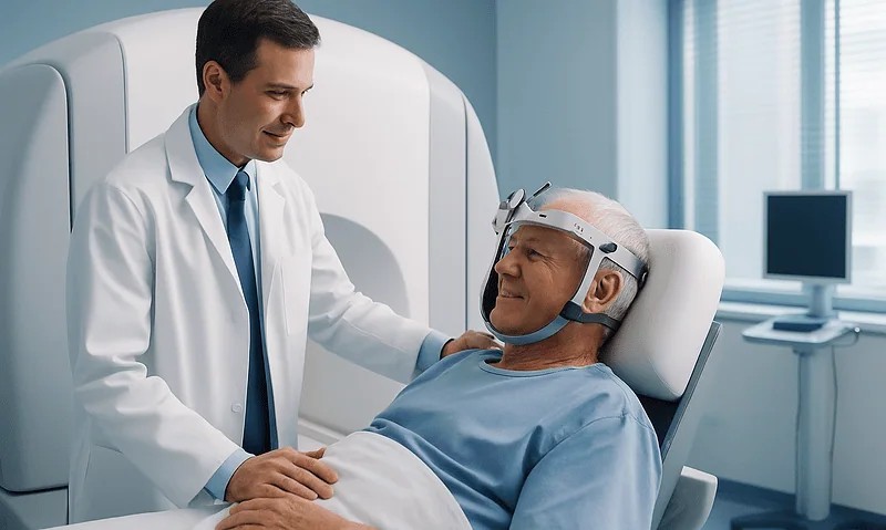
What Happens During Gamma Knife Radiosurgery: Frame or Mask, Planning and the Session Itself
During Gamma Knife radiosurgery, a lightweight head frame or a custom face mask holds the head still, detailed MRI or CT images are taken,…

How Gamma Knife Focuses Many Weak Beams Into One Precise Target Without an Incision
Gamma Knife is a form of stereotactic radiosurgery that aims roughly 190 to 200 thin beams of gamma radiation from different angles at one…