

How Gamma Knife Focuses Many Weak Beams Into One Precise Target Without an Incision

Key Takeaways
- Gamma Knife uses roughly 190 to 200 individually weak gamma-ray beams whose energy adds up only where they cross, which is why healthy tissue along each beam's path is largely spared.
- Nothing is removed during radiosurgery; abnormal cells are left unable to divide and shrink or stop growing over months, while abnormal vessels can take a couple of years to close.
- Head immobilization with a pinned frame or molded mask, not the radiation source, is what delivers accuracy to within about a millimeter.
- Targets are usually small, commonly under about 3 to 4 centimeters, and large tumors causing pressure or masses needing a biopsy are generally treated in other ways.
- The most common side effects are short-lived headache, nausea, fatigue and pin-site tenderness, while delayed swelling and radiation necrosis can appear months to years later, which is why follow-up scans continue long term.
- Life expectancy after Gamma Knife depends on the underlying diagnosis rather than the procedure, and generalized survival figures cannot be applied to any individual.
Gamma Knife is a form of stereotactic radiosurgery that aims roughly 190 to 200 thin beams of gamma radiation from different angles at one small spot inside the head. Each beam is too weak to harm the tissue it passes through, but where they all cross, the combined energy is strong enough to stop abnormal cells or vessels from growing. No cut is made; a frame or mask holds the head still.
The letter from the neurosurgeon’s office says “radiosurgery,” and the word does not sit easily. Surgery means a scalpel. Radiation means weeks of daily visits and a scorched, tired feeling. So which is it? A man in his sixties, newly told about a small benign tumor pressing on his hearing nerve, reads the paragraph three times and then types the question almost everyone in his position types: how does gamma knife work, and how can anything called a knife leave no scar?
The answer is a small piece of physics that has been in clinical use for decades, and once it clicks, the rest of the conversation with the care team gets much easier. This explainer walks through the mechanism, the day itself, what the following weeks usually feel like, the honest drawbacks, and the questions worth bringing to the appointment.
One note before we begin: Gamma Knife is a proprietary name for one type of machine. The principles here apply to stereotactic radiosurgery in general, and your team will tell you which system they use.
How does gamma knife work? Many weak beams, one strong crossing point
Picture a darkened room and two hundred people, each holding a dim flashlight, standing in a circle around a single chair. One flashlight barely lights the chair. All of them pointed at the same spot make it glow. Everyone else in the room stays in near darkness. That is the whole idea.
Inside the machine sit roughly 190 to 200 small sources of cobalt-60, a radioactive form of cobalt that gives off gamma rays, a high-energy form of light that passes through tissue (Cleveland Clinic). Each source sends a narrow beam through a metal channel called a collimator, which shapes and aims the beam. The channels are arranged on a curved helmet or dome so that every beam converges on one point in space, known as the isocenter, the spot where the beams meet.
Any single beam deposits so little energy on its way in that healthy brain along its path receives only a small exposure. At the isocenter, the beams overlap and the energy adds up sharply. Abnormal cells in that zone have their DNA damaged badly enough that they lose the ability to divide; the walls of abnormal blood vessels slowly thicken and close.
Precision, not strength, is what makes this possible. The beams are aimed at a target mapped in three dimensions from MRI or CT scans, and the head is held so still that the crossing point stays where it was planned, typically to within about a millimeter (Mayo Clinic). Radiosurgery, then, is a slightly misleading name: nothing is removed. The abnormal tissue is left in place and its biology is changed.
What actually happens on treatment day, step by step
Most people are surprised by how ordinary the day feels. You arrive awake, usually having eaten lightly if your team allows, and you go home the same day in the majority of cases (Mayo Clinic).
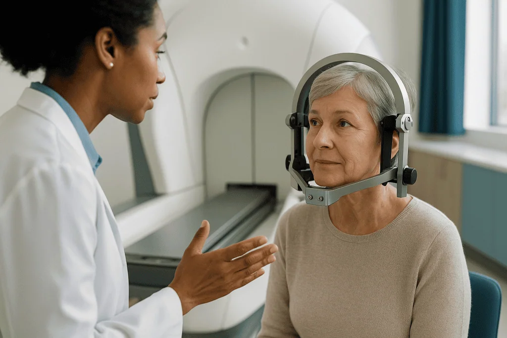
The first step is immobilization. Depending on the system and the target, a lightweight frame is attached to the head with four small pins after the scalp is numbed with local anesthetic, or a custom molded plastic mask is fitted over the face. The frame or mask has one job: to make sure the head is in exactly the same position during scanning and during treatment.
Next comes imaging. With the frame or mask in place, you have an MRI, a CT, or for blood vessel abnormalities an angiogram, which is an X-ray of the vessels after a dye is injected. A physicist, a radiation oncologist and a neurosurgeon then sit at a planning computer and outline the target in three dimensions, choosing how many crossing points, or “shots,” are needed to cover its shape. Planning commonly takes an hour or more while you wait (Cleveland Clinic).
Treatment itself is quiet. You lie on a couch, the frame or mask locks into the machine, and the couch slides your head into the dome. There is no sensation from the beams. You can talk to staff through a microphone, and many people doze. Depending on the size and complexity of the target, this part lasts from under half an hour to a few hours (Mayo Clinic). Afterward the frame comes off, pin sites are dressed, and you are observed for a short while.
Why the frame or mask matters more than the beams
Ask a radiosurgery physicist what keeps them up at night and the answer is rarely the radiation. It is movement. The machine can only be as accurate as the position of the head it is aiming at.
Stereotactic simply means using a fixed three-dimensional coordinate system to locate a point in the body. The frame creates that system. Once it is pinned to the skull, every structure inside the head can be given an address, three numbers, relative to the frame. The scan is taken with the frame on, the plan is written in frame coordinates, and the machine positions the frame so that the chosen address sits at the isocenter. If the frame does not move, the target does not move.
Frameless systems use a molded mask plus a small infrared or optical camera that watches the nose or forehead position many times a second. If the head drifts beyond a set tolerance, the beams pause automatically. Both approaches are in routine use; teams choose based on the target, the number of sessions and patient comfort (Johns Hopkins Medicine).
Why does a millimeter matter? Some targets sit against the optic nerves, the brainstem or the cochlea, the snail-shaped organ of hearing. The steep drop-off in energy just outside the isocenter is what protects those structures, and it only works if the isocenter is where the plan says it is. This is also why you will be asked to keep still, why sedation is sometimes offered to people who struggle with confinement, and why the fitting step can feel fussy. It is the fussiness that keeps the beams honest.
Gamma knife vs surgery vs conventional radiotherapy: what genuinely differs
People often arrive thinking these are three doses of the same thing. They are three different tools, and the treating team weighs them against the specific problem, not against each other in the abstract.

| Feature | Stereotactic radiosurgery (e.g., Gamma Knife) | Open neurosurgery | Conventional fractionated radiotherapy |
|---|---|---|---|
| Incision | None | Yes, through scalp and skull | None |
| Sessions | Usually one, sometimes up to five | One operation | Daily sessions over several weeks |
| Anesthesia | Local at pin sites; sedation if needed | General | None |
| Tissue removed | No | Yes | No |
| Sample for pathology | No | Yes | No |
| Time to effect | Weeks to months, sometimes years | Immediate | Weeks to months |
| Typical target size | Small, well-defined | Any size reachable safely | Larger or diffuse areas |
| Usual hospital stay | Same-day discharge common | Days | Outpatient |
Two rows deserve a closer look. Open surgery is the only option that provides a tissue sample, which matters when the diagnosis is uncertain or when the mass is causing pressure that needs relieving now. Radiosurgery cannot do either; it changes tissue slowly and leaves it in place (Mayo Clinic).
Conventional radiotherapy spreads a lower daily exposure over many sessions, a method called fractionation, which gives healthy cells time to repair between visits. That approach suits larger or less sharply bordered targets where a steep edge is not achievable. Radiosurgery trades fractionation for geometry: fewer sessions, a sharper edge, a smaller target. None of these is “better”; each is right for a different anatomy and a different goal, which is why multidisciplinary tumor boards exist.
What does gamma knife treat? The conditions where it is commonly considered
The common thread is a small, well-defined target that scans can outline clearly. Reference sources list several groups of conditions (Mayo Clinic; Cleveland Clinic; MedlinePlus).
- Benign brain tumors. Meningiomas, which grow from the membranes covering the brain; vestibular schwannomas, also called acoustic neuromas, which arise on the balance and hearing nerve; and pituitary adenomas, growths on the hormone gland at the base of the skull.
- Brain metastases. Deposits that have spread from a cancer elsewhere in the body, particularly when they are limited in number and size.
- Arteriovenous malformations (AVMs). Tangles of abnormal vessels where arteries connect directly to veins without the usual capillaries in between, carrying a risk of bleeding.
- Trigeminal neuralgia. A condition of sudden, severe facial pain caused by irritation of the trigeminal nerve, the main sensory nerve of the face; radiosurgery targets the nerve root to interrupt pain signals (NHS).
- Selected other uses. Certain seizure disorders and tremor conditions are treated in some centers, generally when medicines have not helped and other options are limited.
Notice what is missing from the list: large tumors, tumors with fuzzy edges that blend into normal brain, and anything that needs urgent decompression. Radiosurgery is not a whole-brain treatment and it does not remove pressure. For metastases, it is frequently one part of a broader plan that also includes systemic therapy directed by an oncologist. For AVMs, the goal is slow closure of the abnormal vessels, and the team will explain that the bleeding risk does not fall until that closure is complete, which can take a considerable time (Mayo Clinic).
Whether any individual sits in one of these groups is a judgment for the treating team, based on imaging, symptoms and overall health.
Who is usually offered radiosurgery, and who is usually asked to wait
Selection is where the real expertise lives, and it is worth understanding the logic even though the decision is not yours alone to make.
People commonly offered radiosurgery share a few features. The target is small, often described as under roughly 3 to 4 centimeters, about the width of two fingers, because the geometry that protects healthy tissue only works well at that scale (Cleveland Clinic). The target sits in a place where open surgery would carry a high risk to speech, movement or vision. The person may have heart, lung or clotting conditions that make general anesthesia unwise. Or a previous operation has left a small remnant that is now growing.
Others are usually asked to wait, or steered elsewhere. A large tumor causing pressure symptoms needs that pressure relieved, and radiation cannot do it quickly. A mass of uncertain nature may need a biopsy first, because treating something without knowing what it is can close off later options. Someone whose scans show many scattered deposits may be better served by a whole-brain approach or systemic therapy. Pregnancy prompts a careful conversation about timing. And a small, symptom-free benign tumor in an older adult may simply be watched with periodic scans, since treatment of any kind carries risks and the growth may never cause trouble.
“Wait” is not a dismissal. Active surveillance, meaning scheduled scans without treatment, is a legitimate plan for many slow-growing lesions, and it keeps every option open (Mayo Clinic). If you are told to wait, ask what would change the decision and how often you will be scanned.
How painful is gamma knife surgery? An honest account of the day
The radiation itself cannot be felt. There is no heat, no tingling, no sound from the beams. People who expected something dramatic usually report that the most memorable sensation of the day was boredom.
The frame is a different matter, and it is fair to be straightforward. The four pin sites are injected with local anesthetic first, which stings briefly, in the way a dental injection does. As the pins are tightened many people feel firm pressure and a sense of tightness across the head rather than sharp pain. Some describe a dull, pressure-type headache while the frame is on that fades once it is removed (Cleveland Clinic). A mild sedative is often available for people who find the idea distressing.
After removal, the pin sites can feel tender or bruised for a day or two, and a small amount of swelling or a little bleeding at the forehead sites is common. Occasionally there is numbness or a tingling patch of scalp near a pin that settles over weeks. Mask-based systems avoid pins altogether, at the cost of a snug plastic fit some find claustrophobic.
In the hours after treatment, headache, nausea and tiredness are the most frequently reported complaints, and they are usually mild and short-lived (Mayo Clinic). Your team will tell you which over-the-counter pain relievers are acceptable for you; do not assume, since some people are asked to avoid particular ones because of bleeding risk or interactions with existing prescriptions.
The honest summary: expect a few uncomfortable minutes during frame placement, a long and dull wait, and a day or so of tenderness. Severe pain is not a normal part of the experience and should be reported.
What happens to a tumor after Gamma Knife?
This is the question that follows most people home, and the answer requires a shift in expectation. Nothing happens on the day. The tumor is still there on the drive home and on the scan a week later. The change unfolds over months.
Radiation works by breaking strands of DNA inside cells. Many tumor cells are not killed outright; they are left unable to divide successfully, so when they next try, they fail and die. Because cells in a slow-growing benign tumor divide infrequently, the visible effect lags. In benign tumors such as meningiomas and vestibular schwannomas, the usual goal is control, meaning the tumor stops growing, and many either stay the same size or shrink gradually over months to years (Mayo Clinic). Some never shrink at all yet never grow again, which is considered a satisfactory result.
Metastases from cancers elsewhere tend to respond faster because their cells divide more often, and shrinkage is often visible within weeks to months (Cleveland Clinic). Arteriovenous malformations follow the slowest path: the vessel walls thicken and the channel closes over a period that commonly stretches across a couple of years, and the team will schedule imaging to check for closure.
Two things can confuse the picture on follow-up scans. First, a treated tumor may briefly appear larger or brighter because of swelling before it settles; radiologists who read radiosurgery scans routinely expect this. Second, the center of a treated lesion can lose its blood supply and change appearance without changing size. Neither is automatically bad news, which is why the same team that treated you should interpret the follow-up images rather than an isolated report.
Gamma knife recovery time: what the following days and weeks usually look like
Recovery from radiosurgery is quieter than recovery from an operation, but it is not nothing, and knowing the typical arc helps you tell ordinary from concerning.
The first 24 to 48 hours. Most people go home the same day, usually with someone to drive them (Mayo Clinic). Pin sites may ooze slightly and feel sore. A headache, mild nausea and a wave of tiredness are common. Many people rest for a day and return to desk work or routine activities within a day or two, though some centers advise avoiding driving until the following day if a sedative was given.
The first two weeks. Pin-site scabs fall away. Some people notice a small patch of hair thinning where beams entered near the scalp, particularly if the target was close to the surface; this is usually temporary (Cleveland Clinic). Fatigue can come and go for a couple of weeks. If you were prescribed a short course of a steroid medicine to limit swelling, the team will explain how it is tapered off; do not alter the schedule without asking.
Weeks to months. Symptoms caused by the original problem, such as facial pain or hearing changes, may improve, stay the same or occasionally worsen briefly as swelling develops around the target before settling. For trigeminal neuralgia, relief may take weeks to months to arrive (NHS). The first follow-up scan is typically scheduled a few months out, then at intervals your team sets.
Almost nobody needs wound care beyond keeping pin sites clean, and there are no stitches to remove. The main task of recovery is patience: the treatment is finished, but its work has just begun.
What are the drawbacks of gamma knife surgery? Gamma knife side effects explained
A treatment with no incision can sound risk-free. It is not, and a candid list serves you better than reassurance.
Common and usually short-lived. Headache, nausea, fatigue, scalp tenderness and small pin-site bruises in the first days. A temporary patch of hair loss when the target lies near the surface (Cleveland Clinic).
Less common, delayed. Swelling, or edema, in the brain tissue around the target, appearing weeks to months later and sometimes causing new headaches, weakness or seizures; this is often managed with a course of steroid medicine directed by the team. Radiation necrosis, which is death of a small area of tissue at or near the treated spot, can develop months to a few years after treatment and occasionally requires further treatment (Mayo Clinic). Damage to nearby structures depends on location: hearing loss after treatment of a vestibular schwannoma, facial numbness after treatment for trigeminal neuralgia, or vision or hormone changes with targets near the optic nerves or pituitary.
Rare. The development of a new radiation-associated tumor many years later is described in the medical literature; sources characterize this risk as very small, and it is weighed against the risk of leaving the original problem untreated (Mayo Clinic).
Structural limitations. Radiosurgery cannot relieve pressure, cannot provide a diagnosis, and does not act quickly. Its effect on an arteriovenous malformation takes time, during which the bleeding risk remains. A tumor may fail to respond and require another approach later.
None of these drawbacks make radiosurgery a poor choice; they make it a choice, one that belongs in a conversation with the team weighing your particular anatomy against the alternatives.
Is gamma knife safe in the long run, and why follow-up scans never really end
Because the treatment changes tissue slowly, its safety story is written over years, not days. Long-term follow-up is part of the treatment, not an optional extra.
The main long-term concerns are the delayed effects described above: radiation necrosis, late swelling and, depending on the target, gradual changes to hearing, vision or hormone function. The reason imaging continues for years is that these effects can be caught early, when they are easier to manage, and because a treated tumor that begins to grow again is best noticed while still small. Vestibular schwannomas and meningiomas in particular are followed with MRI at intervals the team sets, often stretching over many years (Mayo Clinic).
Hormone monitoring matters when the target sits near the pituitary gland. Radiation can reduce the gland’s output slowly, sometimes years after treatment, so blood tests may be added to the follow-up schedule and an endocrinologist, a hormone specialist, may join the team (Cleveland Clinic).
What about radiation exposure to the rest of the body? The beams are confined to the head, and the design of the machine means the rest of the body receives very little. You are not radioactive afterward, and there is no restriction on contact with children or pregnant family members (MedlinePlus).
Living with a treated lesion also means living with scan appointments and, sometimes, a scan result that needs a second look. Ask your team who will call you with results, what they consider a normal post-treatment appearance, and what would prompt them to act. Knowing the plan in advance takes much of the anxiety out of the waiting room.
Life expectancy after gamma knife surgery: why the honest answer is "it depends on the condition, not the treatment"
Search this phrase and you will find numbers. Treat them with caution. Radiosurgery is a tool applied to many very different problems, and asking about life expectancy after Gamma Knife is a little like asking about life expectancy after an MRI. The answer lives in the diagnosis, not the machine.
For someone with a benign meningioma or a vestibular schwannoma, the condition itself is rarely life-limiting, and the purpose of treatment is to protect function, hearing, balance, vision, and to prevent growth. For someone with trigeminal neuralgia, the aim is pain control and quality of life. For an arteriovenous malformation, the goal is to lower the long-term risk of bleeding. Radiosurgery in these settings is about how you live rather than how long.
For brain metastases, the picture is shaped mainly by the primary cancer, its type, how well it is controlled elsewhere in the body, and the person’s overall health. Radiosurgery to the brain is one component of that broader care, usually chosen to control specific deposits while systemic treatment continues (Cleveland Clinic). Any figures you encounter online are averages drawn from mixed groups and cannot be applied to an individual.
This explainer will not offer a survival number, and you should be wary of any source that does without stating exactly which condition and which study it comes from. The person who can give you a meaningful answer is your oncologist or neurosurgeon, with your scans and history in front of them. Ask them directly. It is a fair question, and a good team will answer it plainly, with the uncertainty included.
What people often get wrong about gamma knife radiosurgery
Some misunderstandings come up in almost every first consultation. Clearing them away leaves room for the questions that matter.
“It is a laser.” No light or heat is involved. The beams are gamma rays, invisible and silent, and nothing is burned or vaporized.
“The tumor is gone when I leave.” Nothing is removed. The tissue stays where it is and changes over months, and some benign tumors never shrink even when they are fully controlled (Mayo Clinic).
“One session means I am finished.” Follow-up imaging continues for years, and a minority of lesions need further treatment.
“No incision means no risk.” Delayed swelling, radiation necrosis and effects on nearby nerves are real, if uncommon (Cleveland Clinic).
“It works for any brain tumor.” Size, shape, location and the need for a diagnosis all rule targets in or out. Large or diffuse tumors are usually treated differently.
“I will be radioactive.” The sources stay inside the machine. You carry no radiation home (MedlinePlus).
“Gamma Knife and radiotherapy are the same thing.” Both use radiation, but radiosurgery relies on a single sharp crossing point in one or a few sessions, while conventional radiotherapy spreads smaller daily exposures across weeks. Different tools, different jobs.
“If it does not hurt, it is not doing anything.” The absence of sensation during treatment tells you nothing about its effect. DNA damage is silent.
Underneath most of these myths is a single intuition: that treatment should look and feel like fixing something. Radiosurgery does not fix; it redirects biology, and it asks for patience in return.
Questions to ask your care team before deciding
The consultation goes better when you arrive with questions written down. These are the ones that consistently produce useful answers.
- Why are you recommending radiosurgery for me rather than observation, open surgery or fractionated radiotherapy, and what would make you change that view?
- What is the goal for my specific target: stopping growth, shrinking it, closing a vessel, or relieving pain?
- Will this be one session or several, and will a frame or a mask be used?
- Which healthy structures sit closest to the target, and what side effects could that produce?
- How soon might I notice any change in my symptoms, and how soon will you be able to see a change on scans?
- What is the follow-up schedule, and who will contact me with results?
- If I am given a steroid or other medicine afterward, how will it be tapered and who do I call with questions about it?
- What symptoms in the first weeks would you want to hear about the same day?
- If the lesion does not respond, what are the next options?
- How will this treatment fit with the other care I am receiving, such as chemotherapy or immunotherapy?
- Is there anything about my other health conditions or medicines that affects the plan?
Bring someone with you if you can. Two sets of ears catch more, and a second person can take notes while you listen. Ask whether the team offers a written summary of the plan. And if any answer leaves you unsure, it is entirely reasonable to ask for time to think or for a second opinion; a good team will not be offended.
When to call your doctor after gamma knife treatment: red-flag signs
Most of what you feel in the first days will be mild and expected. A short list of symptoms should never be waited out.
Seek emergency care immediately for a sudden, severe headache unlike any you have had before; a seizure, particularly if you have never had one; new weakness, numbness or drooping on one side of the face or body; sudden trouble speaking, understanding or seeing; or a change in alertness such as unusual drowsiness, confusion or difficulty waking. These can signal bleeding or significant swelling and need same-day assessment (Mayo Clinic).
Call your care team the same day for a headache that steadily worsens over hours or days or does not ease with the pain relief you were advised to use; vomiting that is repeated or prevents you keeping fluids down; a fever, or redness, warmth, spreading swelling or pus at a pin site, which can indicate infection; new or worsening balance problems or falls; new hearing loss, ringing, double vision or facial numbness; or any symptom that frightens you. If you were given a steroid medicine, report new mood changes, marked thirst or frequent urination, since these can be related and may need the prescriber’s attention.
Symptoms that appear months after treatment matter too. Radiation-related swelling and necrosis can emerge long after the day itself, so a new headache pattern, weakness or seizure at any point warrants a call, even if your last scan was reassuring (Cleveland Clinic).
Keep the team’s contact number where you can find it, and do not worry about being a nuisance. Radiosurgery teams expect these calls, and the quiet, uncertain ones are often the most worth making.
Frequently asked questions
How does gamma knife work without cutting the skull?
Gamma rays pass through bone and tissue, so no opening is needed. Roughly 190 to 200 thin beams enter from different angles, each too weak to damage what it passes through, and converge on a single point mapped from MRI or CT scans. At that crossing point the energy is high enough to disable abnormal cells or slowly close abnormal vessels. A frame or mask keeps the head fixed so the crossing point lands exactly where planned.
What are the drawbacks of gamma knife surgery?
The main drawbacks are that nothing is removed, no tissue sample is obtained, and the effect takes months. Short-term side effects include headache, nausea, fatigue and pin-site soreness. Less commonly, delayed brain swelling or radiation necrosis can appear months to years later, and structures near the target, such as the hearing or optic nerves, can be affected. Large or diffuse lesions are usually not suitable.
What happens to a tumor after Gamma Knife?
The tumor remains in place and its cells are damaged so they can no longer divide successfully. Benign tumors often stop growing and may shrink slowly over months to years, or stay the same size while remaining controlled. Metastases tend to respond faster. On early scans a treated lesion can briefly look larger or brighter from swelling before settling, which is why the treating team should read follow-up images.
What is the average life expectancy after gamma knife surgery?
There is no single figure, because radiosurgery treats very different conditions. For benign tumors, trigeminal neuralgia and vascular malformations, the condition is usually not life-limiting and treatment aims to protect function. For brain metastases, outlook depends mainly on the primary cancer and overall health, with radiosurgery forming one part of broader care. Your oncologist or neurosurgeon, with your records, is the right person to discuss this.
How painful is gamma knife surgery?
The radiation itself cannot be felt. If a frame is used, the four pin sites are numbed with local anesthetic, which stings briefly, followed by a feeling of pressure and sometimes a dull headache while the frame is on. Pin sites may feel bruised for a day or two afterward. Mask systems avoid pins but can feel snug. Severe pain is not expected and should be reported to the team.
What is the typical gamma knife recovery time?
Most people go home the same day and resume routine activities within a day or two, according to reference sources. Headache, tiredness and nausea usually settle within days, pin-site scabs clear over about two weeks, and any small patch of hair thinning is generally temporary. Improvement in the underlying problem takes longer, often weeks to months, and follow-up scans continue at intervals set by the team.
What does gamma knife treat most often?
Reference sources list benign brain tumors such as meningiomas, vestibular schwannomas and pituitary adenomas; limited numbers of brain metastases; arteriovenous malformations; and trigeminal neuralgia. Some centers also use it for selected seizure or tremor disorders when other options are limited. The shared requirement is a small, clearly outlined target on imaging. Suitability for any individual is decided by the treating team.
Gamma knife vs surgery: how do doctors choose?
They weigh the size, location and nature of the lesion against the person’s overall health. Open surgery is favored when tissue must be removed quickly to relieve pressure or when a diagnosis is needed from a sample. Radiosurgery is favored for small targets in hard-to-reach places or for people who cannot safely have general anesthesia. Often the two are combined, with radiosurgery treating a remnant after an operation.
What are the most common gamma knife side effects in the first week?
Headache, mild nausea and fatigue are the most frequently reported, along with tenderness, slight swelling or minor bleeding at the pin sites if a frame was used. A small patch of hair thinning can appear when the target is near the scalp and is usually temporary. Some people notice tingling or numbness near a pin site that fades over weeks. Anything severe or worsening should be reported.
Am I radioactive after gamma knife treatment?
No. The cobalt-60 sources stay sealed inside the machine, and the beams stop when the couch withdraws. Your body does not retain radiation, so there are no restrictions on being near children, pregnant family members or pets. This differs from some other treatments, such as certain radioactive implants or swallowed isotopes, which do carry temporary precautions. Your team can confirm there are none in your case.
References
This article is for general information only and is not a substitute for professional medical advice. Please consult a qualified doctor about your individual situation.

More from the Blog

Second Opinions in Neurodegenerative Diseases: When a Diagnosis Deserves Another Look
A neurodegenerative disease second opinion is a fresh review of your history, examination and test results by another neurologist, often one who focuses on…

Neuroinfectious Diseases Treatment: How Meningitis and Encephalitis Care Is Planned
A meningitis encephalitis treatment plan starts within minutes of suspicion, usually before tests confirm the cause. Doctors give intravenous antibiotics, and often an antiviral,…

Who May Be a Candidate for Peripheral Nerve Surgery? Injury, Compression and Tumor Cases
Peripheral nerve surgery is considered for three groups: people with a nerve that has been cut, torn or crushed and shows no sign of…

Can You Keep Working With Multiple Sclerosis? Fatigue, Heat and Pacing Strategies Explained
Many people with multiple sclerosis continue working, often for years, though the path varies with symptoms, job demands and disease course. Fatigue and heat…

Can Home and Work Routines Be Adapted in Motor Neuron Disease? Mobility Aids, Meals and Rest
Yes. Most people with motor neuron disease can adapt home and work routines for a considerable time by pacing activity, changing how meals are…

Questions to Ask Before Agreeing to a Cerebrovascular Disease Treatment Plan
Before agreeing to a cerebrovascular disease treatment plan, ask what the imaging showed (clot, bleed, narrowing or small vessel damage), why this option was…