

What Happens During Gamma Knife Radiosurgery: Frame or Mask, Planning and the Session Itself
Key Takeaways
- Gamma Knife delivers around 200 fine beams of cobalt-60 radiation that converge on one point, so the target receives an intense effect while tissue a few millimeters away receives very little.
- Adults are awake throughout; the only anesthetic is a local injection at the four scalp sites where the head frame's pins rest, and mask-based treatment needs no injection at all.
- The treatment portion typically lasts from under an hour to about four hours, with one to two extra hours of imaging and planning beforehand that most patients spend waiting.
- Nothing visible happens to the target on the day: benign tumors are judged over months to years, metastases over months, and arteriovenous malformations can take two years or more to close.
- Swelling around the treated area can appear weeks to months later and is the main reason for delayed headaches or returning symptoms, which is why follow-up MRI scans are scheduled at intervals.
- Radiosurgery cannot provide a tissue diagnosis or relieve pressure quickly, so people with large, symptomatic lesions or an uncertain diagnosis are often offered surgery or biopsy first.
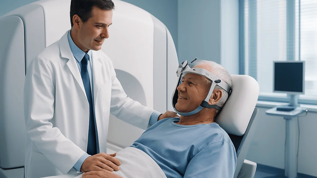
During Gamma Knife radiosurgery, a lightweight head frame or a custom face mask holds the head still, detailed MRI or CT images are taken, and a physics and neurosurgery team plans where roughly 200 fine beams of radiation will cross inside the brain. The patient then lies awake on a couch while the beams are delivered, usually in a single outpatient session, and typically goes home the same day.
The night before, most people picture a knife. The word is right there in the name, and it does the imagination no favors. Then a nurse phones to confirm the appointment and says, almost in passing, that there is no incision, no operating theater and no anesthetic beyond a numbing injection in the scalp. That is usually the moment the fear changes shape, from what will they cut to what, exactly, will they do.
This article answers that second question step by step: what happens during Gamma Knife radiosurgery, from the moment the frame is fitted or the mask is molded, through the hour or two of imaging and planning that most patients never see, to the quiet of the treatment room itself. It also covers the days afterward, which is where much of the real uncertainty lives.
Everything here is drawn from mainstream medical sources, not marketing. The choices, including whether this treatment is right for you at all, belong to your treating team.
What happens during Gamma Knife radiosurgery, in plain language
Gamma Knife is a form of stereotactic radiosurgery: a way of delivering a high, tightly focused amount of radiation to a small target inside the brain in one or a few sessions, guided by three-dimensional imaging. The phrase stereotactic simply means located precisely in space using coordinates. Nothing is cut. The word surgery survives because the intent is surgical, to destroy or disable a specific target, even though the tool is radiation rather than steel.
The machine contains many small sources of radioactive cobalt-60, a metal that emits gamma rays as it decays. Each source sends a thin beam through the skull. On its own, any single beam is too weak to do much to the tissue it passes through. The trick is geometry. Around 200 beams, according to Johns Hopkins Medicine, are aimed so that they intersect at one point. Where they cross, the combined effect is intense; a few millimeters away, it falls off sharply.
Think of sunlight through a magnifying glass. Spread across a page, the light is harmless. Focused to a point, it scorches. The rest of the brain is the page.
A typical day therefore has three phases. The first is immobilization, using either a frame pinned to the skull or a molded mask, so the head cannot shift even a millimeter. The second is imaging and planning, where MRI or CT scans are loaded into software and the team shapes the radiation to fit the target. The third is the treatment itself, when the patient lies on a couch that slides into the machine while the beams are delivered.
Mayo Clinic notes that the treatment portion may take less than an hour or up to about four hours, and that most people go home the same day. The whole visit, including planning, often runs longer.
Who is Gamma Knife usually for, and who is usually asked to wait
The technique suits targets that are small, well defined on imaging and deep or awkward enough that open surgery carries real risk. Mayo Clinic and MedlinePlus list the common indications: brain metastases (cancer that has spread to the brain from elsewhere), meningiomas (usually slow-growing tumors of the brain’s covering), vestibular schwannomas (also called acoustic neuromas, benign tumors on the hearing and balance nerve), pituitary tumors, arteriovenous malformations (tangles of abnormal blood vessels that can bleed) and trigeminal neuralgia (a nerve condition causing severe facial pain). Some people with tremor or certain seizure disorders are also evaluated for it.

Size matters more than most people expect. Because the beams converge on a point, the method works best when the target is compact. Larger lesions receive a less even distribution and expose more healthy tissue, so the team may propose staged treatment, conventional fractionated radiotherapy, surgery or a combination.
Several groups are commonly asked to wait or to consider other options first:
- People whose tumor is causing pressure symptoms, such as severe headache, vomiting or rapid neurological decline, because radiation does not relieve pressure quickly and surgery may be needed first.
- People whose diagnosis is uncertain, since radiosurgery provides no tissue sample and a biopsy may come first.
- People with widespread disease across the brain, where a whole-brain approach or systemic therapy might be discussed instead.
- People who are acutely unwell, pregnant, or unable to lie still, for whom timing or method needs adjusting.
None of these is an absolute rule. Multidisciplinary teams weigh imaging, symptoms, overall health and the person’s own priorities. The honest summary is that Gamma Knife is one tool among several, and being told it is not the right one is not a rejection but a judgment about fit.
Frame or mask: how the head is kept perfectly still
Radiosurgery lives or dies on precision. If the head moves, the point where the beams cross moves with it, and a treatment meant for a tumor lands partly on normal brain. Two approaches solve this.
The traditional method is a rigid head frame. It is a lightweight metal ring, described by Mayo Clinic as attached to the skull with four pins, two on the forehead and two at the back. A local anesthetic is injected into the scalp at each site first. The pins sit against the bone under the skin; they do not enter the skull cavity. Once fitted, the frame becomes part of the imaging and the treatment: markers on the frame appear on the scans, giving the planning software a fixed coordinate system, and the frame later locks into the machine’s couch. The result is that the target’s position relative to the frame is known to within a fraction of a millimeter.
The newer alternative is a thermoplastic mask. A sheet of mesh plastic is warmed until pliable, laid over the face and molded to its contours, then hardened. It fastens to the couch and is paired with a small infrared marker that rests on the nose and is tracked continuously by a camera. If the head drifts beyond a preset tolerance, the machine pauses.
Neither is simply better. The frame offers the most rigid fixation and is the standard for single-session treatment of very small targets such as the trigeminal nerve. The mask avoids pins and makes it practical to divide treatment into several sessions, an approach called fractionation, which may be preferred for larger lesions or those close to sensitive structures like the optic nerves. Your team chooses based on target size, location and your circumstances, and will tell you which to expect.
Is Gamma Knife painful? What the frame fitting actually feels like
The radiation itself has no sensation. You cannot see, hear or feel gamma rays passing through tissue, and patients frequently describe the treatment portion as the most uneventful part of the day. The honest answer to the pain question therefore concerns the frame.
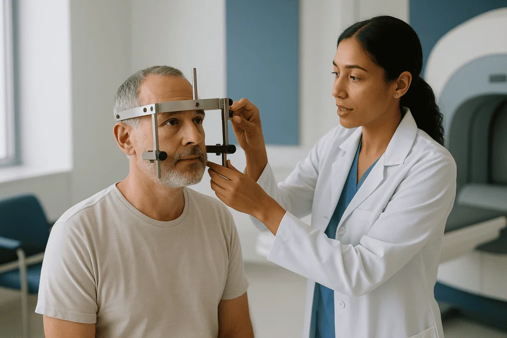
Fitting begins with the anesthetic injections, four small stings comparable to a dental injection. Most people rate this as the least comfortable moment. As the pins are tightened, there is a sensation of firm, even pressure around the skull, sometimes described as a very tight headband or the feeling of wearing a heavy helmet. It is odd rather than sharp. Some people notice a brief change in how their face feels or a mild headache from the pressure, which eases as they adjust.
Once fitted, the frame stays on for the entire visit, including the waiting time while the plan is prepared. You can sit up, walk to the bathroom with help and drink through a straw. Eating is possible but awkward. Many centers give a mild sedative or pain reliever if requested; whether and what to give is the clinical team’s decision and depends on your health and other medicines.
Removal at the end of the day takes a minute or two. The pin sites may bleed a little and feel tender; MedlinePlus notes that swelling or bruising around them is common and usually settles within days. A dull headache on the evening of treatment is frequent and typically short-lived.
The mask route avoids pins entirely. Its discomfort is different: a snug, warm plastic pressing on the face, a feeling some find claustrophobic. The mask is porous and you can breathe freely, but people who dislike enclosed spaces should say so in advance so the team can plan for it.
Imaging and planning: what the team does while you wait
Once the frame or mask is in place, you are taken for scanning. Most centers use MRI (magnetic resonance imaging, which maps soft tissue in fine detail using magnets and radio waves), sometimes with a contrast agent given through a vein to make tumors and vessels stand out. CT (computed tomography, a rapid series of X-ray images) may be added because it shows bone and frame coordinates with high geometric accuracy. For arteriovenous malformations, a catheter angiogram may be included to trace the abnormal vessels.
The images are then fused, meaning the software overlays them so that every point in one scan corresponds exactly to the same point in the others. This is the part patients rarely see and often underestimate. A neurosurgeon and a radiation oncologist outline the target on each slice, and they also outline the structures they want to protect: the optic nerves and chiasm, the brainstem, the cochlea, the pituitary stalk.
A medical physicist, a specialist in the physics of radiation, then builds the plan. Rather than one large sphere of converging beams, the software stacks many small spheres, called shots, of different sizes to fill an irregular shape, much as a sculptor might pack marbles into a mold. Each shot can have some beams blocked to steer radiation away from a nearby nerve. The physicist checks the resulting map of radiation intensity against accepted limits for each protected structure and, in fractionated cases, divides it across sessions.
This typically takes one to two hours, during which you wait in a quiet room, often with a companion. The plan is independently verified before treatment begins. If the frame was used, you never leave the coordinate system, so nothing needs re-registering. With a mask, a brief positioning scan on the treatment couch confirms alignment before the first beam.
What happens during the Gamma Knife session itself
The treatment room is unremarkable: a couch, a large rounded machine at one end, dim light, often music if you want it. You lie on your back. The frame is bolted to a docking station on the couch, or the mask is clipped down and the infrared marker positioned on your nose. A camera confirms your position. Staff then leave, because the room is shielded and no one else may be inside while the sources are exposed.
You are never alone in practice. A microphone and speaker keep you in two-way contact, and a camera lets the team watch you throughout. You can speak at any time, and treatment can be paused within seconds.
The couch moves you head-first into the machine. From your point of view, this feels like being slid gently into a large, open-ended dome; the space around your head is wider than an MRI tunnel and many people find it less confining. The machine then moves the couch in small increments to place each planned shot at the exact focal point of the beams. Between shots there are soft mechanical sounds, clicks and hums, and slight movements. Some people count them; many doze.
Nothing about the radiation is perceptible. There is no heat, no light, no tingling. A small number of people notice a faint metallic taste or a fleeting sense of a smell when beams pass near the olfactory region, a harmless quirk that fades immediately.
Mayo Clinic describes the treatment portion as lasting from under an hour to about four hours, depending on how many shots the plan contains and how complex the target is. When the last shot is complete, the couch withdraws, the team returns, and the frame or mask is released.
Are you awake during Gamma Knife?
Yes, almost always. Adults are awake for the frame fitting, the imaging, the waiting and the treatment. General anesthesia would add risk without adding benefit, because there is no incision, no manipulation of brain tissue and nothing that hurts once the frame is on. Being awake also allows you to tell the team immediately if anything feels wrong.
What awake means in practice varies. Some centers offer a mild oral sedative before the frame is fitted to take the edge off anxiety; others rely on local anesthetic alone and a calm explanation. If you have a history of panic in enclosed spaces, a strong gag reflex, or a movement disorder that makes lying still difficult, tell the team at the planning visit. These are exactly the situations where sedation is considered, or where a frame might be chosen over a mask because it holds the head regardless of small muscle tremors.
Children are the main exception. Young children cannot reliably tolerate a frame or hold still for a long treatment, so pediatric radiosurgery is often performed under general anesthesia with an anesthesiologist present throughout. The pathway is planned well in advance and families are walked through it separately.
Being awake also shapes the day for companions. Because there is no recovery from anesthesia, most people can talk normally within minutes of the frame coming off, walk unaided and eat. Mayo Clinic notes that patients usually go home the same day, though a responsible adult should drive if any sedative was given. A few centers observe patients overnight after complex plans or when nausea or headache is troublesome; that is a local judgment, not a sign that something has gone wrong.
Frame versus mask versus conventional radiotherapy: how the options compare
People are often surprised to learn that several kinds of radiation treatment can be offered for the same brain lesion. The table below summarizes the practical differences from the patient’s side of the couch. It describes typical patterns drawn from Mayo Clinic, MedlinePlus and the National Cancer Institute; your own plan may differ.
| Feature | Frame-based Gamma Knife | Mask-based radiosurgery | Conventional fractionated radiotherapy |
|---|---|---|---|
| How the head is held | Metal frame pinned to skull under local anesthetic | Molded thermoplastic mask with infrared tracking | Molded mask, no pins |
| Number of visits | Usually one treatment day | One to a small number of sessions, commonly up to five | Daily sessions over several weeks |
| Radiation per session | High, focused | High, focused; may be divided | Lower per session, larger volume |
| Best suited to | Small, sharply defined targets | Small to moderate targets, or those near sensitive nerves | Larger or diffuse targets |
| Anesthesia | Local to scalp; awake | None; awake | None; awake |
| Same-day discharge | Typical | Typical | Typical |
A few points deserve emphasis. First, fractionation is not a lesser option. Dividing radiation across sessions gives healthy tissue time to recover between exposures, which is why it is often preferred when a target sits against the optic pathway or brainstem. Second, the choice between frame and mask is partly about the target and partly about you: how long you can lie still, how you feel about pins, how far you are traveling. Third, open surgery remains the comparison that matters most for many people, because it removes tissue immediately, provides a diagnosis and relieves pressure, at the cost of an operation and hospital stay. Radiosurgery does none of those things; it stops growth or closes vessels gradually. Which trade-off is right is a conversation, not a formula.
What happens after Gamma Knife: the first hours and days
The frame comes off, the pin sites are cleaned and dressed, and you are usually observed for a short while before discharge. If contrast was used for imaging, you may be encouraged to drink fluids. Many people feel a strange lightness once the frame is gone, followed within an hour or two by a dull headache that responds to ordinary measures your team suggests.
The pin sites are the main physical reminder. They can ooze slightly on the first evening and feel bruised for a few days. MedlinePlus advises keeping them clean and dry as instructed; a small scab forms and drops off within about a week or two. Numbness or a tingling patch on the forehead near the front pins is not unusual and generally fades over weeks, though a small area of altered sensation can occasionally persist.
Fatigue is the most common complaint in the first days, sometimes out of proportion to how little seemed to happen. Part of this is the long day, part the emotional release, and part a genuine biological response to radiation. Nausea can occur, particularly when the target is near the brainstem, and the team will have told you what to do if it does.
Mayo Clinic notes that most people return to normal activities within a day or two. Driving is usually fine once you feel alert and no sedative is in your system, but ask. Work, exercise and travel plans rarely need more than a brief pause. Hair loss, when it happens, is limited to a small patch near where the beams entered most densely, and tends to appear a few weeks later rather than immediately.
Nothing visible happens to the target during this period. That is normal, and it is the subject of the next two sections.
Gamma Knife side effects in the weeks and months that follow
Radiation acts slowly. It damages the DNA of cells in the target so that they fail when they next try to divide, and it gradually thickens and closes the small blood vessels feeding a tumor or malformation. Tumor cells that divide slowly, as most benign brain tumors do, may take a long time to reach that point. This is why side effects, like benefits, unfold over months rather than days.
The most discussed delayed effect is swelling, called edema, in the brain tissue around the target. MedlinePlus and Mayo Clinic both list it. It can develop weeks to months after treatment and may cause headache, worsening of the symptoms the lesion originally caused, or new symptoms depending on location. Often it is seen only on a follow-up scan and causes nothing you would notice. When it does cause symptoms, teams commonly use a short course of a corticosteroid, an anti-inflammatory medicine, to settle it; whether that is needed, and for how long, is a decision for your prescribing clinician.
Less common is radiation necrosis, a patch of tissue damaged beyond recovery that can mimic tumor regrowth on scans. Distinguishing the two sometimes requires specialized imaging or, rarely, surgery.
Other effects depend on what sits next to the target. Treatment near the hearing nerve can affect hearing or balance over time; near the optic apparatus, vision; near the pituitary, hormone production, which may show up years later as fatigue or changes in weight or menstrual cycles. Trigeminal neuralgia treatment can leave facial numbness.
A theoretical long-term risk is that radiation could contribute to a new tumor decades later. The NHS describes this as rare for focused radiotherapy. Your team will have weighed it against the risk of leaving the lesion alone, and you are entitled to ask how.
Gamma Knife recovery time and when results actually show
Recovery in the everyday sense is quick; recovery in the sense of knowing whether it worked is not. Holding those two ideas together is the hardest part of the months that follow.
Mayo Clinic is direct about the timeline. For benign tumors such as meningiomas and vestibular schwannomas, the goal is usually to stop growth rather than to make the tumor disappear, and shrinkage, when it occurs, is measured over months to years. Cancerous tumors and metastases tend to respond faster, often within a few months, because their cells divide more rapidly. Arteriovenous malformations close gradually as vessel walls thicken, a process Mayo describes as taking two years or more, during which the risk of bleeding remains and some people need repeat imaging or a second treatment. Trigeminal neuralgia pain may improve within weeks for some people and take several months for others.
Follow-up therefore centers on imaging. A first MRI is commonly scheduled a few months after treatment, then at intervals that lengthen if things look stable. For vestibular schwannomas, hearing tests are added; for pituitary targets, blood tests for hormones. Scans in the first year can look worse before they look better, because swelling and the changes of dying tissue can enlarge the apparent size of a lesion. Experienced teams expect this and will tell you when a change is meaningful.
A practical note on travel: because there is no wound and no anesthetic, flying or long car journeys in the following days are usually acceptable, but confirm with your team, particularly if edema is a concern for your target’s location, and make sure follow-up imaging is arranged with a clinician who can act on the results.
What people often get wrong about Gamma Knife
The name causes the first misunderstanding. There is no knife, no blade and no incision. The term was coined to convey surgical precision, and it has confused people ever since. Nothing enters the skull except radiation.
The second is that it is a form of chemotherapy or makes you radioactive. Gamma rays pass through the body and are gone the instant the source is shielded; you can hold a baby that evening. Cobalt-60 stays inside the machine.
The third is that a single-session treatment means a single-session result. Because radiation works by preventing cells from dividing successfully, the target does not vanish on the day. A scan the following week would look essentially unchanged. People who expect immediate proof often feel anxious or cheated; those who understand the mechanism can settle into the waiting.
The fourth is that radiosurgery is always gentler than surgery and therefore always preferable. It avoids an operation, which matters enormously for some people, but it cannot relieve pressure quickly, cannot provide a diagnosis and carries its own delayed risks of swelling and nerve effects. For a large tumor causing symptoms, an operation may be the safer path.
The fifth is that it is only for cancer. Many, arguably most, people treated have benign conditions: vestibular schwannomas, meningiomas, vascular malformations, facial pain.
The sixth is that a follow-up scan showing the lesion at the same size means failure. For benign tumors, stability is often the intended outcome. Mayo Clinic frames the goal as controlling growth, not erasing the lesion, and the two should not be confused.
Finally, some assume they will be unconscious. Adults are awake, and that is by design.
Questions to ask your care team before Gamma Knife
A planning consultation moves quickly, and it is easy to leave with the logistics clear and the reasoning vague. The questions below are the ones patients most often wish they had asked. Take them in writing and ask for the answers in plain language.
- Why is radiosurgery being recommended for me rather than surgery, conventional radiotherapy or watchful waiting, and what would change that recommendation?
- Will I have a frame or a mask, and why that choice for my target?
- Is my treatment planned as a single session or divided into several, and what drives that decision?
- Which structures near the target are you most concerned about protecting, and what symptoms would tell me they had been affected?
- What is the realistic goal: stopping growth, shrinkage, closing a vessel, easing pain? Over what timeframe would you expect to see it on scans?
- What side effects are common in the first week, and which ones should prompt a phone call rather than patience?
- Who do I contact after hours if something worries me, and what is the number?
- When is my first follow-up scan, and who will review it with me?
- If my current medicines include blood thinners, steroids or seizure medicines, do any need adjusting around the treatment day, and who will tell me exactly what to do?
- Could I need a second treatment, and how would you decide?
It also helps to ask what the team considers a poor outcome and how often they see it, framed in ranges rather than promises. Clinicians who work with this technology every day are generally comfortable with the question. Write down the answers; the frame day is long, and you will want to reread them later.
When to call your doctor after Gamma Knife
Most people need nothing beyond the follow-up appointments already booked. A short list of warning signs, drawn from MedlinePlus and Mayo Clinic guidance on stereotactic radiosurgery, marks the exceptions.
Call the team the same day, or seek urgent care if you cannot reach them, if you notice:
- A severe or steadily worsening headache, especially one that is different from the pressure headache of the frame and does not ease with the measures you were given.
- New or worsening weakness or numbness in the face, an arm or a leg, difficulty speaking or understanding speech, or a droop on one side of the face.
- New confusion, unusual drowsiness that is hard to rouse from, or a change in personality noticed by others.
- A seizure, or any seizure that is longer, different or more frequent than your usual pattern if you already have epilepsy.
- Repeated vomiting, or nausea that prevents you keeping down fluids for more than a day.
- Sudden change in vision, double vision, or new loss of hearing or balance beyond what the team told you to expect.
- Pin sites that become increasingly red, hot, swollen, painful or leak pus, or a fever without another obvious cause.
- For those treated for a vascular malformation, any sudden severe headache described as the worst of your life, a stiff neck or collapse, which can signal bleeding and warrants emergency care.
Fatigue, mild headache, tenderness at the pin sites and a small patch of hair loss weeks later are expected and do not need a call. When in doubt, ring anyway; radiosurgery teams would far rather hear about a false alarm than miss swelling that responds well when treated early. Keep the after-hours number somewhere you can find it without your glasses.
Frequently asked questions
Is Gamma Knife radiation painful?
The radiation itself cannot be felt at all; there is no heat, tingling or sound from the beams. Discomfort, when it occurs, comes from the head frame: four brief local anesthetic injections in the scalp followed by a sensation of firm pressure while the pins are tightened. Pin sites can feel bruised for a few days afterward. Mask-based treatment avoids pins but can feel snug or confining on the face.
Are you awake during a Gamma Knife?
Yes, adults are awake for the whole process, from frame fitting through imaging, planning and the treatment itself. A mild sedative is sometimes offered for anxiety, but general anesthesia is not needed because nothing is cut and the radiation is painless. You stay in two-way voice contact with the team and can pause treatment at any time. Young children are the exception and are usually treated under general anesthesia.
What happens after a Gamma Knife?
The frame or mask is removed, pin sites are dressed, and most people go home the same day after a short observation period. A dull headache and tiredness on the first evening are common, and Mayo Clinic notes that normal activities usually resume within a day or two. Follow-up MRI scans are then scheduled, typically starting a few months later, because the treatment’s effect on the target develops slowly.
What is the life expectancy after gamma knife surgery?
The procedure itself does not determine life expectancy; the underlying condition does. Someone treated for a benign meningioma, a vascular malformation or trigeminal neuralgia has a very different outlook from someone treated for brain metastases from an advanced cancer, and within each group the picture depends on age, overall health and other treatments. No single figure applies. Your treating team can discuss what the evidence shows for your specific diagnosis.
How long does the Gamma Knife session take?
According to Mayo Clinic, the treatment portion may take less than an hour or up to about four hours, depending on the number and complexity of the planned shots. The full visit is longer because the frame or mask must be fitted, imaging performed and the plan built and checked, which commonly adds one to two hours. Most centers advise setting aside most of a day.
What are the most common Gamma Knife side effects?
Early effects include fatigue, headache, tenderness or mild swelling at the pin sites and occasionally nausea, all usually short-lived. A small patch of hair loss may appear weeks later where beams entered most densely. Delayed effects, listed by MedlinePlus and Mayo Clinic, include swelling around the target that can cause headache or a return of symptoms, and, less commonly, damage to nearby nerves affecting hearing, vision, facial sensation or hormone production.
How long is Gamma Knife recovery time?
Physical recovery is brief; Mayo Clinic notes that most people return to normal activities within a day or two, with pin-site tenderness fading over about a week. Recovery in the sense of seeing results is much slower. Benign tumors are assessed over months to years, cancerous lesions over months, and arteriovenous malformations may take two years or more to close, so follow-up imaging continues long after you feel completely well.
Can Gamma Knife be repeated if the first treatment does not work?
Sometimes. If follow-up imaging shows continued growth, incomplete closure of a vascular malformation or a new metastasis elsewhere in the brain, a second treatment may be considered. The decision depends on how much radiation nearby structures have already received, the size and location of the new target and your overall situation. Teams weigh this case by case and may prefer surgery or another approach the second time.
Do I need to stop my medicines before Gamma Knife?
Do not change anything on your own. Most medicines continue as usual, but blood thinners, steroids and seizure medicines are the ones teams most often want to review around the treatment day, because of pin-site bleeding, swelling and the risk of seizures. Your prescribing clinician will tell you exactly what to take and when. Bring a full list, including supplements, to the planning visit.
Does Gamma Knife make me radioactive or unsafe around children?
No. The cobalt-60 sources stay sealed inside the machine, and gamma rays pass through the body without leaving anything behind. Once the couch withdraws and the sources are shielded, there is no radiation in or on you. You can be around children, pregnant people and pets immediately, and you do not need to take any special precautions at home.
References
This article is for general information only and is not a substitute for professional medical advice. Please consult a qualified doctor about your individual situation.

More from the Blog

How Does Recovery Differ After Brain Aneurysm Coiling and Clipping? Timelines Side by Side
After an unruptured brain aneurysm is treated, coiling recovery is usually shorter: most people go home within one to two days and ease back…

Who Is a Candidate for TMS? Why Implants, Metal in the Head and Seizure History Matter
TMS is usually considered for adults with depression that has not improved after standard treatment, and for a few other approved conditions. Candidacy depends…

Everyday Life With Neurosarcoidosis: Infection Precautions While on Immunosuppression
People living with neurosarcoidosis who take corticosteroids or other immunosuppressive medicines carry a higher risk of infections, including some that a healthy immune system…

Is Alzheimer’s Disease Managed Differently When It Starts Before Retirement Age?
Yes, in practical ways. The disease biology, diagnostic tests and medicine classes are largely the same whether Alzheimer's begins at 52 or 82, but…

Preparing for Transcranial Magnetic Stimulation: Medication List, Implants and First Mapping
Preparing for TMS usually means three things: giving your care team a complete list of every prescription, over-the-counter medicine and supplement you take; disclosing…

When Do Movement or Coordination Changes Call for a Huntington’s Disease Evaluation?
Movement or coordination changes call for a Huntington's disease evaluation when they are new, persistent and unexplained, especially involuntary fidgety or flowing movements, growing…