

Quick answer
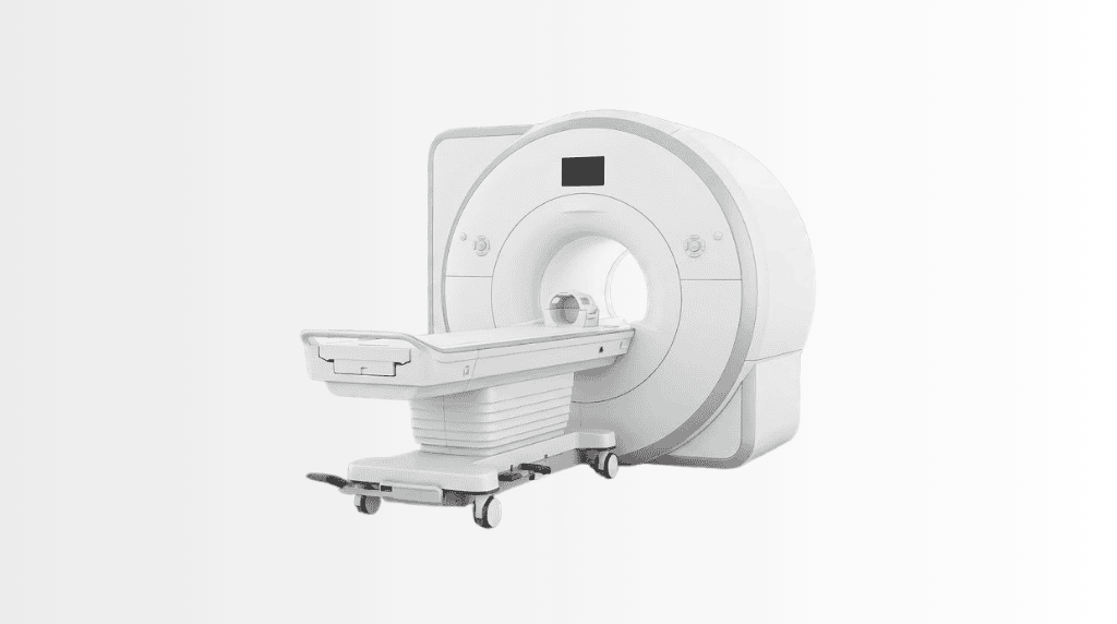
Intraoperative 3 Tesla MRI provides detailed magnetic resonance images during selected brain and spine operations, helping surgeons assess the procedure before surgery is completed. At Acibadem in Turkey, it is integrated into the operating workflow to support real-time surgical decisions when clinically appropriate.
Intraoperative 3 Tesla MRI
Intraoperative 3 Tesla MRI is an advanced imaging system used inside or alongside the operating theatre during selected brain and spine surgeries. It allows the surgical team to obtain highly detailed magnetic resonance images while the procedure is still in progress.
Rather than waiting until after surgery to assess the result, neurosurgeons can review new images during the operation when clinically appropriate. This can help confirm the surgical outcome, assess the treatment area and support informed decisions before the procedure is completed.
A 3 Tesla MRI uses a powerful magnetic field to produce high-resolution images of soft tissues, including the brain, spinal cord, nerves and surrounding structures. MRI does not use ionising radiation. For patients undergoing complex neurosurgical care, this technology can provide an additional level of imaging guidance at a critical stage of treatment.
At Acibadem International, intraoperative 3 Tesla MRI is used by experienced Neurosurgery teams as part of an individual treatment plan. Its use is determined by your diagnosis, the location of the condition, the planned procedure and your overall medical needs.
What Is Intraoperative 3 Tesla MRI?
“Intraoperative” means “during surgery.” Intraoperative 3 Tesla MRI is therefore an MRI scan performed while a patient is undergoing an operation.
Conventional MRI is usually performed before or after surgery in a separate imaging department. During brain and spine procedures, however, the anatomy may change as treatment progresses. Tissue can shift, fluid levels can change and the relationship between the surgical area and nearby important structures may be different from what was seen on earlier scans.
Intraoperative MRI gives the surgical team updated imaging at that moment. The images can help surgeons assess whether the planned target has been treated as intended and whether further surgical action may be beneficial or necessary.
The term “3 Tesla” refers to the strength of the MRI magnet. A 3 Tesla system can provide detailed images that are particularly valuable in evaluating complex soft-tissue anatomy. This high-resolution imaging may support precise surgical planning and intraoperative decision-making in carefully selected cases.
How Intraoperative 3 Tesla MRI Works
MRI creates images using a strong magnetic field and radiofrequency energy. The scanner detects signals from the body’s tissues and converts them into detailed images. Different MRI sequences can show different types of tissue and can help distinguish normal structures from areas affected by a tumour, vascular condition, inflammation, injury or degenerative change.
During a procedure, the patient is positioned using MRI-compatible equipment designed for the operating environment. At the appropriate stage of surgery, the team performs the scan according to a carefully coordinated workflow. The neurosurgeon, anaesthesia team, radiology professionals and operating theatre staff work together to maintain patient safety and continuity of care.
The newly acquired images are reviewed by the surgical team. Depending on the findings, the surgeons may proceed as planned, consider additional treatment within the same operation or confirm that the intended surgical objective has been achieved. The scan is one important source of information; it is always interpreted alongside the patient’s diagnosis, surgical findings and clinical condition.
Why updated imaging can be valuable during surgery
- It can provide a current view of the treatment area after part of the procedure has been completed.
- It may help identify residual abnormal tissue or clarify whether an area requires further assessment.
- It can support surgical navigation when normal anatomy has shifted during the operation.
- It may help the team protect important structures involved in movement, speech, sensation, vision or spinal function.
- It can help confirm treatment results before the patient leaves the operating theatre.
Not every brain or spine operation requires intraoperative MRI. Your specialist will explain whether it may add clinical value in your individual case.
Clinical Applications
Intraoperative 3 Tesla MRI is most often considered for complex neurosurgical procedures where real-time confirmation of anatomy and treatment results may be especially helpful. Its role varies by patient, diagnosis and surgical objective.
| Clinical application | How intraoperative 3 Tesla MRI may support care |
|---|---|
| Brain tumour surgery | May help assess the surgical area during tumour removal and support decisions about the extent of resection when clinically appropriate. |
| Glioma and other infiltrative brain lesions | Can provide updated images when boundaries between abnormal and healthy brain tissue are complex or change during surgery. |
| Skull base and deep brain lesions | May support orientation around delicate structures, including nerves and blood vessels, in selected procedures. |
| Epilepsy surgery | May assist with confirming the targeted treatment area in carefully planned operations for drug-resistant epilepsy. |
| Pituitary and sellar region surgery | Can be used in selected cases to assess the treatment area near important endocrine, visual and neurological structures. |
| Spinal cord and intradural spinal lesions | May help evaluate selected tumours or lesions affecting the spinal cord, nerve roots or tissues within the spinal canal. |
| Complex revision neurosurgery | Can offer updated imaging where prior surgery, scar tissue or altered anatomy makes surgical assessment more challenging. |
The technology is not a replacement for surgical expertise, pathology, neurological monitoring or other imaging methods. It is integrated into a wider treatment strategy designed by the multidisciplinary team.
Conditions It May Help the Team Assess
Depending on the procedure, intraoperative 3 Tesla MRI may be used to support surgery for benign and malignant brain tumours, metastatic lesions, gliomas, meningiomas, pituitary tumours, selected epilepsy-related brain abnormalities and certain spine or spinal cord tumours.
It may also be considered for lesions located close to areas of the brain that control speech, movement, sensation, memory or vision. In these cases, the aim is not simply to remove or treat abnormal tissue. The surgical team also works to preserve neurological function as far as possible.
For spine surgery, its use may be considered for selected conditions involving the spinal cord, intradural tumours, nerve-related lesions and other complex abnormalities. The decision depends on the anatomical location, imaging findings, surgical approach and expected clinical benefit.
Your specialist will review your MRI scans, pathology results where available, neurological symptoms, previous treatments and overall health before recommending the most suitable approach.
Benefits and Advantages for Patients
The key value of intraoperative 3 Tesla MRI is that it brings high-resolution imaging into the surgical workflow. It can give the team more information before the operation is completed, potentially reducing uncertainty in selected complex procedures.
For many patients, particularly those with brain tumours or lesions near important functional areas, the ability to assess the surgical result during the operation may support a more personalised balance between treatment effectiveness and preservation of healthy tissue.
| Patient benefit | What it may mean for your care |
|---|---|
| Detailed imaging during surgery | Your surgical team can review current MRI images rather than relying only on scans obtained before the procedure. |
| Support for precise surgical decisions | Updated images may help guide decisions about the treatment area and next surgical steps where appropriate. |
| Assessment before surgery is completed | The team may be able to evaluate the operative result while you are still in the operating theatre. |
| High-resolution soft-tissue detail | 3 Tesla MRI can provide clear views of the brain, spinal cord and nearby structures in selected cases. |
| No ionising radiation | MRI uses magnetic fields and radiofrequency energy rather than X-rays or CT radiation. |
| Integrated multidisciplinary care | Neurosurgeons, radiologists, anaesthesiologists, nurses and other specialists coordinate the imaging and surgical process. |
Benefits cannot be guaranteed, and the use of intraoperative MRI does not automatically mean that a larger amount of tissue will be removed or that every clinical risk can be eliminated. The safest and most effective surgical plan is always tailored to the individual patient.
What to Expect During a Procedure Using Intraoperative MRI
Before you travel or arrive
International patients can share medical reports, imaging files, pathology results and previous treatment information for specialist review. Our international patient team helps coordinate the process and can guide you on the documents needed for your consultation and treatment planning.
Your case is assessed by the relevant specialist team. If intraoperative 3 Tesla MRI may be appropriate, the neurosurgeon will explain its role in the planned operation, as well as possible alternatives, expected benefits and important limitations.
Before surgery, you may need blood tests, anaesthesia assessment and additional imaging or neurological evaluation. You should tell your medical team about all medicines, allergies, implants, prior surgeries and medical conditions.
On the day of surgery
Most procedures performed with intraoperative MRI take place under general anaesthesia. You will be asleep and will not feel the scan or the surgery. The anaesthesia team monitors your breathing, heart rhythm, blood pressure, oxygen level and other vital signs throughout the procedure.
Because MRI uses a powerful magnet, special safety checks are essential. The team will confirm whether you have any metal implants, medical devices, clips, cochlear implants, pacemakers, medication pumps or retained metal fragments. Many modern implants are MRI-conditional, but each device must be assessed individually.
The operation begins according to the planned surgical approach. At a suitable stage, the surgical team may pause to obtain intraoperative MRI images. The duration and number of scans vary depending on the type of procedure and the clinical questions that need to be answered.
After the scan and surgery
Once images are reviewed, the surgeon decides how to proceed based on the findings and the treatment plan. The operation is then completed, and you are transferred to the recovery area or, when needed, to a specialised intensive care setting for close monitoring.
After brain or spine surgery, your recovery plan may include neurological checks, pain management, physiotherapy, medication adjustment and follow-up imaging. The length of hospital stay depends on the procedure, your condition and your recovery progress.
Your care coordinator can help organise follow-up appointments, medical reports, discharge documentation and continuity of care after you return home. Where needed, information can be shared with your referring doctor, subject to your consent and applicable privacy requirements.
Safety Considerations
Intraoperative 3 Tesla MRI is used under strict MRI and operating theatre safety protocols. Because the technology combines advanced imaging with surgery and anaesthesia, it requires careful planning, specialist training and coordinated teamwork.
Before the procedure, every patient is screened for MRI compatibility. The team also checks all surgical instruments, monitoring equipment and anaesthesia devices to ensure they are suitable for the MRI environment. Only approved MRI-safe or MRI-conditional equipment is used according to established protocols.
As with any surgery, there are risks related to anaesthesia, infection, bleeding, blood clots, neurological changes and the specific procedure being performed. The use of intraoperative MRI does not remove these risks. Its purpose is to provide additional imaging information that may support safer and more informed surgical decisions in appropriate cases.
Your surgeon and anaesthesiologist will discuss the risks that are most relevant to you. Be sure to ask questions about your diagnosis, surgical goals, expected recovery and the reason intraoperative MRI is being considered.
Quality Standards and Accreditation
Acibadem International supports international patients through coordinated, multidisciplinary care in JCI-accredited hospital settings. JCI accreditation reflects a commitment to established quality and patient safety standards, including clinical processes, infection prevention, medication safety, patient identification and continuity of care.
Complex neurosurgical procedures require more than advanced technology. They depend on experienced teams, careful case selection, high-quality imaging, modern operating theatre infrastructure, anaesthesia expertise, critical care capability and structured post-operative follow-up.
Intraoperative 3 Tesla MRI is used as part of this wider clinical pathway. The technology is most valuable when it is applied thoughtfully, interpreted by the right specialists and linked to an individual treatment plan.
Why Intraoperative 3 Tesla MRI Matters for International Patients
Travelling abroad for brain or spine surgery can feel overwhelming, especially when treatment involves complex technology and several specialist appointments. International patients often need clear information before travelling, coordinated scheduling and support throughout their treatment journey.
Our international patient team can help you understand the proposed care pathway, arrange appointments with the relevant specialists and support communication in your preferred language where available. This coordination is particularly important when treatment planning involves review of imaging, surgical consultations, anaesthesia assessment, hospital admission and post-operative follow-up.
Before you travel, you can submit your available reports and imaging for preliminary medical review. This helps the specialist team understand your condition and advise whether further evaluation is needed. Final recommendations are made after a full clinical assessment and may change based on new examinations, imaging findings or test results.
During your stay, the care team works to coordinate clinical appointments and keep you informed about the next steps. After discharge, you can receive medical documentation to support ongoing care with your doctor at home.
Frequently Asked Questions
Is intraoperative 3 Tesla MRI safe?
Intraoperative MRI is used by trained teams who follow detailed MRI and surgical safety protocols. MRI does not use ionising radiation, but the powerful magnetic field means that careful screening for implants, devices and metal exposure is essential. Your team will explain any considerations specific to your condition and medical history.
Will every brain or spine surgery require intraoperative MRI?
No. Intraoperative 3 Tesla MRI is used selectively. It is most helpful in procedures where updated imaging during surgery may influence clinical decisions. Your neurosurgeon will recommend it only if it is considered appropriate for your case.
Does intraoperative MRI guarantee complete tumour removal?
No. The aim of surgery depends on many factors, including the tumour type, its location, its relationship to important brain or spinal structures and your neurological condition. In some cases, preserving function may mean that the safest plan is not to remove all visible tissue. Your surgeon will discuss realistic treatment goals with you.
Will I feel the MRI scan during surgery?
Most patients undergoing brain or spine surgery with intraoperative MRI are under general anaesthesia. You will be asleep during the operation and scan. Your anaesthesia team will monitor you continuously.
Can my insurance cover treatment involving intraoperative MRI?
Coverage depends on your insurance policy, diagnosis, hospital network and authorisation requirements. You may share your insurer details and policy information with the international patient team so that available options can be reviewed before treatment whenever possible.
How can I begin the assessment process?
You can submit your medical reports, imaging studies and previous treatment information for review. A specialist team can assess whether an intraoperative 3 Tesla MRI-supported procedure may be relevant to your treatment plan and advise on the next clinical steps.
Related Medical Units
Available at these Hospitals


















Related Treatments
Speak with our medical team
Find out how this technology could support your diagnosis or treatment.