

Quick answer
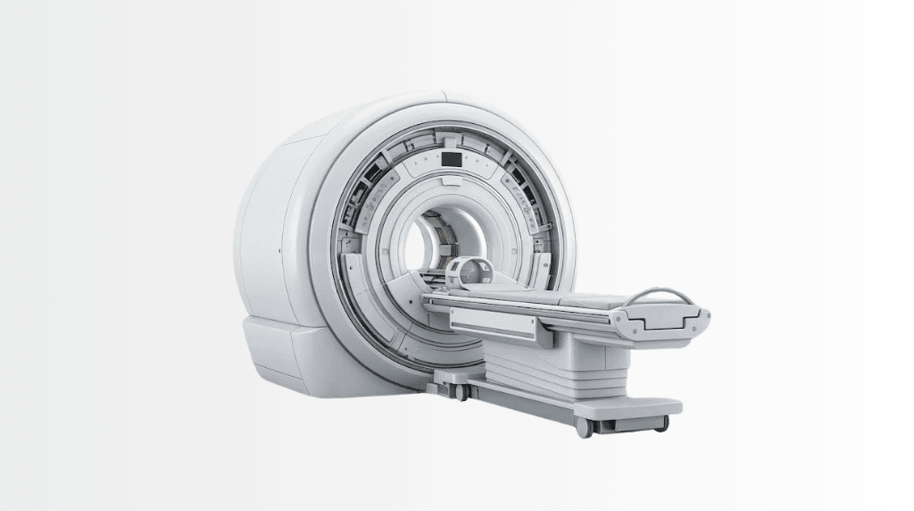
3 Tesla MR is a high-field MRI technology that uses magnetic fields and radio waves to create detailed images without ionizing radiation. At Acibadem in Turkey, it supports diagnosis, treatment planning and follow-up for neurological, musculoskeletal, cardiovascular and body conditions after appropriate MRI safety screening.
A 3T MRI runs at double the magnetic field strength of the standard 1.5 Tesla scanner, and in imaging, field strength is signal, and signal is choice: spend it on finer detail, faster scans, or both. For the brain’s small structures, the prostate’s zones, joints’ cartilage layers and the subtleties functional imaging chases, 3 Tesla is where modern answers live. This page explains what a 3T MRI genuinely does better, where 1.5T remains the right tool, and what the examination is like.
What does 3 Tesla mean?
Tesla measures magnetic field strength; 3T is roughly 60,000 times Earth’s field. A stronger field aligns more hydrogen nuclei in your tissues, producing more signal for the scanner to work with. Radiologists spend that surplus deliberately: sub-millimetre spatial resolution where anatomy is fine, shorter acquisitions where patients struggle to lie still, and the signal-hungry advanced sequences, spectroscopy, functional MRI, high-resolution diffusion, that lower fields cannot feed well. No radiation is involved at any field strength.
Where 3T MRI genuinely matters
Neuroimaging leads the list: small lesions in epilepsy work-ups, early demyelination in multiple sclerosis, pituitary and inner-ear detail, and pre-surgical functional mapping. Prostate MRI is a 3T flagship: the multiparametric protocols behind modern prostate diagnosis and MRI-fusion biopsy read best at high field. Musculoskeletal imaging gains cartilage-level clarity for joints, labrum and small ligaments. Breast MRI, liver protocols and vascular studies also draw on the extra signal. The honest counterpart: for many routine questions a modern 1.5T answers perfectly well, and some situations, certain implants, some abdominal artefact profiles, actually favour 1.5T. The radiologist matches field to question; that is the job.
The examination: what to expect
An MRI visit at any field: metal screening first, bring implant cards; most modern implants are MRI-conditional but each is checked; then 20–60 minutes in the scanner depending on protocol, lying still while sequences run. The machine knocks loudly (hearing protection is standard), the tunnel is snug, and claustrophobia is manageable with preparation, talk to the team beforehand. Contrast (gadolinium) is used only where it adds diagnostic value, screened against kidney function and stated in advance. No radiation, no recovery time.
Why the reading matters as much as the magnet
A 3T image is only as good as its protocol and its reader. High-field imaging demands sequence tuning, artefact control at 3T is a craft, and subspecialty reading turns detail into decisions: the neuroradiologist for the epilepsy protocol, the prostate reader scoring PI-RADS, the musculoskeletal radiologist grading cartilage. Across Acibadem’s radiology practice, protocols are standardised and reports are written into a group file every campus can read, so a scan done once serves every consultation after it.
3T MRI at Acibadem
The group runs 3 Tesla scanners across its network, corporate-verified 3T installations include Ataşehir and Kartal hospitals in Istanbul and Sistina in Skopje, with intraoperative 3T capability serving neurosurgery, alongside a wider high-field fleet. Records-first applies to imaging too: prior scans travel ahead, protocols answer defined questions, and repeat imaging happens only for a stated reason.
The advanced sequences: what extra signal buys
High field’s dividend is paid in sequences ordinary strength cannot feed well. Functional MRI (fMRI) maps which cortex activates when you move a hand or name words, pre-surgical cartography that lets neurosurgeons plan around language and motor territory before opening anything. Diffusion tensor imaging traces the white-matter cables themselves, drawing the tracts a tumour has displaced rather than destroyed. Spectroscopy reads tissue chemistry non-invasively, metabolite signatures that help separate tumour from radiation change. Perfusion mapping grades blood flow through lesions; susceptibility imaging finds microbleeds invisible elsewhere; and high-resolution 3D acquisitions let one scan be re-sliced in any plane at reading time. None of these are exotic add-ons at a 3T-equipped centre: they are the standard vocabulary of modern neuro, prostate and oncological protocols, and the honest reason “an MRI” at high field can answer questions an older scan of the same body part could not.
A practical patient’s guide to the MRI visit
What genuinely helps, in order: bring implant documentation, pacemakers, clips, stimulators, cochlear devices and pumps each have specific conditions, and the safety screen is the one queue worth every minute; arrive with prior imaging or have it sent ahead, since comparison multiplies a report’s value; declare claustrophobia early, position coaching, mirrors, music and, where appropriate, mild sedation or a wider-bore scanner turn most “I can’t” into a completed study; leave metal-threaded clothing and cards outside the room; and for contrast studies, recent kidney values smooth the day. During the scan, stillness is your entire contribution, sequences restart when movement blurs them, so a comfortable position negotiated at the start is time saved twice. Reports at Acibadem land in the group file with the images themselves, which means the next consultation, any campus, any specialty, opens your scan rather than a summary of it.
Frequently Asked Questions
What is functional MRI used for?
Mapping active brain territory, language, movement, before surgery, so operative plans respect the cortex your daily life depends on; a 3T-strength application par excellence.
Can MRI tell tumour from radiation change?
Advanced sequences, spectroscopy and perfusion, add chemical and blood-flow evidence to the picture; the combination, read by neuroradiology, often settles what structure alone cannot.
I am claustrophobic, will I manage the scan?
Usually yes, with preparation, coaching, music, mirrors and where appropriate sedation or a wider-bore system; tell the team when booking, not on the table.
What is the difference between 1.5T and 3T MRI?
Double the field strength means roughly double the signal, spent on finer detail, faster scans or advanced sequences; for many routine questions a modern 1.5T remains perfectly sufficient.
Is a 3T MRI safe?
Yes: MRI uses magnetic fields and radio waves, no radiation; safety turns on metal screening, which is why implants are checked individually.
Can I have a 3T MRI with an implant?
Many modern implants are approved for 3T, some only for 1.5T, bring your implant card and the team matches you to the right scanner.
How long does a 3T MRI take?
Typically 20–60 minutes depending on the protocol; higher field often shortens individual sequences.
Does 3T help with prostate MRI?
Yes, multiparametric prostate protocols and PI-RADS reading are a 3T flagship application, feeding MRI-fusion biopsy decisions.
Is 3T better for brain imaging?
For fine questions, epilepsy work-ups, early MS lesions, pituitary and inner-ear detail, functional mapping, high field is where the answers live.
Will I need contrast?
Only where it adds diagnostic value, stated in advance and screened against kidney function; many 3T studies run without it.
Is the 3T scanner louder or tighter?
Noise is comparable and hearing protection is standard; bore sizes vary by machine rather than field strength, claustrophobia strategies are discussed beforehand.
Why did my doctor order 1.5T instead of 3T?
Because the field is matched to the question, some implants, artefact profiles and routine indications are genuinely better served at 1.5T.
Is 3T MRI safe in pregnancy?
MRI without contrast is used in pregnancy when clinically justified: the decision belongs to your physician and radiologist together.
Do I need to fast before an MRI?
Only for specific abdominal protocols: your appointment instructions say; most MRI needs no preparation beyond metal screening.
Can my previous MRI be compared with a new 3T scan?
Yes, priors are integrated wherever usable, and comparison is often exactly why the higher-field study was requested.
Related Medical Units
Available at these Hospitals


















Related Treatments
Speak with our medical team
Find out how this technology could support your diagnosis or treatment.