Barium Enema Procedure: An Evidence-Based Patient Guide
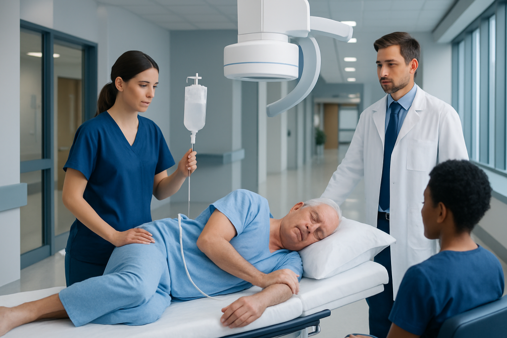
A barium enema uses liquid contrast inserted through the rectum to make the colon visible on X-ray images. Thorough bowel cleansing is essential because stool can obscure images or resemble an abnormality.
Key Takeaways
- A barium enema uses liquid contrast inserted through the rectum to make the colon visible on X-ray images.
- Thorough bowel cleansing is essential because stool can obscure images or resemble an abnormality.
- Most people experience pressure, fullness, and cramping rather than severe pain; the test does not usually require sedation.
- Colonoscopy is often preferred when tissue samples or polyp removal may be needed, but the best test depends on the clinical question.
- Brief constipation and pale stools can occur after the examination; urgent symptoms after the test should be assessed promptly.
A barium enema procedure is a diagnostic imaging test that uses barium contrast and X-rays to outline the large intestine (colon). It is now used less often than colonoscopy or CT colonography, but may still be appropriate in selected situations when a clinician needs information about the bowel’s shape or structure.
Overview: what is a barium enema procedure?
A barium enema procedure is an imaging examination of the large bowel, also called the colon. During the test, a radiology professional gently introduces barium sulfate contrast through a small tube placed in the rectum. Barium coats the lining of the bowel and makes its outline easier to see on X-ray images.
The examination may be performed as a single-contrast study, using barium alone, or as a double-contrast study, where air is also introduced to expand the colon and provide more detailed views of the lining. The test can help identify changes in the shape, width, or surface of the colon, such as narrowing, large polyps, diverticular disease, or signs of inflammation.
Modern practice more commonly uses colonoscopy or CT colonography for many bowel concerns. However, a barium enema may still be considered if another examination is incomplete, unavailable, unsuitable, or if the medical team needs a particular structural view of the colon. The radiologist and referring clinician choose the most suitable examination based on the person’s symptoms, medical history, and prior test results.
Who may need this examination?
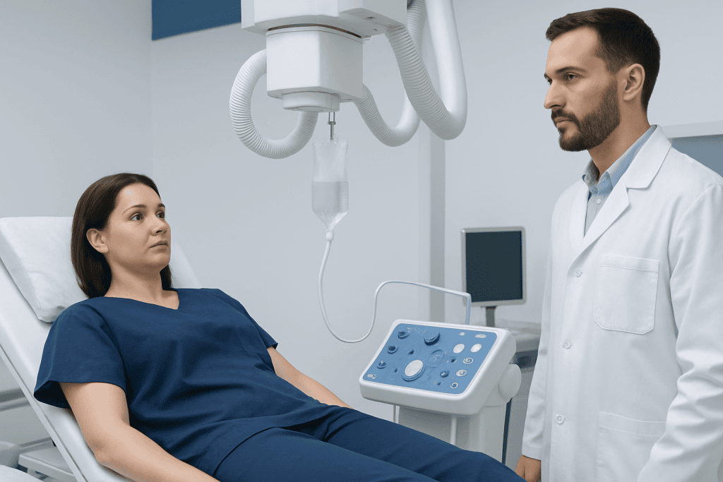
A clinician may request a barium enema when symptoms or prior findings suggest that the large bowel needs further assessment. These can include a persistent change in bowel habits, unexplained constipation, abdominal discomfort, bleeding from the rectum, or an abnormality seen on another imaging test. The procedure is diagnostic: it helps guide next steps but does not itself treat the cause.
It may be particularly helpful when a complete colonoscopy has not been possible because of a narrowed segment of bowel, difficult anatomy, or inadequate passage of the scope. If the images show a concerning finding, a person may still need colonoscopy, biopsy, or another investigation to confirm the diagnosis and, where necessary, provide treatment.
Before scheduling the test, the care team should know about pregnancy or possible pregnancy, severe abdominal pain, recent bowel surgery, inflammatory bowel disease, bowel obstruction, known perforation, and allergies or prior reactions to contrast materials. A water-soluble contrast study rather than barium may be selected in certain situations, especially when a leak or perforation is a concern.
People with known or suspected bowel conditions may require tailored assessment. For example, symptoms associated with diverticulitis or inflammatory disease should be evaluated by a clinician before any bowel imaging is arranged.
How should I prepare my bowel for a barium enema?

Barium enema patient preparation is important because the colon needs to be as empty as possible. Residual stool can hide a finding or create shadows that are difficult to distinguish from a polyp or other bowel change. The imaging centre will provide specific instructions, and these should take priority because preparation schedules can vary.
Common barium enema procedure preparation includes following a low-residue or clear-liquid diet for a defined period before the examination, taking a prescribed bowel-cleansing laxative, and sometimes using a rectal preparation. People are generally asked to drink approved clear fluids to reduce the chance of dehydration, unless they have been given fluid restrictions for a heart, kidney, or other medical condition.
Patients should tell the team about diabetes, kidney disease, heart failure, swallowing difficulties, constipation, and all regular medicines. Some medicines may need individual timing advice around the bowel preparation, particularly diabetes medicines, iron supplements, blood thinners, and medicines that affect fluid balance. A person should not stop prescribed medication unless the clinician who manages it advises this.
For useful barium enema patient education, it helps to arrange transport if the preparation causes urgency or weakness, wear comfortable clothing, and bring a list of medicines and relevant health information. Patients should contact the imaging department in advance if they cannot complete the preparation or develop significant vomiting, dizziness, or worsening abdominal pain.
Barium enema procedure step by step
On arrival, the patient is usually asked to change into a gown and remove metal items that could affect the X-ray images. A radiographer or radiology nurse reviews the preparation, asks safety questions, and explains what will happen. An initial X-ray may be taken to check whether the bowel appears adequately clean.
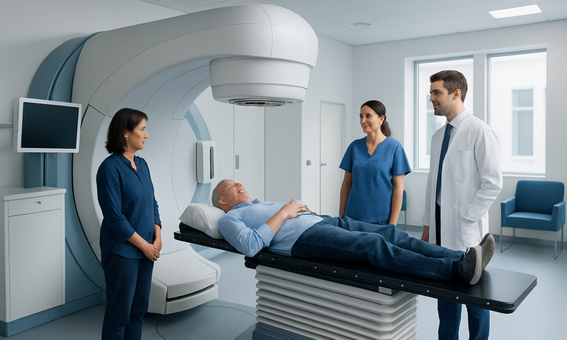
The patient lies on an X-ray table, often first on one side. A lubricated, narrow tube is carefully placed a short distance into the rectum. Barium flows through the tube into the colon, and the radiologist watches the contrast move through the bowel using fluoroscopy, a type of real-time X-ray. In a double-contrast examination, air may then be introduced to gently expand the colon.
During this barium enema procedure step by step, the patient may be asked to turn onto the back, stomach, or sides, or to hold a position briefly. These movements help coat different portions of the colon and allow images from several angles. Clear communication is encouraged; the patient should say if discomfort becomes difficult to manage.
The tube is removed once the required images have been obtained, and the patient is usually allowed to use the toilet to pass the contrast. A final image may be taken afterward. The examination often takes around 30 to 60 minutes, although timing varies. Sedation is not usually needed, so people are generally awake and able to return home after the test.
How painful is a barium enema?
A barium enema is commonly uncomfortable but should not be severely painful. As the colon fills with barium and, in some cases, air, many people feel fullness, pressure, an urge to have a bowel movement, or temporary cramping. The sensations usually ease after the contrast is passed.
Slow breathing, relaxing the abdominal muscles, and telling the radiology team promptly about discomfort can help. The staff can explain what sensations are expected and may adjust positioning or the flow of contrast where appropriate. The procedure can be paused or stopped if there is significant pain or another safety concern.
Severe, sharp, or worsening pain is not something a patient should ignore during the test. It can indicate that the examination needs reassessment. People who are particularly worried about discomfort should discuss this before the appointment; the team can explain whether a different diagnostic approach may be more suitable.
Is it better to do a colonoscopy or a barium enema first?
For many people with bowel symptoms or a need for colorectal cancer assessment, colonoscopy is often the first-choice test because it allows the clinician to see the bowel lining directly. During the same procedure, a specialist can take a biopsy or remove many types of polyps. A barium enema cannot take tissue samples or remove abnormalities.
That said, the order is not the same for everyone. A barium enema may be considered when colonoscopy cannot be completed, when it is not medically suitable, or when a clinician needs additional information about the overall contour of the bowel. CT colonography is another imaging option that may be discussed in appropriate circumstances.
The decision should reflect the person’s symptoms, age, prior investigations, bowel preparation tolerance, risk factors, and whether an intervention is likely to be needed. If a barium enema identifies a possible polyp, narrowing, or other suspicious change, colonoscopy is commonly recommended afterward to clarify the finding and obtain tissue if needed.
Patients with persistent bowel symptoms can discuss colonoscopy assessment with a gastroenterology specialist. The most appropriate sequence of tests should be determined by a qualified clinician rather than chosen solely on the basis of convenience or discomfort.
Is barium enema better than colonoscopy?
Barium enema is not generally considered better than colonoscopy overall; the two examinations provide different types of information. Colonoscopy directly examines the lining of the colon and can enable biopsies and removal of polyps, making it especially valuable when cancer, polyps, inflammation, or unexplained bleeding need investigation.
A barium enema can offer a broader X-ray outline of the colon and may be useful when colonoscopy is incomplete or unsuitable. It is typically performed without sedation and does not involve passing a scope through the entire colon. However, it is less able to detect some small or flat lesions and cannot provide a tissue diagnosis.
Both tests require bowel preparation, and both have limitations and potential complications. The best option depends on why the examination is being considered. A clinician may also recommend CT colonography in selected patients, particularly when a non-endoscopic view of the bowel is appropriate.
Procedure videos can make unfamiliar steps easier to visualise, but a barium enema procedure video cannot replace personalised instructions. Patients should rely on guidance from their radiology and gastroenterology teams for preparation, safety, and follow-up.
Recovery, benefits, risks, and when to seek medical care
Most people can resume usual light activities shortly after a barium enema. Drinking extra fluids, if medically appropriate, can help the body clear the barium. Stools may look white, pale, or chalky for a day or two, and constipation can occur. The care team may recommend a laxative if needed, but patients should follow the instructions provided rather than self-treating aggressively.
The benefit of the test is that it can provide useful information about the colon without sedation or endoscopy. Risks are uncommon but include constipation or impaction from retained barium, leakage of contrast outside the bowel, and, very rarely, a tear in the bowel wall. Radiation is used, but the imaging team keeps exposure as low as reasonably achievable while obtaining clinically useful images.
When to seek medical care: Patients should contact a clinician urgently after the examination if they develop severe or increasing abdominal pain, marked abdominal swelling, fever, vomiting, inability to pass stool or gas, heavy rectal bleeding, fainting, or symptoms that are concerning to them. They should also seek advice if they cannot pass the barium or have persistent constipation despite following post-test instructions.
Results are interpreted by a radiologist and sent to the referring clinician, who explains what they mean and whether more testing is needed. Acibadem International’s multidisciplinary specialists and JCI-accredited hospitals can assess bowel concerns and coordinate diagnostic care for international patients when appropriate.
Frequently asked questions
What does a barium enema show?
A barium enema outlines the large intestine on X-ray images. It can help show narrowing, changes in bowel shape, diverticula, larger polyps, or other structural abnormalities. It cannot confirm a diagnosis that requires a tissue sample.
How long does a barium enema procedure take?
The imaging portion commonly takes about 30 to 60 minutes, although this varies according to the type of study and individual anatomy. Patients should allow additional time for check-in, preparation, and post-procedure instructions.
Can I eat after a barium enema?
Many people can return to their usual diet after the examination unless the care team gives different instructions. Drinking fluids is often encouraged to help pass the barium, provided there is no medical reason to limit fluids. The imaging centre’s discharge advice should be followed.
Why are barium enemas used less often now?
Colonoscopy can directly inspect the bowel, remove many polyps, and obtain biopsies, while CT colonography can provide detailed cross-sectional imaging in some settings. These alternatives have reduced the role of barium enema, but it can still be useful in selected clinical circumstances.
Will I be asleep for a barium enema?
Sedation is not usually required because the examination does not involve passing an endoscope through the colon. The patient remains awake and can communicate with the radiology team throughout the test. Temporary pressure or cramping may occur while the bowel is filled.
What happens if the barium enema result is abnormal?
The referring clinician will review the radiology report in the context of the patient’s symptoms and medical history. Further testing, often colonoscopy or another imaging study, may be recommended to clarify the finding. An abnormal image does not by itself establish a final diagnosis.
References
- American College of Radiology
- Radiological Society of North America
- National Institute of Diabetes and Digestive and Kidney Diseases
- National Health Service
- Mayo Clinic
This article is for general information only and is not a substitute for professional medical advice. Please consult a qualified doctor about your individual situation.
Persistent digestive symptoms? Get evaluated in Turkey
JCI-accredited · board-certified surgeons · reply within 24h