Meningioma MRI: Preparation, Procedure and Results
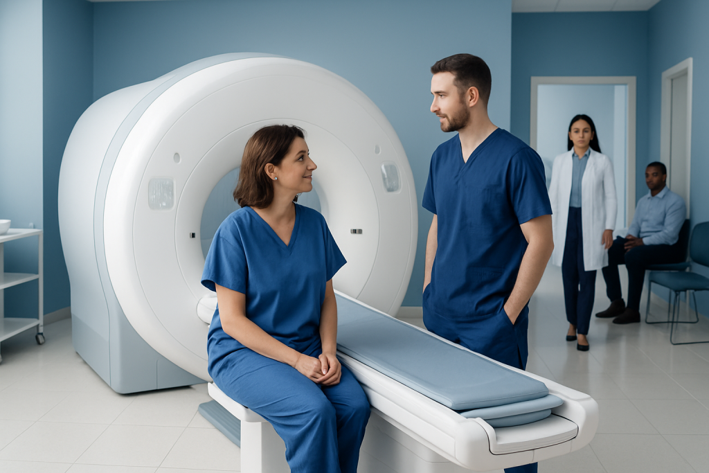
MRI is the preferred imaging examination for suspected or known meningioma because it provides detailed views of the brain, tumor and surrounding structures. Most meningiomas are slow-growing and noncancerous, but their impact depends mainly on size, location, growth rate and effects on nearby tissue.
Key Takeaways
- MRI is the preferred imaging examination for suspected or known meningioma because it provides detailed views of the brain, tumor and surrounding structures.
- Most meningiomas are slow-growing and noncancerous, but their impact depends mainly on size, location, growth rate and effects on nearby tissue.
- A contrast-enhanced MRI can help clinicians distinguish a meningioma from other conditions and plan surgery, radiation therapy or observation.
- Preparation is usually simple, but patients should report implants, kidney problems, pregnancy, allergies and anxiety about enclosed spaces before the scan.
- Follow-up MRI schedules are individualized and may be used after diagnosis, during active surveillance, or after treatment.
A meningioma MRI is the main imaging test used to detect a suspected meningioma, define its location and size, and guide monitoring or treatment planning. It is painless and does not use ionizing radiation, although some scans involve an intravenous contrast agent for clearer detail.
Overview: What Is a Meningioma MRI?
A meningioma MRI is a magnetic resonance imaging scan of the brain performed to look for, assess or monitor a meningioma. It uses a strong magnetic field and radio waves to create detailed images of the brain and its coverings. MRI does not use X-rays or other ionizing radiation, and it is usually the most informative scan when a meningioma is suspected.
Many examinations include a gadolinium-based contrast agent given through a small intravenous line. Contrast can make the borders, blood supply and relationship of a suspected tumor to nearby brain tissue, nerves and blood vessels easier to see. The scan can help a specialist decide whether observation is appropriate or whether further evaluation and treatment should be considered.
A meningioma begins in the meninges, the protective layers surrounding the brain and spinal cord. Although many are benign and grow slowly, even a noncancerous tumor may need attention if it presses on important structures. MRI findings must always be interpreted alongside a person’s symptoms, medical history and examination.
Is a Meningioma a Big Deal?
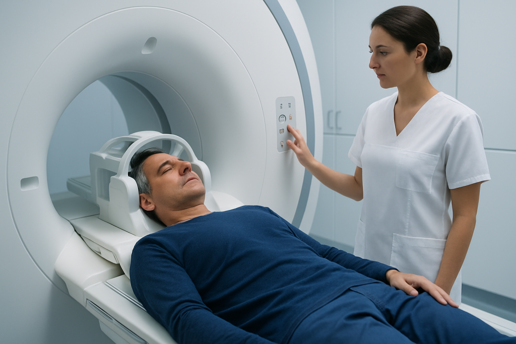
A meningioma can be important, but it is not automatically an emergency or a sign of cancer. Many meningiomas are found incidentally during imaging for an unrelated concern and remain stable for years. Others can gradually cause symptoms because they occupy space inside the skull or affect nearby nerves, blood vessels or brain regions.
The clinical significance depends more on location, size, growth pattern and symptoms than on the diagnosis alone. For example, a small tumor near the optic nerves, brainstem or major blood vessels may require closer assessment than a larger tumor in a less sensitive area. MRI helps show these relationships in detail.
Care may range from periodic imaging to surgery or focused radiation treatment. A neurologist, neurosurgeon, neuroradiologist and radiation oncology team may work together when treatment planning is needed. A clear diagnosis and regular follow-up can help people make informed, timely decisions without assuming the worst.
How a Meningioma MRI Works and Who May Need One
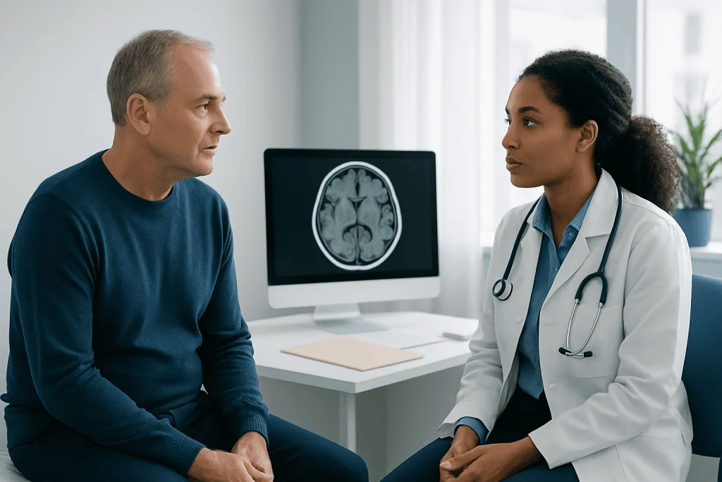
During MRI, the body is placed inside a scanner containing a powerful magnet. Radiofrequency signals interact with water molecules in tissues, and a computer converts the resulting signals into detailed cross-sectional images. Different image sequences highlight different tissue characteristics, including swelling, fluid, blood products, calcification patterns and enhancement after contrast.
A meningioma MRI protocol commonly includes several views and sequences before and after contrast when contrast is appropriate. The exact meningioma brain MRI protocol varies with the clinical question. For example, imaging near the skull base, optic nerves, spine or major venous channels may require additional high-resolution sequences or specialized vascular imaging.
Clinicians may request MRI for persistent or unexplained neurological symptoms, a finding on a CT scan, evaluation before treatment, or routine surveillance of a known tumor. It may also be used after surgery or radiation therapy to establish a baseline and check for residual tumor, recurrence or treatment-related change.
Most people can have MRI safely, but screening is essential. People should tell the imaging team about pacemakers, neurostimulators, cochlear implants, aneurysm clips, metal fragments, joint replacements, insulin pumps, medication patches, pregnancy or impaired kidney function. Some devices are MRI-compatible under specific conditions, while others require an alternative imaging approach.
What to Avoid Before a Brain MRI?
Most people can eat, drink and take regular medicines before a brain MRI unless the imaging center provides different instructions. If sedation is planned for severe claustrophobia or difficulty remaining still, fasting instructions may be necessary. It is best to confirm practical details in advance, particularly when contrast or sedation may be used.
Before entering the scan room, patients should avoid bringing metal or electronic items into the MRI environment. This includes jewelry, watches, hearing aids, hairpins, removable dental appliances, phones, keys, credit cards and some cosmetics that may contain metallic particles. The staff will provide a safety checklist and secure storage for belongings.
Patients should not stop prescribed medicines on their own. However, they should inform the team about all medicines, supplements and allergies, as well as prior reactions to contrast agents. People with kidney disease may need a kidney function review before receiving gadolinium contrast, and pregnant patients should discuss whether MRI and contrast are necessary at that time.
For comfort, it can help to wear loose clothing without metal fasteners and to request ear protection, as MRI scanners make loud tapping sounds. Anyone who feels anxious in enclosed spaces should mention this before the appointment. Options may include extra coaching, a support person where permitted, relaxation techniques or clinician-directed medication.
Step by Step: What Happens During the Procedure?
On arrival, an MRI technologist reviews safety questions and explains the examination. Patients change into an MRI-safe gown if needed and remove all metal-containing belongings. If contrast is planned, a small cannula is placed into a vein, usually in the arm or hand.
For a brain scan, the patient lies on a padded table with the head positioned in a specialized coil that receives the MRI signals. Gentle supports may be used to help keep the head still. The table then moves into the scanner; the patient can communicate with the technologist through an intercom and is usually given an alarm device to use if assistance is needed.
The scan is performed in a series of image sets, each lasting several minutes. It is important to remain as still as possible because movement can blur the images. If contrast is used, it is injected partway through the examination, followed by additional images. The full appointment commonly takes around 30 to 60 minutes, though complex studies may take longer.
Once imaging is complete, the technologist removes the intravenous line if one was used. A radiologist reviews the images and prepares a report for the referring clinician. Urgent findings are communicated promptly through the appropriate clinical process.
What Are the Typical MRI Findings for Meningioma?
On MRI, a typical meningioma appears as a well-defined mass attached to the dura, the outer protective membrane around the brain. Many are located outside the brain tissue itself and may have a broad area of contact with the dura. After gadolinium contrast, meningiomas often show strong, relatively uniform enhancement, meaning they become brighter on post-contrast images.
Radiologists also assess features such as tumor size, shape, internal texture, calcification, fluid-related swelling in adjacent brain tissue, pressure on nearby structures and involvement of bone or venous channels. A “dural tail,” or enhancement of dura next to the mass, can support the diagnosis but is not unique to meningioma.
MRI can suggest a meningioma but cannot always determine its exact grade or predict behavior with certainty. In some situations, other tumors or inflammatory conditions can look similar. If surgery is performed, laboratory examination of tissue provides the definitive diagnosis and tumor grade.
Results should be reviewed with the clinician who ordered the scan. They can explain whether the imaging supports observation, whether another scan or specialist review is needed, and how the findings relate to symptoms. A report describing a lesion does not by itself determine the treatment plan.
What Is the Most Common Location for Meningioma?
Meningiomas can develop anywhere there are meninges, but they most commonly occur inside the skull along the brain’s outer coverings. Frequent sites include the convexity, which is the surface beneath the skull, and the parasagittal region near the large midline venous channel called the superior sagittal sinus.
They can also arise at the skull base, near the sphenoid bone, around the olfactory groove, near the optic pathways, or in the region behind the brain known as the posterior fossa. Less commonly, meningiomas occur around the spinal cord. The location is a major factor in the symptoms a person may experience and in the safest treatment approach.
For instance, a tumor near the visual pathways may affect vision, while one near areas controlling movement or language may cause different concerns. Location also influences whether a tumor can be completely removed surgically, monitored safely, or treated with precisely targeted radiation. MRI is especially valuable for mapping these anatomical relationships.
Results, Follow-Up, Benefits and Risks
Results may be available within a few days, depending on the imaging center and whether urgent review is needed. The report usually describes the location, measurements, imaging characteristics and effects on nearby structures. For a known meningioma, comparing the new MRI with prior scans is often the most useful way to identify whether the tumor is stable, shrinking or growing.
A meningioma MRI follow up plan is individualized. A small tumor without symptoms may be monitored with scans at intervals chosen by the treating team, while follow-up may be more frequent after surgery, radiation therapy or evidence of growth. The goal is to balance close observation with avoiding unnecessary testing.
The main benefits of MRI are detailed soft-tissue imaging, no ionizing radiation and the ability to evaluate a tumor over time. Risks are generally low, but the scanner can be uncomfortable for people with claustrophobia, loud noise may be bothersome, and remaining still can be challenging. Gadolinium contrast is usually well tolerated, though allergic-like reactions are possible and rare serious complications require appropriate screening.
When a meningioma needs treatment, options may include observation, neurosurgery, stereotactic radiosurgery or other radiation approaches depending on the individual situation. Acibadem International’s multidisciplinary specialists at JCI-accredited hospitals evaluate and treat meningioma for international patients, coordinating imaging findings with neurosurgical and radiation treatment planning.
When to Seek Medical Care
Anyone with new or persistent neurological symptoms should speak with a qualified doctor, particularly if symptoms are worsening or affecting daily activities. Symptoms that can warrant assessment include ongoing headaches that change in pattern, seizures, new weakness or numbness, balance problems, changes in vision or hearing, confusion, memory concerns, or personality changes. These symptoms can have many causes, and evaluation helps identify the appropriate next step.
Emergency medical care is important for sudden severe headache, a first seizure, sudden weakness on one side of the body, loss of consciousness, new difficulty speaking, or abrupt vision loss. These symptoms may be caused by conditions other than meningioma and need urgent assessment.
People with a diagnosed meningioma should attend planned appointments and report new symptoms rather than waiting for the next scheduled MRI. Their care team can decide whether an earlier examination, additional testing or a change in management is appropriate.
Frequently asked questions
Does an MRI always need contrast to diagnose a meningioma?
Contrast-enhanced MRI is often preferred because many meningiomas enhance clearly and their boundaries can be easier to define. However, non-contrast MRI can still provide useful information, especially when contrast is not appropriate. The radiologist and referring clinician choose the protocol based on the clinical question and the person’s health history.
How long does a meningioma MRI take?
A routine brain MRI commonly takes about 30 to 60 minutes. The timing can vary depending on whether contrast is used, whether specialized sequences are needed, and how easily the patient can remain still. Additional preparation and safety screening add time to the appointment.
Can a meningioma be missed on MRI?
MRI is highly sensitive for many meningiomas, particularly when contrast is used, but very small lesions or tumors in complex anatomical areas can be more difficult to assess. Image quality, scan technique and comparison with prior imaging can also affect interpretation. If concerns remain, a clinician may recommend repeat imaging, a specialized MRI protocol or another test.
What happens if MRI shows a meningioma?
The next step depends on the tumor’s size, location, appearance, growth and whether it is causing symptoms. Some people need only scheduled MRI monitoring, while others benefit from review by a neurosurgeon or radiation oncology team. Treatment decisions are individualized and do not depend on imaging alone.
Can I drive myself home after a brain MRI?
Most people can drive and return to usual activities immediately after a standard MRI, including one with contrast. If sedation or anxiety medication was used, driving is not safe and an adult should arrange transport home. The imaging center will provide specific instructions based on the medicines used.
How often is MRI repeated for a meningioma?
There is no single schedule that suits everyone. Follow-up timing depends on whether the tumor is newly found, stable, growing, treated or causing symptoms, as well as its location and imaging features. The treating specialist will recommend an interval that reflects the individual risk and care plan.
References
- National Institute of Neurological Disorders and Stroke
- American Brain Tumor Association
- Radiological Society of North America
- European Association of Neuro-Oncology
- National Cancer Institute
This article is for general information only and is not a substitute for professional medical advice. Please consult a qualified doctor about your individual situation.
Joint and spine care in Turkey — expert assessment & treatment
JCI-accredited · board-certified surgeons · reply within 24h
Add us as a Preferred Source to see more of our trusted health content across Google Search, AI Overviews and Discover.

More from the Health Library



Related Specialists

Gizem Gürzoğlu
Physical Medicine & Rehabilitation
Prof. Dr. Hakan Turan Çift
Orthopedic Surgery & Traumatology
Dr. Murat Yildirim
Physical Medicine & Rehabilitation
Dr. Leyla Şen
Radiation Oncology