

Phyllodes Tumors of the Breast
Learn about phyllodes tumors of the breast, including common symptoms, possible causes, how doctors confirm the diagnosis, and typical treatment options.
Quick answer
Phyllodes tumors of the breast are rare growths arising from the breast's connective tissue. Most are benign, some are borderline, and a minority are malignant. They usually appear as a firm, fast-growing lump in women in their 40s and 50s. Diagnosis relies on imaging and biopsy, and treatment is surgical removal with clear margins, since all types can recur.
What is phyllodes tumors of the breast?
Phyllodes tumors of the breast are rare growths that develop in the connective tissue (also called stromal tissue) of the breast, rather than in the milk ducts or glands where most common breast cancers begin. The name comes from a Greek word meaning “leaf-like,” because under a microscope the tumor cells often grow in a pattern that looks like leaves. Doctors sometimes call them phylloides tumors or cystosarcoma phyllodes, although the older term “cystosarcoma” is misleading because most of these tumors are not cancerous.
Phyllodes tumors are grouped into three types based on how the cells look under the microscope:
- Benign phyllodes tumors are not cancerous and make up the majority of cases.
- Borderline phyllodes tumors have some features that are more concerning but are not clearly cancerous.
- Malignant phyllodes tumors are cancerous and have the potential to spread to other parts of the body, although this is uncommon.
All three types share one important trait: they can grow back in the same area after surgery, which is called local recurrence. This is why careful surgical removal and follow-up matter even when the tumor is benign.
Phyllodes tumors account for a very small share of all breast tumors. They occur almost exclusively in women and are most often found in women in their 40s and early 50s, which is on average about a decade later than fibroadenomas, a much more common type of benign breast lump. They can, however, occur at any adult age and, rarely, in adolescents. Men are affected only in exceptional cases. In many hospital systems, including the Breast Health Department at Acibadem, these tumors are managed by a team that includes breast surgeons, radiologists, and pathologists.
Phyllodes tumors of the breast symptoms
The most common phyllodes tumors of the breast symptoms relate to a lump that can be felt. Many people discover the lump themselves, or it may be noticed during a routine breast examination or on imaging done for another reason. Common features include:
- A firm, smooth, or slightly bumpy lump in the breast that can usually be moved under the skin
- A lump that grows noticeably over weeks or months, sometimes quite quickly
- A lump that is usually painless, although some people report tenderness or a feeling of pressure
- Visible enlargement or a change in the shape of one breast when the tumor is large
- Skin over the lump that looks stretched, shiny, or thin
- Enlarged veins visible under the skin over the lump
- In rare cases, skin breakdown or an open sore (ulceration) over a very large tumor
Rapid growth is one of the features that often distinguishes a phyllodes tumor from a fibroadenoma. A fibroadenoma tends to stay the same size or grow slowly, while a phyllodes tumor may change size over a short period. That said, size and growth alone cannot tell the two apart, and many phyllodes tumors are first suspected to be fibroadenomas.
Symptoms do not reliably reveal whether a phyllodes tumor is benign, borderline, or malignant. Malignant tumors are more likely to be large at diagnosis, but a large lump can also be benign. Only examination of tissue under a microscope can determine the type. Nipple discharge and swelling of lymph nodes under the arm are uncommon with phyllodes tumors, because these tumors rarely spread through the lymphatic system.
Causes and risk factors
The exact phyllodes tumors of the breast causes are not known. Researchers believe the tumor begins when cells in the stromal (connective) tissue of the breast acquire genetic changes that allow them to grow and divide in an uncontrolled way. Some phyllodes tumors are thought to develop within an existing fibroadenoma, while others appear to arise on their own.
Several factors are associated with a higher chance of developing a phyllodes tumor, although most people with these factors never develop one:
- Age: most cases are diagnosed in women in their 40s and 50s.
- Li-Fraumeni syndrome: a rare inherited condition caused by a change in the TP53 gene, which raises the risk of several types of tumors, including phyllodes tumors.
- History of a fibroadenoma: a small number of phyllodes tumors are thought to develop from or alongside a fibroadenoma.
- Female sex: the tumor is extremely rare in men.
Unlike many common breast cancers, phyllodes tumors are not clearly linked to hormone use, reproductive history, breastfeeding, diet, or lifestyle. Having a phyllodes tumor does not appear to be caused by anything a person did or failed to do. Because the causes are not well understood, there is no proven way to prevent these tumors.
Diagnosis
Phyllodes tumors of the breast diagnosis can be challenging because these tumors often look like fibroadenomas on imaging and even on small biopsy samples. Doctors usually follow a stepwise approach:
- Clinical breast examination: the doctor feels the lump to assess its size, shape, texture, and whether it moves freely.
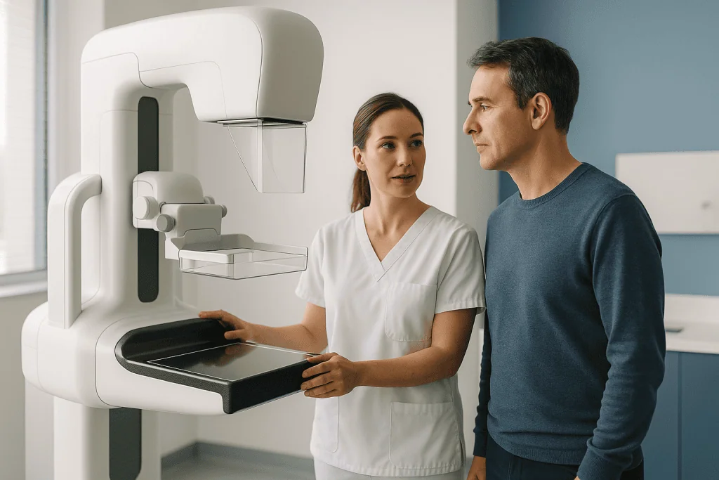
- Mammogram: an X-ray of the breast. A phyllodes tumor often appears as a well-defined, round or lobed mass, which is similar to how a fibroadenoma looks.
- Breast ultrasound: uses sound waves to create images. It may show a solid mass, sometimes with small fluid-filled spaces (cysts or clefts) inside, which can raise suspicion for a phyllodes tumor.
- Breast MRI (magnetic resonance imaging): not always needed, but may be used to assess the size of a large tumor or to plan surgery.
- Core needle biopsy: a hollow needle removes small cylinders of tissue for examination by a pathologist, a doctor who studies tissue under a microscope. This is the main way a phyllodes tumor is first identified, although small samples can miss the diagnosis.
- Excisional biopsy or surgical removal: removing the entire lump so the pathologist can examine the whole tumor. In many cases this is the only way to confirm the diagnosis and to classify the tumor as benign, borderline, or malignant.
When the pathologist examines the tissue, they look at several features to grade the tumor. These include how densely packed the stromal cells are, how many cells are dividing (called mitotic activity), whether the cells look abnormal (called atypia), whether the tumor edge is smooth or pushes irregularly into surrounding tissue, and whether there is stromal overgrowth, meaning connective tissue that crowds out the glandular parts. The combination of these features determines the type.
Because imaging and needle biopsy cannot always distinguish a phyllodes tumor from a fibroadenoma, doctors may recommend removing a lump that is growing quickly, is large, or has uncertain biopsy results, even when it is expected to be benign. If a malignant phyllodes tumor is found, additional imaging such as a chest CT scan may be considered to check whether it has spread, since the lungs are the most common site if spread does occur.
Treatment options
Phyllodes tumors of the breast treatment centers on surgery. Because even benign tumors can come back, and because the type cannot be fully known until the whole tumor is examined, most phyllodes tumors are removed rather than watched.
- Wide local excision (lumpectomy): the surgeon removes the tumor together with a rim of normal breast tissue around it, known as a margin. The aim is a clear margin, meaning no tumor cells at the edge of the removed tissue, which lowers the chance of local recurrence. This is the most common approach and preserves most of the breast.
- Re-excision: if the pathology report shows tumor cells close to or at the edge of the removed tissue, a second operation may be recommended to remove more tissue. This is common when a phyllodes tumor was initially thought to be a fibroadenoma and removed without a margin.
- Mastectomy: removal of the entire breast. This may be recommended when the tumor is very large relative to the breast, when clear margins cannot be achieved with a smaller operation, or in some cases of recurrent or malignant tumors. Breast reconstruction can be discussed as part of planning.
- Lymph node surgery: usually not needed. Phyllodes tumors rarely spread to lymph nodes, so removal of lymph nodes under the arm is generally not part of treatment unless the nodes appear abnormal.
- Radiation therapy: uses high-energy beams to destroy remaining tumor cells. It is not used for benign tumors. For borderline and malignant tumors, some doctors may recommend radiation after surgery, particularly for large tumors, recurrent tumors, or when margins are narrow, in an effort to reduce the chance of local recurrence. Its role continues to be studied.
- Chemotherapy: medicines that kill fast-growing cells. It is not used for benign or borderline tumors. For malignant phyllodes tumors that have spread to distant parts of the body, chemotherapy of the type used for sarcomas (cancers of connective tissue) may be considered, although responses vary and evidence is limited.
- Hormone therapy: medicines that block estrogen are a standard part of treatment for many common breast cancers, but they are not effective for phyllodes tumors and are not recommended.
- Observation: watchful waiting is rarely advised once a phyllodes tumor is suspected, because the diagnosis and type usually cannot be confirmed without removing it. In selected situations, such as a very small lesion in a person who cannot safely have surgery, a doctor may discuss close monitoring.
Recovery after wide local excision is usually similar to recovery after any lumpectomy, with soreness and bruising that improve over one to a few weeks. Recovery after mastectomy takes longer. Your surgical team will explain wound care, activity limits, and when to resume normal routines.
Living with phyllodes tumors of the breast and outlook
The outlook for most people with a phyllodes tumor is good. Benign tumors, which make up the majority, do not spread to other parts of the body, and complete removal with clear margins is often the only treatment needed. Borderline tumors also rarely spread, although they carry a somewhat higher chance of coming back locally. Malignant phyllodes tumors have the potential to spread, most often to the lungs or bones, but this happens in a minority of cases, and many people with malignant tumors are treated successfully with surgery alone.
The main long-term concern for all three types is local recurrence, meaning the tumor grows back in the same breast, usually within the first few years after surgery. The risk is lowest for benign tumors removed with wide margins and highest for malignant tumors or tumors removed with narrow or involved margins. When a tumor recurs, it is usually treated with another operation. Occasionally a recurrent tumor is a higher grade than the original, which is another reason regular follow-up is recommended.
Follow-up typically includes clinical breast examinations and imaging at intervals set by your doctor, more frequently in the first few years. Between visits, it is reasonable to become familiar with how your breast looks and feels after surgery so that any new change is easier to notice. Having had a phyllodes tumor is not thought to substantially raise the risk of developing common breast cancer, but routine breast screening appropriate for your age continues to apply.
Emotionally, being told you have a rare tumor can be unsettling, especially when doctors cannot say with certainty what type it is until after surgery. Many people find it helpful to ask their care team to explain the pathology report in plain language, to clarify what the margin status means, and to outline the follow-up plan. Support from family, counselors, or patient support groups can be valuable during the waiting periods that are common in this condition.
Frequently asked questions
Are phyllodes tumors of the breast cancer?
Most are not. The majority of phyllodes tumors are benign, meaning non-cancerous. A smaller share are borderline, and a minority are malignant, meaning cancerous. Only examination of the removed tumor under a microscope can determine which type it is. Even benign phyllodes tumors are usually removed surgically because they can grow large and can come back.
What are the first symptoms of phyllodes tumors of the breast?
The first and often only symptom is a firm, smooth, movable lump in the breast that is usually painless. What often sets it apart is growth: many people notice that the lump has become clearly larger over a few weeks or months. Skin changes such as stretching or visible veins tend to appear only if the tumor becomes large.
How is phyllodes tumors of the breast diagnosis confirmed?
Diagnosis usually begins with a breast examination, a mammogram, and an ultrasound, followed by a core needle biopsy. Because needle samples can look like a fibroadenoma, the diagnosis is often confirmed only after the entire lump is surgically removed and examined by a pathologist, who also determines whether it is benign, borderline, or malignant.
What is the standard phyllodes tumors of the breast treatment?
Surgery to remove the tumor with a margin of healthy tissue is the standard treatment for all types. Mastectomy may be recommended for very large tumors or when clear margins cannot otherwise be achieved. Radiation therapy may be considered for some borderline or malignant tumors. Chemotherapy is reserved for the uncommon situation in which a malignant tumor has spread, and hormone therapy is not effective.
Can phyllodes tumors come back after surgery?
Yes. All three types can recur in the same breast, most often within the first few years. The chance is lower when the tumor is benign and was removed with wide clear margins, and higher when margins were narrow or the tumor was malignant. Recurrent tumors are usually treated with another operation, and follow-up examinations and imaging are recommended to detect any recurrence early.
What causes phyllodes tumors of the breast?
The cause is not known. They arise from the connective tissue of the breast and are thought to result from genetic changes in those cells. They are more common in women in their 40s and 50s and in people with the rare inherited Li-Fraumeni syndrome. They have not been clearly linked to hormone use, reproductive history, or lifestyle, and there is no known way to prevent them.
How is a phyllodes tumor different from a fibroadenoma?
Both are lumps made of connective and glandular tissue, and they can look alike on imaging and needle biopsy. Phyllodes tumors tend to occur in slightly older women, grow faster, and become larger, and they can recur or occasionally become malignant, whereas fibroadenomas are always benign and often do not need removal. A pathologist distinguishes them by examining the density and activity of the connective tissue cells.
When to see a doctor
Any new breast lump or any change in an existing lump should be evaluated by a doctor. Most lumps turn out to be benign, but only an examination and appropriate tests can determine the cause. This applies whether or not you have previously had a breast tumor.
Arrange to be seen promptly if you notice any of the following warning signs:
- A new lump in the breast or under the arm
- A known lump that is growing quickly or has changed in texture
- Skin over the breast that is stretched, thinned, reddened, dimpled, or has developed an open sore
- A visible change in the size or shape of one breast
- Newly prominent veins over one area of the breast
- Unexplained nipple discharge, especially if bloody or from one breast only
- Persistent breast pain that is localized to one area and does not go away
- After surgery for a phyllodes tumor: a new lump at or near the scar, increasing redness, swelling, fever, or fluid leaking from the wound
- After a diagnosis of malignant phyllodes tumor: new persistent cough, shortness of breath, chest pain, or bone pain, which should be reported so that spread can be ruled out
If you have already been diagnosed with a phyllodes tumor, keep the follow-up schedule your care team recommends and report any of these changes between visits rather than waiting for the next appointment.
Medically reviewed by the Acıbadem International Medical Board — September 13, 2026
See our medical review board →
Update history
- PublishedSeptember 13, 2026
- Medical review approvedSeptember 13, 2026
- Last content updateSeptember 13, 2026