

Chronic Wound Care for Diabetic Foot Ulcers: Blood Sugar, Offloading and Daily Foot Checks

Key Takeaways
- Between 19 and 34 percent of people with diabetes develop a foot ulcer during their lifetime, and roughly half of those ulcers become infected.
- Offloading counts per step, not per outing: activity-monitor studies found people wore removable devices for only a minority of their daily steps.
- The percentage a wound has shrunk by about four weeks predicts whether it is likely to close by twelve, so that visit is a decision point.
- High glucose slows healing at the cellular level by impairing infection-fighting white cells, collagen-producing fibroblasts and new capillary growth.
- A sterile probe that touches bone at the base of a wound raises concern for osteomyelitis, which usually needs prolonged antibiotics and sometimes bone removal.
- Around 40 percent of healed ulcers recur within one year and about 65 percent within five, which is why a closed ulcer is best described as a foot in remission.
Diabetic foot ulcer care rests on three pillars working at once: keeping pressure off the wound (offloading), keeping blood glucose in the range your team sets so tissue can rebuild, and having the wound cleaned, debrided and dressed regularly by trained clinicians. Daily foot checks catch new breaks in the skin early. Most ulcers need weekly professional review, and any new redness, odor, fever or spreading swelling should be reported the same day.
The blister came from a new pair of walking shoes. That was the story he told the nurse, anyway, though he could not remember feeling it form. He had noticed a damp spot on his sock two weeks earlier, rinsed the small open sore under the tap, and covered it with a strip bandage. By the time he took the sock off in the clinic, the wound was the width of a quarter and the skin around it had turned the color of a ripe plum.
That gap between injury and noticing is the whole problem, and it is why diabetic foot ulcer care looks so different from treating an ordinary scrape. Nerve damage silences pain. Narrowed arteries slow the supply of oxygen. High glucose blunts the immune cells that should be clearing bacteria. A wound that would close in ten days on a healthy foot can sit open for months.
What follows is an honest account of what good care actually involves: the parts that happen in a clinic chair, the parts that happen in your kitchen every morning, and the parts nobody enjoys, like giving up the shoes you love for a boot you hate.
What is a diabetic foot ulcer, and why doesn't it hurt?
A diabetic foot ulcer is an open sore, usually on the sole, heel or side of a toe, that breaks through the full thickness of the skin in someone with diabetes. It rarely starts as anything dramatic. A callus thickens over a bony point, the tissue underneath bleeds and softens, and the skin eventually gives way.
Three things conspire to keep it open. The first is peripheral neuropathy, nerve damage from prolonged high glucose that reduces or removes sensation in the feet. Pain is the body’s alarm system; without it, a person keeps walking on a wound they cannot feel, grinding it wider with every step. The second is peripheral artery disease, a narrowing of the arteries that carry blood to the legs, which starves the wound of the oxygen and nutrients healing requires. The third is glucose itself, which in high concentrations impairs the white blood cells that fight infection and slows the cells that lay down new tissue.
The scale of the problem is larger than most people expect. A review in the New England Journal of Medicine estimated that between 19 and 34 percent of people with diabetes will develop a foot ulcer at some point in their lives, and that roughly half of those ulcers become infected. Nerve damage also changes the shape of the foot over years, pulling toes into claw positions and shifting weight onto spots never designed to bear it.
None of this means an ulcer is a personal failure. It means the foot has lost its early-warning system, and care has to supply that warning from the outside.
How diabetic foot ulcer care works: three pillars at once
Think of a wound as a construction site. It needs the right materials delivered (blood, oxygen, glucose in a usable range), it needs the debris cleared (dead tissue and bacteria), and it needs the work not to be trampled every day (pressure relief). Diabetic foot ulcer care is built around all three, and neglecting any one tends to undo the other two.
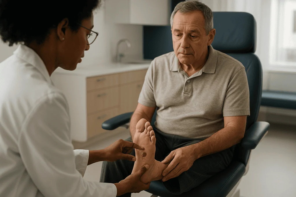
Wound-bed management is the clinic side. A clinician removes dead or thickened tissue, a process called debridement, assesses depth and any exposed tendon or bone, checks for infection and chooses a dressing that keeps the base moist while protecting the edges. This is typically repeated weekly.
Offloading is the mechanical side. The wound must stop bearing weight. That means a cast, a boot or a specialized shoe insert, and it means genuinely wearing it, including for the four steps to the bathroom at night.
Metabolic and vascular care is the whole-body side. Your diabetes team adjusts glucose management, often intensifying it during healing; a vascular specialist may measure blood flow and, where arteries are significantly narrowed, discuss procedures to reopen them.
The NIH’s diabetes institute describes a foot ulcer as a wound that should be seen by a health professional promptly rather than managed at home, precisely because the depth and blood supply cannot be judged by eye. The team usually includes a podiatrist, a diabetes nurse or educator, a wound specialist and, when needed, a vascular surgeon and an infectious disease physician. Decisions about which dressing, which device and when to escalate sit with that team, informed by what they see at each visit.
Who is treated straight away, and who is asked to wait for advanced options
Nobody with an open foot ulcer is asked to wait for basic care. Cleaning, debridement, offloading and glucose review begin at the first visit. What varies is who moves quickly to more intensive interventions and who is asked to give standard care time to work.
Same-day or urgent escalation is usual when the wound reaches bone or joint, when there are signs of spreading infection such as redness tracking up the foot, when part of the foot has turned dusky or black, or when foot pulses cannot be felt. These findings point toward deep infection or critical blood-flow loss, situations where the CDC and NIH both note the risk of amputation rises sharply, and where hospital admission and vascular imaging are often arranged promptly.
Advanced therapies are a different matter. Options such as negative-pressure wound therapy (a sealed dressing attached to a gentle vacuum), bioengineered skin substitutes, or hyperbaric oxygen are generally considered only after a wound has failed to respond to good standard care over several weeks, and only when circulation is adequate. The evidence for each is mixed; guidelines describe them as adjuncts for selected wounds, not routine first steps. Someone whose ulcer is shallow, uninfected and shrinking with a cast and weekly debridement is usually asked to continue exactly that.
People with advanced kidney disease, very poor circulation not yet addressed, or active infection are typically asked to wait for elective procedures until those problems are treated, because a skin graft placed on tissue with no blood supply cannot survive. That sequencing is not delay for its own sake. It reflects which problem is the bottleneck.
Blood sugar and wound healing: what high glucose actually does to a wound
The relationship between blood sugar and wound healing is not vague encouragement to “eat better.” It operates at the level of individual cells, and it matters at every stage of repair.
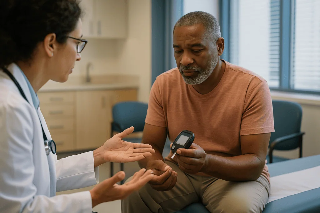
Neutrophils, the white blood cells that arrive first to engulf bacteria, work less efficiently when glucose is persistently elevated; their ability to move toward the wound and destroy what they find is measurably impaired. Fibroblasts, the cells that produce collagen scaffolding, divide more slowly. New capillaries, which must sprout into the wound bed to deliver oxygen, form less readily. Red blood cells become stiffer and less able to squeeze through the smallest vessels. Over years, high glucose also drives the nerve and artery damage that caused the ulcer in the first place.
None of this reverses overnight, and no single reading decides the outcome. What your team will look at is the overall pattern: your hemoglobin A1C, a blood test reflecting average glucose over about three months, alongside any home or continuous monitoring data. The NIH and CDC both list glucose management among the core steps for preventing and healing foot problems.
Illness and infection push glucose higher, so an infected ulcer often sends readings up even when nothing else has changed. Your prescribing clinician may adjust glucose-lowering medicines, which work through different mechanisms (increasing insulin, improving the body’s sensitivity to it, or helping the kidneys excrete glucose), and may temporarily use insulin during acute infection. Those choices, and any changes to what you already take, belong to that clinician. What you can do is keep monitoring, bring the numbers to each visit, and mention any dip in appetite, because eating less while medicines stay the same can cause lows.
Offloading a diabetic foot ulcer: why the boot matters more than the dressing
Offloading means redirecting body weight away from the wound so that pressure and friction stop reopening it. Ask most wound specialists what single factor most often stalls a plantar ulcer and the answer is not the dressing choice. It is that the person kept walking on it.
The mechanics are simple. The sole of a walking foot experiences peak pressures several times body weight at push-off. Tissue under that load is repeatedly compressed, blood flow to the wound edge is squeezed off, and the fragile new cells at the margin are sheared away. A dressing, however sophisticated, cannot counteract that.
The device with the strongest evidence for plantar forefoot ulcers is the total contact cast, a below-knee cast molded to the whole sole so that pressure is spread across the entire limb. It cannot be removed, which is both its strength and its inconvenience. Removable options exist, and they are widely used because they allow inspection and washing, but activity-monitor studies cited in the NEJM review found people wore removable walkers for only a minority of their daily steps. A device sitting beside the bed protects nothing.
| Offloading option | Removable? | Typical use | Common trade-offs |
|---|---|---|---|
| Total contact cast | No | Plantar forefoot or midfoot ulcers without active infection or poor circulation | Heavy, needs weekly changes, harder to bathe, cannot check wound daily |
| Removable cast walker | Yes | Similar wounds when a cast is unsuitable or daily inspection is needed | Effective only when worn for every step |
| Made non-removable (walker wrapped or locked) | No | Bridges the two approaches | Same bathing limits as a cast |
| Healing sandal or post-operative shoe with insert | Yes | Shallow, well-controlled ulcers; heel wounds; later stages | Less pressure relief than a cast |
| Crutches, walker frame or wheelchair | Yes | Short distances, heel ulcers, when other devices cannot be used | Fall risk, upper-body strain |
Which device suits you depends on wound location, circulation, balance and living situation, and the team will weigh those together.
Debridement and dressings: what happens in the chair each week
A typical visit follows a rhythm. The dressing comes off. The clinician photographs or measures the wound in length, width and depth, because a tape measure is a more honest witness than memory. Then comes debridement, the removal of dead tissue, callus and debris from the wound and its rim.
Debridement sounds alarming and usually is not painful, because the same neuropathy that hid the wound also numbs the procedure. It matters for several reasons. Dead tissue is a buffet for bacteria. The thick callus that rings many plantar ulcers acts like a rigid collar, concentrating pressure onto the wound and hiding its true edges. Removing it lets the clinician see how deep the wound really goes and whether bone or tendon is exposed. It also converts a stalled, chronic wound edge back into something closer to a fresh injury, which tends to restart the healing cascade. The NIH describes regular debridement by a health professional as part of standard ulcer treatment.
Dressings are chosen to keep the wound bed moist but not wet, to absorb drainage, and to protect the surrounding skin from maceration, the softening that happens when skin sits in moisture. Categories include hydrogels for dry wounds, foams and alginates for wet ones, and antimicrobial dressings when the team is concerned about surface bacteria. Evidence comparing one dressing type to another is limited and often of low quality; guidelines generally advise choosing on wound characteristics and comfort rather than assuming any product heals faster.
Between visits, you or a family member may change the dressing according to the team’s instructions. Keep the wound clean with what they recommend, avoid soaking the foot, and never trim callus or skin yourself.
Infection and osteomyelitis: how your team tells a dirty wound from a dangerous one
Every open wound has bacteria on its surface. Infection is different: it means bacteria have invaded living tissue and the body is fighting back, with redness, warmth, swelling, new drainage or odor. The NEJM review estimated that roughly half of diabetic foot ulcers develop infection at some point, and infection is the most common route from ulcer to hospital.
Clinicians grade infection by extent. Redness confined to a narrow rim around the wound with no deeper involvement is mild. Redness spreading more widely, or involvement of deeper structures such as tendon or joint, is moderate. Fever, chills, fast heart rate or confusion mark severe infection, where the whole body is affected. The distinction guides whether oral or intravenous antibiotics are used, whether admission is needed, and how urgently a surgeon should look.
Osteomyelitis, infection of the bone, is the complication teams worry about most. It should be suspected when a wound is deep, has been open for many weeks, sits over a bony prominence, or when a sterile probe passed gently into the wound touches bone. Blood tests can add support; imaging such as X-rays or MRI may show bone changes; a bone sample sent for culture is the most definitive test. Treatment typically combines a prolonged antibiotic course chosen by the team, often guided by culture results, with removal of infected bone where needed.
Antibiotics are chosen by class and adjusted to what the cultures grow. Your team decides the agent and duration; taking leftover antibiotics from a cupboard, or stopping a prescribed course because the foot “looks better,” both undermine that plan. Report any new symptom rather than waiting for the next scheduled visit.
Why your circulation is checked before anything else is promised
Before a wound can heal, blood has to reach it. That is why one of the first things a specialist does is feel for the two main foot pulses, one on top of the foot and one behind the inner ankle, and why so many appointments involve a blood-pressure cuff around your ankle.
The ankle-brachial index compares blood pressure at the ankle with blood pressure at the arm; a lower ankle reading suggests narrowed leg arteries. In diabetes, calcified arteries can make this test read falsely normal, so teams often add a toe pressure measurement or a test of oxygen levels in the skin. Ultrasound of the leg arteries shows where narrowing sits. If flow is significantly reduced, a CT or catheter-based angiogram maps the arteries in detail.
Why does this come first? Because the answer changes everything downstream. A wound with good blood supply will usually respond to offloading and debridement. A wound with critically poor supply may not respond to anything until flow is restored, and aggressive debridement of tissue that cannot heal can make the wound larger. Skin substitutes and grafts fail on ischemic tissue. Even infection behaves differently, because antibiotics travel in blood.
Where arteries are narrowed, a vascular specialist may discuss angioplasty (a balloon inflated inside the artery, sometimes with a stent), or bypass surgery that reroutes blood around a blockage. Each carries risks the specialist will lay out, and not everyone is a candidate. The Mayo Clinic notes that poor circulation, alongside nerve damage and infection, is one of the main drivers of diabetes-related amputation, which is exactly why teams check it before promising a timeline.
The daily diabetic foot check: what to look for and how to do it
The daily diabetic foot check is not a screening test you interpret yourself. It is a habit of looking, so that anything new gets reported to someone who can. The NIH’s diabetes institute and the CDC both recommend checking your feet every day, ideally at the same time, such as after a shower or before bed.
Choose good light. Sit somewhere you can bring your foot close, or place a mirror on the floor for the sole. If your eyesight or flexibility makes this hard, a family member can look, or a phone camera on the floor can substitute for a mirror. Look at the top, the sole, the heel, the sides and between every toe. Run a clean hand over the surface as well, since fingers notice warmth and swelling that eyes miss.
What you are noticing, and what deserves a call, includes any break in the skin, a blister, a new red or dark patch, a spot that feels warmer than its neighbor, swelling on one side only, drainage on a sock, or a change in the smell of a wound you already have. Thickened calluses, ingrown nails and cracks between toes also matter because each is a doorway.
The rest of the routine is unglamorous. Wash feet daily in lukewarm water tested with your elbow or a thermometer rather than your foot, dry carefully between toes, and moisturize the tops and soles but not between toes. Wear socks without tight seams and shoes indoors and out. Shake out shoes before putting them on. Never use heating pads or hot-water bottles on numb feet, and leave nail and callus care to a podiatrist if sensation is reduced. A professional foot exam at least once a year, with your shoes and socks off, is the recommended minimum even when nothing seems wrong.
What the first weeks of treatment usually look like
The first visit is the longest. Expect measuring, probing, pulse checks, an ankle pressure test, possibly an X-ray to look for gas in the tissues or bone changes, and often a swab or tissue sample sent to the laboratory. You will leave with a dressing, an offloading plan, glucose targets or a referral to review them, and a date to return, usually within a week.
Weeks one through four are about establishing whether the wound is on a healing trajectory. Each visit repeats the measure-debride-dress cycle. A cast is changed as swelling settles. Cultures come back and antibiotics are adjusted if infection is present. The NEJM review notes that the percentage reduction in wound area by around four weeks predicts whether a wound is likely to close by twelve weeks; a wound that has barely shrunk by then prompts a rethink rather than more of the same.
Life during this stretch is genuinely disrupted. Driving is usually not advised with a cast on the right foot. Showering requires a waterproof cover. Work that involves standing may need adjusting, and the team can provide documentation. Sleep is affected by the weight of a cast. Naming these frustrations early helps, because the single greatest threat to healing in this phase is abandoning the device.
From week four onward, the path splits. Wounds that are shrinking continue the same plan with visits perhaps spaced out slightly. Wounds that stall are reassessed: Is offloading really happening? Is there infection or bone involvement missed earlier? Has circulation been fully evaluated? Are glucose readings running high? Only after those questions are answered do adjunct therapies enter the conversation.
How long do diabetic foot ulcers take to heal, and why they come back
People want a number, and honest clinicians hesitate to give one. Healing time depends on wound depth, blood supply, infection, glucose control and, above all, how completely the wound is offloaded. Shallow, well-perfused ulcers under a total contact cast often close within several weeks; deeper wounds with compromised circulation can take many months. Twelve weeks is a common benchmark in research for judging whether standard care has worked, which is why the four-week area check described above carries so much weight.
Closure is not the end of the story, and this is the part that surprises people most. The NEJM review reported that roughly 40 percent of ulcers recur within one year of healing, about 60 percent within three years, and around 65 percent within five. The authors suggested the honest term for a healed ulcer is a foot in remission, in the same sense the word is used in cancer care: quiet, but requiring surveillance.
Why so high? Because closure fixes the wound but not what caused it. The neuropathy remains. The bony prominence remains. The habit of walking barefoot to the kitchen remains. Newly healed skin is thinner and less elastic than the original, so it breaks more easily under the same load.
Remission care therefore looks a lot like ulcer care with the wound removed: prescribed footwear with custom insoles that spread pressure, regular podiatry to manage callus, glucose management, daily inspection, and a low threshold for reporting anything new. Some people with recurrent ulcers over a fixed deformity are offered surgery to correct the bony cause; that is a specialist discussion with its own risks and is not routine.
What people often get wrong about diabetic foot ulcer care
“It doesn’t hurt, so it can’t be serious.” The absence of pain is the reason the wound exists, not evidence that it is minor. Depth and infection are judged by examination, not by how it feels.
“Letting it air out helps it heal.” Wounds heal faster in a moist, protected environment. A dry, exposed wound bed forms a crust that cells must burrow beneath, and an uncovered wound on a foot collects bacteria with every step.
“I’ll wear the boot when I go out.” The four steps to the bathroom at night, on hard tile, barefoot, are exactly the steps that reopen a forefoot ulcer. Offloading counts per step, not per outing.
“Soaking in salt water or hydrogen peroxide cleans it.” Soaking softens skin and invites maceration; peroxide and iodine damage the fragile cells trying to rebuild the wound. Use only what the team recommends.
“Antibiotics will heal the ulcer.” Antibiotics treat infection. They do nothing for pressure or blood flow, and using them for an uninfected wound breeds resistance without benefit.
“Once it’s closed, I can go back to my old shoes.” Recurrence rates of 40 percent within a year say otherwise. The shoes that caused the wound will cause it again.
“Special honey, ointments or supplements will speed things up.” Some topical products have small studies behind them; none has evidence strong enough for guidelines to recommend them ahead of offloading, debridement and glucose management. Anyone promising rapid healing from a single product is selling, not treating.
“Amputation is inevitable.” Most ulcers heal without it. The CDC’s message is that daily foot care, prompt reporting and good glucose management substantially reduce the risk, and that early attention is the single best protection.
Questions to ask your care team about your diabetic foot ulcer
Appointments move fast, and the most useful questions are often the ones nobody thinks to ask until the drive home. Bring this list, or a version of it, and write the answers down.
- How deep is the wound, and does it reach tendon, joint or bone?
- What did the circulation tests show, and do I need to see a vascular specialist?
- Is there infection now, and if so, how far does it extend?
- Which offloading device do you recommend for my wound, and what should I do for the steps I take at home?
- How will we know at four weeks whether the plan is working, and what happens if it isn’t?
- What glucose range are we aiming for while this heals, and who adjusts my medicines if readings run high or low?
- Who changes the dressing between visits, with what, and how often?
- What signs should make me call before my next appointment, and which number do I call after hours?
- Can I shower, drive or work with this device? What paperwork can you provide for my employer?
- Once the wound closes, what footwear and follow-up schedule will I need to prevent it coming back?
- Are there any advanced therapies you would consider for me, what is the evidence for them, and what would need to be true first?
Two habits make these conversations more productive. First, bring your glucose log or meter, because the team can act on data they can see. Second, bring the shoes you actually wear, not the pair you think you should wear. A podiatrist reading the wear pattern on a sole learns more in thirty seconds than in ten minutes of description. If something in the plan is not workable in your home or job, say so at the visit; a plan that cannot be followed is worse than a modest plan that can.
When to call your doctor
Diabetic foot ulcers change quickly, and the interval between scheduled visits is long enough for a mild problem to become an emergency. Do not wait for the next appointment if something is new.
Call your care team the same day if you notice redness spreading beyond the immediate edge of the wound or tracking up the foot or leg; new or increasing swelling; drainage that has increased, turned cloudy or developed an odor; a wound that has become deeper or larger; skin around the wound that feels warmer than the other foot; a new break in the skin, blister or dark patch anywhere on either foot; glucose readings running persistently higher than usual with no other explanation; or any part of the device rubbing, cracking or causing a new sore.
Seek emergency care without delay if you develop fever or chills, feel confused or unusually drowsy, notice a fast heartbeat or breathing, see a portion of the foot turn dusky, purple or black, develop sudden severe pain in a foot that is usually numb, or find your glucose is very high with vomiting or you are unable to keep fluids down. These can signal severe infection spreading into the bloodstream, a sudden loss of blood supply, or a metabolic emergency, and each is treated in hospital.
A practical rule: if you would hesitate to describe the foot as “the same or better” than yesterday, that hesitation is itself the reason to call. Teams would far rather hear about a false alarm than see a wound a week too late. Keep the after-hours number somewhere visible, and if you cannot reach your team, an urgent care or emergency department can examine the foot and contact them.
Frequently asked questions
How does offloading a diabetic foot ulcer actually help it heal?
Offloading removes the repeated pressure and shearing that crush new tissue at the wound edge with every step. A total contact cast spreads weight across the whole lower leg so the ulcer bears almost none; removable walkers and healing shoes do the same to a lesser degree. Studies consistently show plantar ulcers close faster when weight is kept off them, provided the device is worn for every step, including short trips indoors.
What is the link between blood sugar and wound healing?
Persistently high glucose impairs the white blood cells that clear bacteria, slows the fibroblasts that build collagen, and hampers growth of the new capillaries a wound needs for oxygen. It also stiffens red blood cells so they pass less easily through small vessels. Bringing glucose into the range your team sets removes these brakes; no single reading decides the outcome, but the overall pattern matters.
How long do diabetic foot ulcers take to heal?
There is no reliable single answer. Shallow ulcers with good blood flow, fully offloaded, often close within several weeks; deeper wounds or those with reduced circulation can take many months. Researchers commonly use twelve weeks as a benchmark for judging standard care, and the amount of shrinkage by about four weeks is a strong predictor of whether that benchmark will be met.
What should a daily diabetic foot check include?
Look at the top, sole, heel, sides and between every toe in good light, using a mirror or a family member for the sole. Feel for warmth or swelling. Anything new, such as a skin break, blister, red or dark patch, drainage on a sock, or a change in an existing wound, should be reported to your team the same day rather than watched.
Can I walk on a diabetic foot ulcer?
Walking on an unprotected plantar ulcer is one of the main reasons wounds fail to heal, because pressure repeatedly damages the wound edge. Your team will prescribe an offloading device and tell you how much activity is acceptable with it in place. Non-weight-bearing wounds on the top of the foot are managed differently, so follow the specific instructions you are given.
How do doctors know if a diabetic foot ulcer is infected?
Infection is diagnosed by examination: redness beyond the wound edge, warmth, swelling, new drainage or odor, sometimes with fever. Clinicians grade it as mild, moderate or severe by how far it spreads and whether the whole body is affected. Tissue cultures identify the bacteria, and imaging or a bone sample is used when infection of the bone, osteomyelitis, is suspected.
Is debridement of a diabetic foot ulcer painful?
Usually not, because the nerve damage that allowed the ulcer to form also reduces sensation at the wound. Debridement removes dead tissue and callus so the true depth can be seen, bacteria have less to feed on, and the wound edge is reset to a more active healing state. If you do have sensation, tell the clinician, who can adjust the technique or offer local numbing.
Why do diabetic foot ulcers come back after healing?
Closure fixes the wound but not its causes. Nerve damage, foot deformity, thin new skin and old footwear habits all persist. A review in the New England Journal of Medicine reported roughly 40 percent recurrence within a year and about 65 percent within five, which is why prescribed footwear, regular podiatry and daily inspection continue after healing.
Do special dressings, honey or supplements heal diabetic foot ulcers faster?
Evidence comparing dressing types is limited and generally low quality, and no topical product or supplement has evidence strong enough for major guidelines to place it ahead of offloading, debridement, infection control and glucose management. Dressings are chosen for wound characteristics and comfort. Be cautious of any product promising rapid healing on its own.
When is a diabetic foot ulcer an emergency?
Seek emergency care for fever or chills, confusion, a racing heart, a part of the foot turning dusky or black, sudden severe pain in a normally numb foot, or very high glucose with vomiting. These may indicate infection spreading to the bloodstream, sudden loss of blood supply, or a metabolic crisis. Spreading redness, new swelling or odor warrant a same-day call to your team.
References
- Diabetes and Foot Problems: NIH National Institute of Diabetes and Digestive and Kidney Diseases
- Diabetes – foot ulcers: MedlinePlus Medical Encyclopedia
- Diabetes and Your Feet: CDC
This article is for general information only and is not a substitute for professional medical advice. Please consult a qualified doctor about your individual situation.

More from the Blog

Arthrofibrosis: When a Knee Gets Stiff After Surgery, and What Helps
Arthrofibrosis is an excessive buildup of internal scar tissue after joint surgery or injury, most often in the knee, that causes persistent stiffness, pain,…

How to Sleep (and Live) with a Kidney Stent
Most people sleep better with a kidney (ureteral) stent by lying on the side opposite the stent or slightly propped up, emptying the bladder…

Tube Feeding vs IV Nutrition in Clinical Nutrition Therapy: When Doctors Use Each Option
Tube feeding (enteral nutrition) delivers liquid formula into the stomach or small intestine through a tube, while IV nutrition (parenteral nutrition) infuses nutrients directly…

Hardware Removal Surgery: When Plates and Screws Should Come Out
Most orthopedic plates and screws are designed to stay in the body permanently and never need removal. Hardware removal surgery is usually considered only…

Flying with a DVT, Compression Stockings and Prevention
Most people can fly with a DVT once they are established on anticoagulant treatment and their symptoms are stable, but the timing is an…

Living With a Central Venous Port: Showering, Swimming, Flushing Schedules and Travel
Living with a chemo port is mostly a matter of protecting the skin over it and keeping the line clear. Once the incision has…