

Tenosynovitis on Ultrasound: What the Scan Shows and How It Guides Splinting or Injection
Key Takeaways
- Tenosynovitis is inflammation of the sheath around a tendon, so on ultrasound the tendon can look normal while the sleeve around it shows fluid, thickening and Doppler blood flow.
- Fluid alone is not a diagnosis; a thin film is normal around several tendons, and radiologists judge it against the other side and against whether the lining is thickened.
- Tendonitis affects the tendon cord and usually needs gradual loading, while tenosynovitis affects the sheath and often settles with rest, splinting and, in some cases, a sheath injection.
- Ultrasound cannot reliably distinguish an infected finger sheath from an inflamed one; that requires clinical assessment and often a fluid sample, and it is an urgent situation.
- Guided injection places corticosteroid in the sheath and away from the tendon and nearby arteries, and can reveal a septum in De Quervain that a landmark injection might miss.
- Mayo Clinic describes around-the-clock splinting for four to six weeks in early De Quervain tenosynovitis and a possible pain flare for up to 48 hours after a corticosteroid injection.
On ultrasound, tenosynovitis appears as fluid or thickened tissue inside the sleeve (sheath) that surrounds a tendon, often with extra blood flow on Doppler imaging, while the tendon itself may look normal. Radiologists use these findings to confirm which tendon compartment is inflamed, judge how active it is, and, when a corticosteroid injection is chosen, place the needle inside the sheath rather than into the tendon.
The mug was the tell. She could lift it fine with her left hand, but wrapping her right thumb around the handle sent a hot wire of pain up the side of her wrist. Six weeks of a new baby, a new laptop and a lot of one-handed lifting had done something to the tendons at the base of her thumb, and her primary care doctor wrote two words on the referral: tenosynovitis ultrasound.
She had questions before she reached the parking lot. What would the scan actually show? Could a probe and some gel really see inflammation? And why did the clinic seem to be deciding between a splint and an injection before she had even lain down?
Those are the right questions, and the honest answers are more interesting than the usual reassurance. Ultrasound is very good at some things here and genuinely limited at others, and knowing which is which changes how you read your own report.
What tenosynovitis actually is: a tendon and its sleeve
Picture a bicycle brake cable. The steel wire is the tendon, the cord that connects muscle to bone. The plastic housing it slides through is the tendon sheath, a thin, fluid-lined sleeve that lets the tendon glide without friction. Tenosynovitis is inflammation of that sleeve. The suffix matters: teno for tendon, synov for the synovial lining that produces lubricating fluid, itis for inflammation.
Not every tendon has a sheath. The Achilles, for example, is wrapped in a looser tissue called a paratenon, so it develops tendinopathy rather than true tenosynovitis. Sheathed tendons cluster where a tendon has to turn a corner or pass under a tight band: the wrist, the fingers, the inner and outer ankle, and the front of the shoulder. Those are exactly the places where a sonographer is most often asked to look.
When the sheath is irritated, three things tend to happen. The lining secretes more fluid than the tendon needs, so the sleeve swells. The lining itself thickens, sometimes into soft, velvety tissue. And small blood vessels open up to feed the inflamed lining. Each of these has a signature on ultrasound, which is why the scan is so useful in this particular condition.
The triggers vary. Repetitive gripping and lifting are the everyday causes, and new-parent thumb pain is common enough that Mayo Clinic lists lifting a child as a classic pattern for De Quervain tenosynovitis at the base of the thumb. Inflammatory conditions such as rheumatoid arthritis can attack sheaths directly. Rarely, bacteria enter through a cut and cause an infected sheath, which is a different emergency altogether and is covered later in this article.
Tendonitis vs tenosynovitis: what's the difference?
People use the words interchangeably, and doctors sometimes do too, but they describe different structures. Tendonitis, more accurately called tendinopathy because inflammation is often minimal, is a problem of the tendon cord itself. The fibers become disorganized, thickened and painful, usually from overload. Tenosynovitis is a problem of the sleeve around the cord. The tendon inside can be perfectly healthy while the sheath is swollen and angry.

Why does the distinction change anything? Because the two respond to different logic. A degenerated tendon needs progressive loading over months to remodel; rest alone rarely fixes it. An inflamed sheath often calms down when the irritation stops, which is why splinting and, in some cases, a corticosteroid injected into the sheath are standard options for tenosynovitis but far less appealing for a tendon whose fibers are already weakened. The NHS notes that tendinopathy symptoms often start to improve within two to three weeks of rest and simple measures, but that guidance applies to the tendon, not to a sheath that keeps refilling with fluid every time you grip.
The two can coexist. Long-standing sheath inflammation can eventually stress the tendon beneath it, and a thickened, degenerated tendon can rub the sheath raw. On a scan, the sonographer separates the layers: is the fluid around the tendon, is the thickening in the sheath, or is the tendon itself enlarged with a fuzzy, disrupted fiber pattern? That single question shapes what your treating team offers next.
A related term you may see in your report is stenosing tenosynovitis. Stenosing means narrowing. It describes a sheath that has thickened so much that the tendon catches as it slides through, the mechanism behind trigger finger and most De Quervain cases.
What are the tenosynovitis ultrasound findings?
Radiologists look for a small cluster of signs, and your report will usually name several of them in order.
Fluid in the sheath. On the screen, fluid is black, or anechoic, meaning it reflects no sound. Seen across the tendon, it forms a dark ring around a bright, round tendon, a picture sometimes called the target or halo sign. Seen along the length, it looks like a dark stripe hugging the fibers. A tiny film of fluid can be normal around some tendons, particularly at the ankle and in the biceps groove, so the sonographer compares with the other side and judges whether the amount is more than the tendon needs.
Sheath thickening. An irritated synovial lining thickens into gray, low-echo tissue that does not compress when the probe presses down. Fluid squishes; thickened synovium does not. That simple compression test is one of the most useful moves in the whole exam.
Increased blood flow. Color or power Doppler is an ultrasound setting that paints moving blood in color. An inflamed sheath often lights up with dots and streaks of flow that a quiet sheath would not show. Doppler signal is read as a sign of active inflammation, not just old swelling.
The tendon itself. A healthy tendon shows a tight, fibrillar pattern like the grain in wood. If the fibers look swollen, dark or interrupted, the report will mention tendinosis or a partial tear alongside the tenosynovitis.
Dynamic findings. Because ultrasound is live, the operator can ask you to move. A tendon that snaps or hesitates as it passes a thickened pulley confirms the stenosing type. In De Quervain, the scan can also show a thin wall, or septum, dividing the first compartment into two tunnels, a detail that matters for injection planning.
What actually happens during a tenosynovitis ultrasound
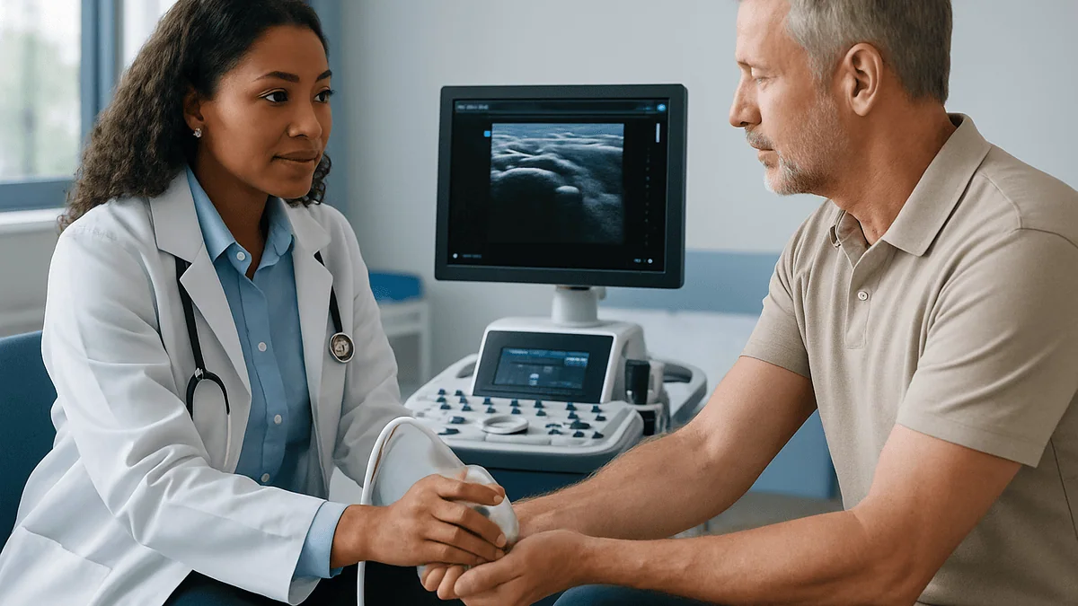
The exam is shorter and lower-key than most people expect. You sit or lie with the painful limb resting on a pillow or small table. The sonographer or radiologist applies warm gel and glides a small, flat probe over the skin. The gel is not medicine; it simply removes the air gap so sound waves can pass into tissue. Nothing enters the body, and there is no radiation, which is one reason ultrasound is used freely during pregnancy and in children.

Expect the probe to press. Compression is diagnostic, as described above, and gentle pressure over the sore spot also confirms that the structure on the screen is the one that hurts. Operators call this sonopalpation, and it is a legitimate part of the exam rather than clumsiness. Tell them if the pressure becomes sharp; they can adjust.
You will likely be asked to move. Wiggle the thumb, bend the fingers, turn the ankle in and out. Watching a tendon slide in real time reveals catching, subluxation (a tendon slipping out of its groove) or a sheath that bunches up at a pulley. The operator will almost always scan the same tendon on your other limb for comparison, because normal varies from person to person.
Many musculoskeletal ultrasound exams take roughly fifteen to thirty minutes, though a focused wrist scan can be quicker. If an injection has been planned for the same visit, the imaging becomes the roadmap: the clinician marks the skin, identifies nearby vessels and nerves with Doppler, and only then prepares the needle. Some services scan and inject in one appointment; others report first and refer you back. Neither approach is wrong, and the choice usually reflects local workflow rather than the severity of your condition.
Can tendonitis be seen on ultrasound, and how does it compare with MRI and X-ray?
Yes, and often more clearly than on any other imaging. Ultrasound resolves tendon fibers at a level of detail that rivals or exceeds MRI in superficial tendons like those of the wrist and ankle. Thickening, loss of the normal fiber pattern, small tears and tiny calcifications all show well. Because the probe can be angled and the limb moved, ultrasound also catches problems that appear only with motion.
MRI has a different set of strengths. It shows the whole region at once, including bone marrow, cartilage and deep tendons that ultrasound cannot reach behind bone. It is less dependent on who holds the probe, which makes it easier to compare scans months apart. On MRI, tenosynovitis appears as bright fluid signal around the tendon on fluid-sensitive sequences, and thickened synovium enhances if contrast is given. The trade-offs are time, cost of access in health-system terms, and the fact that you cannot move during the scan.
X-ray shows bone, not soft tissue, so it cannot diagnose tenosynovitis. It is still ordered in some cases to rule out arthritis, an old fracture or a calcium deposit that might be irritating the sheath.
Where ultrasound falls short is honesty about operators and depth. A skilled musculoskeletal sonographer sees a great deal; an inexperienced one may miss subtle sheath thickening or mistake normal fluid for disease. Tendons deep in the hip or under the shoulder blade are harder to reach. And ultrasound cannot see through a plaster cast or a metal implant. Your treating team weighs these factors when choosing the test. For a painful thumb, wrist or ankle with a clear tender spot, ultrasound is usually the first and often the only imaging needed.
Which tendons are scanned most often, and what each scan looks for
Tenosynovitis has favorite addresses. The table below lists the sheathed tendons most often referred for ultrasound, the everyday name for the condition, and the specific detail the sonographer hunts for at each site. The final column notes how findings typically feed into a splint-or-injection discussion, always as one input among several for the treating team.
| Tendon or compartment | Common name | Key ultrasound detail | How it informs treatment planning |
|---|---|---|---|
| First extensor compartment, wrist (thumb side) | De Quervain tenosynovitis | Sheath thickening over the radial styloid; presence of a dividing septum; Doppler flow | A septum means two tunnels; guided injection can target both, and a thumb spica splint is the usual conservative option |
| Sixth extensor compartment, wrist (little-finger side) | Extensor carpi ulnaris tenosynovitis | Fluid around the tendon; tendon slipping out of its groove on forearm rotation | Instability may favor splinting in a specific position over injection alone |
| Finger flexor tendons at the A1 pulley | Trigger finger (stenosing tenosynovitis) | Thickened pulley; tendon catching on active bending | Guided injection places medicine in the sheath, away from the tendon and digital vessels |
| Tibialis posterior, inner ankle | Posterior tibial tenosynovitis | Fluid and sheath thickening behind the ankle bone; tendon degeneration | Tendon damage shifts emphasis toward bracing and loading rather than injection |
| Peroneal tendons, outer ankle | Peroneal tenosynovitis | Fluid in the shared sheath; split tears; tendon subluxation | Split tears and instability may prompt a surgical opinion |
| Long head of biceps, shoulder | Biceps tenosynovitis | Fluid in the groove, often reflecting shoulder joint fluid | Findings are read alongside rotator cuff status before any decision |
Two patterns are worth noticing. First, the same finding, fluid, means different things at different sites; at the biceps it frequently reflects the shoulder joint rather than the tendon. Second, wherever the tendon itself is damaged, the scan tends to steer the conversation away from a quick injection and toward protection and rehabilitation.
Flexor tenosynovitis ultrasound: can the scan tell infection from inflammation?
This is the one situation where the answer needs to be blunt: ultrasound helps, but it cannot rule infection in or out on its own.
Infectious flexor tenosynovitis is a bacterial infection inside the finger’s flexor tendon sheath, usually after a puncture, bite or cut on the palm side of a finger. Because the sheath is a closed tube, pus builds pressure quickly and can starve the tendon of blood within days. MedlinePlus lists it among the forms of tenosynovitis that need urgent treatment, sometimes including surgical washout.
On the screen, an infected sheath often looks like an inflamed one: fluid around the tendon, thickened lining, Doppler flow. Clues that raise concern include fluid that is cloudy or contains floating debris rather than pure black, a distended sheath that extends the full length of the finger, and, rarely, bright flecks of gas. Ultrasound can also confirm that swelling is inside the sheath rather than in the skin, which helps distinguish a true sheath infection from a simple soft-tissue infection.
But those clues overlap with inflammatory arthritis, gout and even heavy overuse. The definitive test is not imaging; it is drawing fluid from the sheath and sending it to the laboratory, along with the clinical picture: fever, spreading redness, a finger held bent and exquisite pain when it is passively straightened. In emergency departments, bedside ultrasound is increasingly used to speed that decision, not to replace it.
The practical message is simple. If a finger is red, swollen, hot and painful to straighten after a wound, the priority is same-day medical assessment, not a scheduled outpatient scan. Ultrasound may be done in the emergency setting, but it is the hand surgeon’s clinical judgment and the fluid sample that decide what happens next.
Who is usually offered the scan, and who is asked to wait
Not everyone with a sore wrist needs imaging, and a good clinician will say so. De Quervain tenosynovitis and trigger finger are frequently diagnosed by examination alone; Mayo Clinic notes that imaging is not usually needed for a typical De Quervain presentation. Many people are started on splinting and activity changes on the strength of a careful history and a positive provocation test, and are only sent for a scan if that plan stalls.
Ultrasound is more likely to be offered when the diagnosis is uncertain. Pain on the thumb side of the wrist can come from the first compartment, the joint at the base of the thumb, a small ganglion cyst or a nerve branch, and each has a different plan. It is also offered when symptoms have not settled after a reasonable trial of conservative care, when an injection is being considered and the clinician wants to confirm the target, when there is a history of inflammatory arthritis, or when swelling is unusually large, firm or one-sided.
Who is asked to wait? Someone in the first week or two of a clear overuse flare, with no red flags, is often advised to rest, splint and review, because a scan now would confirm what is already obvious and would not change the plan. Similarly, if surgery is already the likely path, some surgeons prefer to operate on clinical grounds, though many still request a scan to check for anatomical variants.
Imaging of children follows the same logic with extra caution about avoiding unnecessary tests, though ultrasound’s lack of radiation makes it the preferred modality when a scan is needed. Whatever your situation, the decision to image, and when, belongs to the treating team, who can weigh your history against what the scan could realistically add.
How a tenosynovitis ultrasound guides splinting
Splinting sounds like the low-tech option, and in a sense it is: a splint stops the tendon sliding through an irritated sheath so the lining can settle. But which splint, how it is positioned and how long to persist are decisions that imaging can sharpen.
The first contribution is confirming the compartment. A thumb spica splint, which holds the thumb and wrist still, is the standard for De Quervain but does nothing for extensor carpi ulnaris pain on the other side of the wrist, which needs a different position. When examination is ambiguous, the scan points to the right sleeve.
The second is grading activity. A sheath with modest fluid and no Doppler flow suggests low-grade irritation, and many clinicians feel comfortable pursuing splinting and activity changes for longer before escalating. Marked thickening with vigorous Doppler signal suggests a more active process, and the treating team may bring an injection into the conversation earlier. This is clinical reasoning rather than a rule written into a guideline, and different teams weigh it differently.
The third is spotting reasons a splint may not be enough on its own. A tendon slipping in and out of its groove, a partial tear or a stenosed pulley that catches the tendon with every movement are all features that shift expectations. A splint can still help, but the team may plan a surgical review in parallel rather than waiting months.
Typical duration is worth knowing. Mayo Clinic describes wearing a splint around the clock for four to six weeks in De Quervain tenosynovitis, with symptoms often improving within that window when treatment starts early. That is a range, not a guarantee, and repeat imaging is rarely needed to decide when to stop; how your hand feels and functions matters more than a follow-up picture.
How ultrasound guided tenosynovitis injection works
The injection most often discussed for tenosynovitis is a corticosteroid, a class of anti-inflammatory medicine placed directly into the tendon sheath. The goal is to bathe the inflamed lining while keeping medicine out of the tendon substance itself, because steroid within tendon fibers is thought to weaken them. Ultrasound guidance exists to solve exactly that geometry problem.
The sequence is straightforward. The clinician rescans the target, identifies the sheath in cross-section and maps nearby structures. Doppler is switched on to find vessels; at the thumb side of the wrist the radial artery runs within millimeters of the first compartment, and in the finger the digital vessels flank the flexor sheath. The skin is cleaned, sometimes numbed, and a fine needle is advanced while the probe watches its tip. When the tip sits in the sheath, the injected fluid is seen spreading around the tendon as a dark ring, confirming placement in real time. If the fluid instead pools in the tendon or outside the sheath, the needle is repositioned before the full amount is given.
Guidance adds two specific advantages in De Quervain. It reveals a septum dividing the compartment, so the clinician can fill both tunnels rather than one. And it shows whether the tendons run in an anatomical variant position that a blind injection would miss.
The honest evidence summary: studies consistently show that ultrasound places medicine in the intended sheath more accurately than landmark-only injection. Whether that accuracy translates into better pain relief for every patient is less settled, and results vary by site and study design. Mayo Clinic notes that the effects of a corticosteroid injection are typically temporary, that injections into the same area are limited in frequency because of concerns about tendon and cartilage weakening, and that they are one option to weigh with your clinician, not a first step for everyone.
What the following days and weeks usually look like
After a diagnostic scan alone, there is nothing to recover from. You wipe off the gel and leave. The report usually reaches the referring clinician within days, and the next conversation is about plan, not about the test.
After a guided injection, the pattern is more predictable than people expect. The numbing medicine, if used, fades within hours and the original pain returns for a while. Mayo Clinic describes a possible flare of pain and inflammation for up to 48 hours after a corticosteroid injection before the anti-inflammatory effect builds. Ice, gentle use and avoiding heavy gripping in that window are standard advice. A small bruise or a brief patch of skin lightening at the needle site can occur and is discussed as a known effect, particularly in people with darker skin.
Improvement, when it comes, tends to arrive over one to two weeks rather than overnight. Many clinicians ask people to continue splinting for part of the day during this period so the newly calm sheath is not immediately re-irritated. Whether a second injection is ever considered is a decision for the treating team based on response, and frequency limits apply.
With splinting alone, the NHS describes tendon-related pain often beginning to ease within two to three weeks of rest and protection, and Mayo Clinic frames four to six weeks as a typical window for De Quervain when treatment begins early. Symptoms that are unchanged after that kind of interval are the usual trigger for review, and possibly for the scan if it has not already been done.
Return to activity is graded. A hand therapist or physiotherapist can show you how to lift, type and carry in ways that keep the thumb in a neutral position, which is often the difference between a flare that settles and one that keeps returning.
What people often get wrong about tenosynovitis and ultrasound
“Fluid on the scan means something is badly wrong.” Small amounts of fluid are normal around several tendons, especially at the ankle and in the biceps groove. What matters is the amount relative to the other side, whether the lining is thickened, and whether Doppler shows active flow. Read the report as a whole, not the single word fluid.
“A normal scan means the pain is not real.” Ultrasound is operator-dependent and can miss subtle sheath thickening. Pain that clearly localizes to a tendon with a negative scan may still be early tenosynovitis, or may come from a nearby joint or nerve. A normal result narrows the search; it does not end it.
“Tendonitis and tenosynovitis are the same thing.” One is the cord, the other is the sleeve. The distinction shapes whether protection, loading or injection makes sense, as covered earlier.
“An injection fixes it for good.” Corticosteroid effects are temporary, and recurrence is common if the underlying load pattern does not change. Mayo Clinic frames injections as one part of treatment alongside splinting and activity modification.
“Ultrasound guidance guarantees a better result.” Guidance improves the accuracy of placement. Evidence that it improves symptoms for every person and every tendon is mixed, and a skilled clinician using landmarks alone may do well at simple sites. Ask what the guidance adds in your specific case.
“If the scan shows a thickened sheath, surgery is inevitable.” Thickening is the expected finding in stenosing tenosynovitis and often settles with conservative care. Surgery to release a tight compartment is an option for persistent cases, not a foregone conclusion written into the image.
“You should push through the pain to keep the tendon strong.” Loading is the right approach for tendon degeneration. For an inflamed sheath, continuing the provoking activity tends to keep the lining irritated. Knowing which problem you have is precisely why the scan is done.
Questions to ask your care team
Walking into a consultation with a few precise questions changes the quality of the conversation. These are the ones that tend to matter most for tenosynovitis, drawn from what people say they wish they had asked.
- Which tendon compartment did the scan identify, and did it show fluid, sheath thickening, Doppler flow or all three?
- Was the tendon itself normal, or is there also tendinosis or a partial tear that changes the plan?
- Was the other side scanned for comparison, and how different was it?
- For a wrist scan: did the report mention a septum in the first compartment, and does that affect how you would inject?
- Based on these findings, what does a reasonable trial of splinting look like for me, and at what point would you want to review?
- If an injection is being considered, what does ultrasound guidance add at this particular site, and what are the known risks, including tendon weakening and skin changes?
- What would make you think this is inflammatory arthritis or infection rather than overuse, and have those been considered?
- Which specific movements or tasks should I change, and can I see a hand or physical therapist to learn how?
- If this does not settle, what are the next steps, and roughly when would surgery to release the compartment enter the conversation?
- Do I need a repeat scan to judge progress, or will you decide based on how my hand works?
Bring the written report if you have it, and do not hesitate to ask the radiologist or sonographer to show you the image on the screen during the exam. Seeing the dark ring of fluid around your own tendon is often the moment the diagnosis stops being abstract, and it makes the splint feel less like a punishment and more like a plan.
When to call your doctor
Most tenosynovitis is uncomfortable, slow and not dangerous. A small number of situations are different, and they should not wait for a scheduled scan or a routine follow-up.
Seek same-day medical care if a finger or hand becomes red, hot and swollen along the length of a tendon, especially after a cut, puncture, bite or splinter; if the finger rests in a bent position and straightening it passively causes severe pain; or if you develop fever, chills or spreading redness up the limb. These features raise concern for infectious flexor tenosynovitis, which MedlinePlus describes as needing urgent treatment to protect the tendon.
Contact your treating team promptly, rather than urgently, if pain is worsening despite splinting, if you notice new numbness, tingling or weakness in the fingers, if the tendon suddenly seems to have lost strength or you cannot lift a finger or thumb that you could lift yesterday, or if a lump along the tendon is growing. A sudden inability to move a digit can signal a tendon rupture, which is rare but changes the plan entirely.
After an injection, call if pain increases sharply after the first two days rather than easing, if the site becomes hot, red or leaks fluid, or if you develop a fever. A short flare in the first 48 hours is expected; a worsening picture after that is not.
And if several weeks of the agreed plan have passed without meaningful change, that in itself is a reason to get back in touch. Persistent symptoms are exactly when a scan, a different splint, an injection or a surgical opinion earns its place, and your treating team is the right group to weigh those options against your history and your hands’ daily demands.
Frequently asked questions
What are the ultrasound findings of tenosynovitis?
The core findings are fluid inside the tendon sheath, thickening of the sheath lining, and increased blood flow on Doppler imaging. Fluid appears as a dark ring around the tendon in cross-section. Thickened synovium is gray and does not flatten when the probe presses on it, which distinguishes it from fluid. The tendon itself may be normal, or may show swelling and a disrupted fiber pattern if tendinosis is also present.
What is the difference between tendonitis and tenosynovitis?
Tendonitis, more accurately tendinopathy, is a problem of the tendon cord itself, usually degeneration from overload. Tenosynovitis is inflammation of the sheath that surrounds the tendon. The tendon can be healthy while its sheath is swollen. The distinction matters because a degenerated tendon generally needs progressive loading, whereas an irritated sheath often responds to rest, splinting and sometimes a corticosteroid placed in the sheath.
Can tendonitis be seen on ultrasound?
Yes. Ultrasound shows tendon fibers in fine detail, so thickening, a fuzzy or disrupted fiber pattern, small tears and calcium deposits are usually visible, particularly in superficial tendons of the wrist, hand and ankle. Because the scan is live, the operator can also watch the tendon move and catch problems that appear only with motion. Deep tendons behind bone are harder to assess and may need MRI.
What are the radiological findings of tenosynovitis on MRI and X-ray?
On MRI, tenosynovitis shows as bright fluid signal surrounding the tendon on fluid-sensitive sequences, with thickened sheath tissue that enhances if contrast is given. X-ray cannot show a tendon sheath at all; it is used only to exclude bone problems such as arthritis, an old fracture or a calcium deposit that might be irritating the sheath. Ultrasound remains the first-line test for most superficial tendons.
Is a flexor tenosynovitis ultrasound enough to rule out infection?
No. Ultrasound can show fluid and thickening in a finger’s flexor sheath, and cloudy fluid with debris raises concern, but infected and inflamed sheaths often look alike. Infection is diagnosed from the clinical picture, including redness, fever and pain on straightening the finger, plus a fluid sample sent to the laboratory. MedlinePlus lists infectious tenosynovitis as needing urgent treatment, so a red, hot, swollen finger warrants same-day care.
Does a tenosynovitis ultrasound hurt?
It is generally comfortable, though the operator deliberately presses on the sore area to confirm the painful structure and to test whether swelling flattens under pressure. That pressure can be briefly uncomfortable but should not be sharp; tell the sonographer if it is. No needles, dye or radiation are involved in a diagnostic scan, and there is nothing to recover from afterward.
How does ultrasound guided tenosynovitis injection differ from a regular injection?
With guidance, the clinician watches the needle tip on screen and sees the injected fluid spread around the tendon inside the sheath, confirming correct placement before the full amount is given. Doppler identifies nearby arteries to avoid, and in De Quervain the scan can reveal a dividing septum so both tunnels are treated. Studies show guidance improves placement accuracy; whether it improves symptom relief for every patient is less settled.
How long do you wear a splint for tenosynovitis?
It depends on the tendon and your response, and the decision sits with your treating team. As a reference point, Mayo Clinic describes wearing a thumb spica splint around the clock for four to six weeks in De Quervain tenosynovitis, with symptoms often improving in that window when treatment starts early. The NHS describes tendon pain often beginning to ease within two to three weeks of rest and protection.
What does fluid around a tendon on ultrasound mean?
It means the sheath contains more lubricating fluid than usual, which is a common sign of irritation, but a thin film is normal around several tendons, especially at the ankle and in the biceps groove at the shoulder. Radiologists compare the amount with your other side and look for sheath thickening and Doppler flow before calling it tenosynovitis. Fluid at the biceps often reflects the shoulder joint rather than the tendon.
Will I need a repeat scan to see if tenosynovitis is improving?
Usually not. Most clinicians judge progress by how your hand or foot feels and functions rather than by repeat imaging, because sheath changes on ultrasound can lag behind symptom improvement. A repeat scan is more likely if symptoms persist despite treatment, if a new problem such as tendon weakness appears, or if an injection or surgical release is being planned and the anatomy needs to be rechecked.
References
This article is for general information only and is not a substitute for professional medical advice. Please consult a qualified doctor about your individual situation.

More from the Blog

Why an Elbow Fracture Often Needs a CT Scan Before Surgery: Planning the Fixation
An elbow fracture often needs a CT scan before surgery because plain X-rays flatten the elbow's three interlocking bones into two views, so hidden…

When Is Tennis Elbow Surgery Considered? What Comes First and What Release Surgery Involves
Tennis elbow surgery is usually considered only after a sustained course of non-surgical care has failed. Mayo Clinic describes roughly 6 to 12 months…

Is It Still Shoulder Impingement? Pain That Does Not Settle With Rehab and When to Go Back
Shoulder impingement not improving after several months of well-performed rehabilitation is a reason to go back, not to push harder. Persistent pain may mean…

Can You Pop a Ganglion Cyst With a Book? Wrist Cyst Myths a Hand Surgeon Corrects
No. Striking a ganglion cyst with a book, or any heavy object, is not a safe or reliable treatment. The blow can bruise skin,…

Tingling After Elbow Surgery: Ulnar Nerve Symptoms That Settle and Those to Report
Tingling after elbow surgery is common and often reflects a bruised, swollen, or recently moved ulnar nerve rather than lasting damage. Pins and needles…
How ACL Reconstruction Is Performed: Graft Harvest, Tunnels and Fixing the New Ligament
ACL reconstruction is a keyhole (arthroscopic) operation in which a surgeon replaces the torn anterior cruciate ligament with a graft, most often a strip…