Am I a Candidate for Private mri scan? Eligibility Criteria
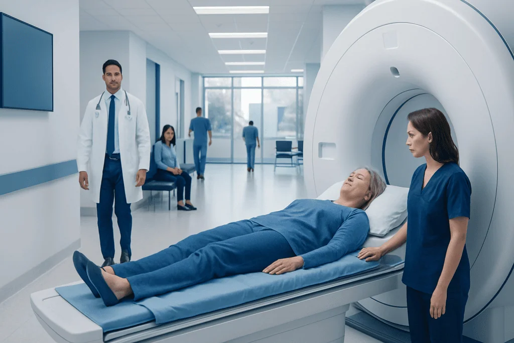
MRI uses strong magnets and radio waves rather than ionising radiation. Eligibility depends on the reason for the scan, safety screening and the type of implant or device a person has.
Key Takeaways
- MRI uses strong magnets and radio waves rather than ionising radiation.
- Eligibility depends on the reason for the scan, safety screening and the type of implant or device a person has.
- Many implants are MRI-conditional rather than automatically unsuitable, but their exact model and settings must be checked.
- Contrast dye is not needed for every MRI; kidney function and previous contrast reactions may affect its use.
- Most people return to normal activities immediately after an MRI, unless sedation or a specific medicine has been used.
Am I a candidate for private MRI scan? Most adults and children can undergo an MRI when it is clinically appropriate, provided a safety screening confirms that any implants, metal exposure, pregnancy considerations, kidney problems and contrast requirements have been assessed. A private MRI appointment may offer timely access, but suitability should always be confirmed by a qualified clinician and imaging team.
Overview: Who May Be Suitable for a Private MRI Scan?
Am I a candidate for private MRI scan? In general, a person may be a candidate if a clinician believes MRI can help investigate symptoms, assess an injury, monitor a known condition or guide treatment, and a pre-scan safety review shows it can be performed safely. MRI is a detailed imaging test that uses a powerful magnetic field and radio waves to create images of organs, soft tissues, blood vessels, joints, the brain and spine.
A private MRI scan is not a different type of MRI; the eligibility and safety principles are the same as in any accredited imaging service. The important questions are whether MRI is the right test for the clinical concern, whether the person can lie comfortably enough for the examination, and whether there are implants, metal fragments or health factors requiring special planning.
Some people may need another imaging test instead, such as ultrasound, X-ray or CT, depending on the symptoms and the information needed. The imaging referral should be based on an individual medical assessment rather than routine scanning alone. For a broader introduction to the service and appointment pathway, see private MRI scan information.
How MRI Works and What It Can Help Assess
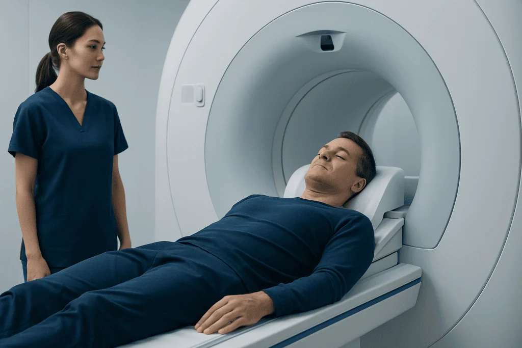
An MRI scanner is a large, tube-shaped machine containing a strong magnet. During the scan, the body is positioned on a table that moves into the scanner. Radiofrequency signals interact with water molecules in the body, and a computer converts these signals into highly detailed images. Unlike CT and X-ray, MRI does not use ionising radiation.
MRI can be particularly useful where soft-tissue detail is important. Depending on the area being examined, it may help assess the brain, spinal cord, nerves, muscles, ligaments, joints, abdominal and pelvic organs, blood vessels, breasts or heart. It may also be used to investigate certain symptoms or follow the course of conditions such as multiple sclerosis, when clinically appropriate.
Images are obtained in several sequences, which may make different tissues or changes more visible. Some scans use an intravenous contrast agent called gadolinium to improve detail in selected tissues or blood vessels. The radiologist and referring clinician decide whether contrast is likely to add useful information.
Eligibility Criteria and the Pre-Scan Safety Assessment

Before booking or attending an MRI, the imaging provider will usually ask the person to complete a detailed screening questionnaire. This pre-assessment form is an essential safety step. It should include information about operations, implants, devices, metal injuries, allergies, kidney disease, pregnancy, current medicines and any difficulty with enclosed spaces.
Most people with joint replacements, dental fillings, braces, surgical clips, plates or screws can have an MRI, but the team still needs to know about them. Some implanted devices, including certain pacemakers, defibrillators, cochlear implants, neurostimulators, infusion pumps and aneurysm clips, may require specific MRI-conditional protocols or may make MRI unsuitable. The device card, manufacturer details and implantation records can help the team confirm this.
People who have worked with metal, such as welders, machinists or metalworkers, or who have had an injury involving metal fragments, should report this before the scan. In some cases, an X-ray may be needed to rule out a metal fragment in or near the eye. A person should never assume that an implant is safe for MRI without verification.
- A clear clinical reason for imaging is needed, based on symptoms, injury, monitoring or treatment planning.
- The person must be able to follow safety instructions and remain still for the required images, with support or adjustments where possible.
- Any implant or device must be identified and checked for MRI compatibility.
- Contrast use, if proposed, requires additional assessment of kidney health and relevant medical history.
Completing the pre-assessment form accurately is the best first step for anyone considering whether they are eligible. If information is uncertain, the MRI team can request records or liaise with the clinician who placed the device.
Special Considerations: Pregnancy, Contrast and Claustrophobia
MRI may sometimes be performed during pregnancy when the expected clinical benefit is important and the examination cannot reasonably wait. Non-contrast MRI is generally preferred when imaging is necessary, particularly in early pregnancy. A pregnant person, or someone who may be pregnant, should tell the referring clinician and MRI team before the appointment so that the indication and timing can be reviewed.
Gadolinium contrast is not required for all MRI examinations. It may be useful for evaluating inflammation, infection, tumours, blood vessels or some post-operative changes. For people with significantly reduced kidney function, the team may review recent kidney blood tests and consider whether contrast should be avoided or whether a different approach is more suitable. Previous reactions to contrast agents should also be discussed, although serious reactions are uncommon.
Feeling anxious in an enclosed space is common and does not automatically rule out MRI. Staff can explain the process, offer communication throughout the scan and, where available, consider practical measures such as music, a companion in the scan room when appropriate, or a wider-bore scanner. In selected cases, a clinician may discuss sedation; this requires advance arrangements and means the person should not drive afterwards.
What Happens During a Private MRI Scan?
On arrival, the person is asked to remove metal objects and electronic items, including jewellery, watches, hearing aids, bank cards, phones and clothing with metal fasteners. They may change into a gown. The radiographer reviews the screening answers again because safety checks remain important on the day of the examination.
The person lies on a padded table, and a specialised receiver coil may be placed around the body part being scanned. Earplugs or headphones are provided because the scanner makes loud tapping and knocking sounds. The radiographer operates the scanner from a nearby control room but can see, hear and speak with the person throughout the test. An alert device is usually available if help is needed.
The scan commonly takes around 20 to 60 minutes, although timing depends on the body area, the number of image sequences and whether contrast is used. Keeping still is important for clear pictures. If contrast is needed, a small cannula is placed in a vein and the agent is given during part of the scan. The person may notice a cool sensation at the injection site, but the procedure itself should not be painful.
Benefits, Limitations and Possible Risks
The main benefit of MRI is its ability to produce detailed images without ionising radiation. This can help clinicians clarify a diagnosis, identify the extent of an injury or condition, plan care and monitor changes over time. MRI can reduce the need for more invasive diagnostic procedures in some situations.
However, an MRI result does not always provide a definite explanation for symptoms. Some findings are incidental and may not require treatment, while a normal MRI does not exclude every possible cause of a problem. Results should be interpreted alongside symptoms, examination findings, medical history and, when needed, other tests.
The principal safety concern is the magnet’s interaction with metal or electronic devices, which is why screening is so thorough. Contrast agents have a low risk of allergic-like reactions, and kidney considerations are assessed in advance. Rarely, a person may feel anxious, warm or uncomfortable because they need to remain still. Promptly telling the radiographer about any discomfort allows the team to respond appropriately.
Recovery, Results and When to Seek Medical Care
Most people can resume normal eating, drinking, work and usual activities immediately after a standard MRI. If contrast has been used, the team may advise normal hydration unless a clinician has given different advice. If sedation was arranged, the person will need recovery monitoring and should follow the instructions about transport, driving, alcohol and returning to work.
A radiologist reviews the images and prepares a report for the referring clinician. The timing and method for receiving results vary by service and by the urgency of the clinical concern. It is helpful to arrange a follow-up conversation with the referring doctor, who can explain what the findings mean in the context of the person’s health and whether further assessment or treatment is needed.
When to seek medical care: A person should seek urgent medical attention rather than wait for a routine MRI if they develop sudden weakness or facial drooping, trouble speaking, new loss of vision, severe chest pain, major breathing difficulty, loss of bladder or bowel control with back pain, or rapidly worsening neurological symptoms. After contrast, urgent help is appropriate for symptoms of a severe allergic reaction, such as difficulty breathing, swelling of the face or throat, or widespread hives.
Acibadem International’s multidisciplinary specialists and JCI-accredited hospitals can assess imaging needs and coordinate MRI evaluation for international patients. A completed pre-assessment form and a discussion with a qualified clinician help ensure that the most appropriate imaging pathway is chosen.
Frequently asked questions
Can anyone book a private MRI scan?
Not everyone will need or be suitable for an MRI, even if they wish to arrange one privately. A clinician should confirm that MRI is appropriate for the concern being assessed, and the imaging team must complete safety screening before the scan.
Can I have an MRI if I have a pacemaker or metal implant?
Possibly, but it depends on the exact device or implant. Many modern devices are MRI-conditional and can be scanned under specified conditions, while others may not be suitable. The MRI team needs the device name, model and implantation details before confirming eligibility.
Is an MRI safe during pregnancy?
MRI may be considered during pregnancy when it is medically needed, particularly when non-contrast imaging can provide the necessary information. Pregnancy should always be disclosed before the appointment so the clinical team can review the best approach and whether timing can be adjusted.
Do I need contrast for an MRI scan?
No. Many MRI examinations are completed without contrast. Contrast is used only when it is likely to improve the diagnostic value of the images, and the team will assess kidney health and relevant medical history before it is given.
What if I am claustrophobic?
Claustrophobia is common and can often be managed with planning and support. The imaging team can explain the procedure, maintain communication during the scan and discuss available comfort measures. If sedation may be needed, it should be arranged in advance with a clinician.
How soon can I return to normal activities after MRI?
Most people return to normal activities immediately after an MRI, including after many contrast-enhanced scans. If sedation has been used, recovery instructions will apply and driving or operating machinery should be avoided for the period advised by the care team.
References
- American College of Radiology
- Radiological Society of North America
- U.S. Food and Drug Administration
- National Health Service
- World Health Organization
This article is for general information only and is not a substitute for professional medical advice. Please consult a qualified doctor about your individual situation.
Explore treatments in Turkey — costs, top hospitals & a free quote
JCI-accredited · board-certified surgeons · reply within 24h