Brain AVM: Symptoms, Bleeding Risk, and Endovascular Treatment Options

A brain AVM is a rare vascular condition in which blood flows directly from arteries to veins without the usual capillary network. Symptoms may include seizures, headaches, weakness, vision or speech changes, or signs of bleeding such as a sudden severe headache.
Key Takeaways
- A brain AVM is a rare vascular condition in which blood flows directly from arteries to veins without the usual capillary network.
- Symptoms may include seizures, headaches, weakness, vision or speech changes, or signs of bleeding such as a sudden severe headache.
- Bleeding risk varies from person to person and is influenced by factors such as previous bleeding, AVM location, drainage pattern, and associated aneurysms.
- Diagnosis usually involves MRI or CT imaging and, when needed, cerebral angiography to map the blood vessels in detail.
- Treatment may include observation, endovascular embolization, microsurgery, stereotactic radiosurgery, or a combination of approaches.
- Urgent medical care is needed for sudden severe headache, seizure, weakness, confusion, vision loss, or any stroke-like symptom.
A brain arteriovenous malformation, or brain AVM, is an abnormal connection between arteries and veins in the brain. Many AVMs can be evaluated safely with modern imaging, and treatment is individualized to reduce symptoms and bleeding risk when appropriate.
Overview
A brain AVM, or brain arteriovenous malformation, is an abnormal tangle of blood vessels in the brain. In a typical circulation pattern, arteries carry oxygen-rich blood to the brain, tiny capillaries slow the flow and deliver oxygen to tissue, and veins carry blood away. In an AVM, some arteries connect directly to veins, creating high-flow channels that can place stress on vessel walls.
Brain AVMs are usually present from birth, although they may not be discovered until later in life. Some are found after a seizure, headache, or bleeding event, while others are detected incidentally during imaging for an unrelated reason. The condition is uncommon, and not every AVM causes symptoms or requires immediate treatment.
The main medical concern is that an AVM can bleed into the brain or surrounding spaces. However, bleeding risk is not the same for every person. A careful evaluation by specialists in neurology, neurosurgery, and interventional neuroradiology helps determine whether observation or treatment is the safest approach.
Brain AVM Symptoms
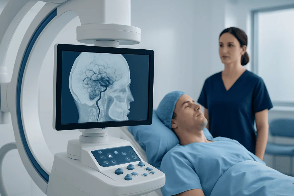
Brain AVM symptoms depend on the AVM’s size, location, blood flow pattern, and whether bleeding has occurred. Some people have no symptoms for years. Others may develop neurological symptoms because the AVM irritates nearby brain tissue, affects blood flow, or causes pressure from enlarged vessels.
Possible symptoms include seizures, recurring headaches, weakness or numbness on one side of the body, difficulty speaking or understanding speech, problems with balance or coordination, vision changes, dizziness, or memory and concentration difficulties. In children and young adults, a first seizure may be the symptom that leads to diagnosis.
If an AVM bleeds, symptoms often appear suddenly. A person may experience a sudden severe headache, nausea or vomiting, loss of consciousness, new weakness, confusion, speech difficulty, or a seizure. These symptoms require emergency medical attention, because bleeding in or around the brain needs rapid assessment and care.
Causes and Risk Factors

The exact cause of most brain AVMs is not fully understood. They are generally considered developmental vascular abnormalities, meaning the abnormal vessel connections form as the brain and blood vessels develop. In most cases, a brain AVM is not caused by lifestyle, stress, diet, or an injury.
Most AVMs occur without a known family pattern. Rarely, vascular malformations can be associated with inherited conditions, such as hereditary hemorrhagic telangiectasia. If there is a personal or family history of multiple vascular malformations, frequent nosebleeds, or similar findings in other organs, a doctor may consider genetic evaluation.
Risk factors that influence the behavior of an AVM are different from risk factors for developing one. Important features include whether the AVM has bled before, whether it is located deep in the brain, whether it drains through deep veins, whether there are associated aneurysms, and the AVM’s size and accessibility. These details are assessed with imaging and specialist review.
Bleeding Risk and What It Means
Bleeding risk is one of the most important parts of brain AVM decision-making. A hemorrhage can be small or large, and its effects depend on where it occurs and how much brain tissue is affected. Some people recover well after treatment and rehabilitation, while others may have lasting neurological symptoms.
The risk of future bleeding is usually higher after an AVM has already bled than in an AVM that has never caused hemorrhage. Certain anatomical features may also raise concern, such as deep location, deep venous drainage, high-pressure flow patterns, venous narrowing, or aneurysms on feeding arteries or within the AVM. These features do not automatically mean treatment is required, but they are important in planning.
Doctors balance bleeding risk against treatment risk. Because the brain controls movement, speech, sensation, vision, thinking, and other vital functions, any intervention must be considered carefully. For some AVMs, the safer option may be monitoring with regular imaging; for others, treatment may offer a meaningful reduction in future risk or symptom burden.
Diagnosis and Imaging Tests
Diagnosis usually begins with a neurological examination and brain imaging. MRI can show the AVM, surrounding brain tissue, old bleeding, and the relationship to important brain areas. CT may be used urgently when bleeding is suspected because it can quickly detect fresh blood in the brain.
CT angiography or MR angiography can outline arteries and veins and help identify the vascular structure. The most detailed test is cerebral digital subtraction angiography, often called catheter angiography. During this procedure, a specialist guides a thin catheter through a blood vessel, injects contrast dye, and takes real-time X-ray images to map blood flow through the AVM.
Angiography helps the care team understand feeding arteries, draining veins, associated aneurysms, and whether endovascular treatment is technically possible. Functional MRI, tractography, or other advanced imaging may be used when an AVM is near areas that control speech, movement, or vision. These tests support safer, more personalized treatment planning.
Treatment Options, Including Endovascular Care
Brain AVM treatment is individualized. The care team considers age, symptoms, prior bleeding, AVM size, location, blood flow, and the person’s overall health and preferences. The main options are observation, endovascular embolization, microsurgical removal, stereotactic radiosurgery, or a staged combination of these treatments.
Endovascular embolization is a minimally invasive catheter-based procedure. A neurointerventional specialist guides a tiny catheter through the blood vessels to the AVM and delivers an embolic material, such as a medical glue-like or liquid agent, to reduce or block abnormal blood flow. Embolization may be used to treat selected AVMs directly, reduce bleeding from a specific weak point, or make surgery or radiosurgery safer and more effective.
Microsurgery aims to remove the AVM through an operation, usually when the AVM is in a surgically accessible location and the expected benefit outweighs the risk. Stereotactic radiosurgery uses focused radiation beams to gradually close the abnormal vessels over time; it is often considered for smaller AVMs or AVMs in locations where open surgery is higher risk. Observation may be recommended when the AVM is low risk, very difficult to treat safely, or found incidentally without symptoms.
No single treatment is best for every brain AVM. Decisions are strongest when made by a multidisciplinary vascular neurology and neurosurgery team experienced in both surgical and endovascular care. The goal is to protect neurological function while addressing symptoms and future bleeding risk as safely as possible.
Prevention, Self-care, and Follow-up
There is no proven way to prevent a brain AVM from forming. Self-care focuses on general brain and vascular health, symptom awareness, and following the treatment plan. People with a known AVM should discuss safe physical activity, work, travel, pregnancy planning, and medications with their doctor, because recommendations vary depending on the AVM’s features.
If seizures have occurred, a neurologist may prescribe anti-seizure medication and provide guidance about driving, swimming, heights, machinery, and other safety issues. Headache management should be reviewed medically, especially if headaches are new, changing, or severe. It is important not to stop prescribed medicines without medical advice.
Follow-up imaging may be needed after observation, embolization, surgery, or radiosurgery. After radiosurgery, closure of the AVM is not immediate, so monitoring continues until imaging confirms the result. After surgery or embolization, angiography may be used to confirm whether the abnormal vessels have been fully treated.
Patients and families often benefit from clear explanations and a written plan for emergency symptoms. At Acibadem International, multidisciplinary specialists in JCI-accredited hospitals evaluate and treat brain AVMs for international patients, including diagnostic imaging, neurosurgical planning, and endovascular procedures when appropriate.
When to See a Doctor
A person should see a doctor if they have unexplained seizures, recurring headaches with neurological symptoms, weakness, numbness, speech difficulty, vision changes, balance problems, or episodes of confusion. These symptoms do not always mean an AVM is present, but they deserve medical evaluation.
Emergency care is needed for sudden severe headache, loss of consciousness, a new seizure, sudden weakness or numbness, facial drooping, trouble speaking, severe dizziness, new vision loss, or symptoms that resemble a stroke. Fast evaluation can identify bleeding, stroke, infection, migraine complications, or other urgent conditions.
Anyone already diagnosed with a brain AVM should keep regular appointments and ask the care team which symptoms require urgent attention. A second opinion from a vascular neurosurgery or neurointerventional team can be helpful when treatment choices are complex or when observation and intervention both seem reasonable.
Frequently asked questions
What is a brain AVM?
A brain AVM is an abnormal connection between arteries and veins in the brain. Blood bypasses the normal capillary network, which can create high-flow pressure inside fragile vessels. Some AVMs never cause symptoms, while others may lead to seizures, headaches, or bleeding.
Is a brain AVM the same as an aneurysm?
No. An aneurysm is a bulging weak area in a blood vessel wall, while an AVM is a tangle of abnormal artery-to-vein connections. However, some AVMs can have associated aneurysms, and this may influence bleeding risk and treatment planning.
How serious is brain AVM bleeding risk?
Bleeding risk varies widely and depends on the AVM's structure, location, drainage pattern, and whether it has bled before. A prior hemorrhage, deep location, deep venous drainage, or associated aneurysms may raise concern. A specialist can estimate risk more accurately after reviewing MRI, angiography, and the person's symptoms.
Can endovascular embolization cure a brain AVM?
In selected cases, embolization may completely close a brain AVM, but it is often used as part of a combined treatment plan. It can reduce blood flow, target high-risk weak points, or prepare an AVM for surgery or radiosurgery. The likelihood of cure depends on the AVM's size, anatomy, and accessibility.
Does every brain AVM need treatment?
No. Some AVMs are monitored rather than treated, especially if they have never bled and are located in areas where treatment could cause significant neurological risk. Observation may include periodic imaging and symptom management. The decision should be individualized with an experienced vascular neurology or neurosurgery team.
What symptoms should be treated as an emergency?
A sudden severe headache, new seizure, loss of consciousness, sudden weakness, facial drooping, speech difficulty, confusion, or vision loss should be treated as an emergency. These symptoms can occur with AVM bleeding or other urgent brain conditions. Immediate medical evaluation is the safest approach.
References
- American Heart Association/American Stroke Association
- National Institute of Neurological Disorders and Stroke
- American Association of Neurological Surgeons
- Society of NeuroInterventional Surgery
- Mayo Clinic
This article is for general information only and is not a substitute for professional medical advice. Please consult a qualified doctor about your individual situation.
Explore treatments in Turkey — costs, top hospitals & a free quote
JCI-accredited · board-certified surgeons · reply within 24h