CT Scan for Pregnant Woman: Preparation, Procedure and Results

CT is often avoided during pregnancy when another test can provide the needed answer, but it should not be delayed when it is medically important. CT scans of body parts away from the abdomen and pelvis generally expose the fetus to very little scattered radiation.
Key Takeaways
- CT is often avoided during pregnancy when another test can provide the needed answer, but it should not be delayed when it is medically important.
- CT scans of body parts away from the abdomen and pelvis generally expose the fetus to very little scattered radiation.
- Ultrasound and MRI are commonly considered first when suitable, although CT may be the best test for certain emergencies.
- Patients should tell the imaging team about a known, possible or recently discovered pregnancy before the scan.
- Most CT scans are completed within minutes, and patients can usually return to normal activities soon afterward.
- An unexpected CT scan before pregnancy was known rarely means there will be harm; the radiology and obstetric teams can review the exposure if needed.
A CT scan for pregnant woman is not usually the first imaging choice, but it can be important when it is needed to diagnose an urgent condition. The care team carefully considers the body area being scanned, pregnancy stage, alternative tests and whether contrast material is needed.
Overview: Is a CT Scan Safe During Pregnancy?
A CT scan for pregnant woman may be recommended when doctors need fast, detailed images to assess a potentially serious illness or injury. Although CT uses ionising radiation and is therefore used thoughtfully in pregnancy, the potential benefit of reaching an accurate diagnosis can outweigh the risk when urgent care is required.
The decision is individual. Clinicians consider what part of the body needs imaging, how far the pregnancy has progressed, the reason for the examination and whether ultrasound or MRI could answer the same clinical question. A scan of the head, chest or limbs usually results in very low fetal exposure because the fetus is outside the direct scan area, while abdominal or pelvic CT needs more careful consideration.
CT is not used to routinely examine the fetus. It is an imaging tool for the pregnant patient, for example when doctors need to investigate major trauma, suspected blood clots in the lungs, certain abdominal emergencies or complications that cannot safely wait for other testing.
How CT Imaging Works and When It May Be Needed
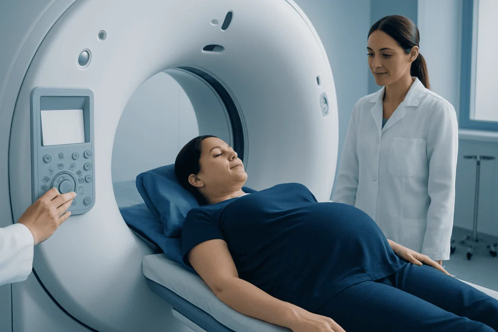
Computed tomography uses a rotating X-ray source and computer processing to create detailed cross-sectional images of the body. These images can show bones, organs, blood vessels, bleeding, inflammation and some injuries more clearly or more quickly than standard X-rays. Depending on the concern, a contrast agent may be given into a vein to make blood vessels and particular tissues easier to evaluate.
During pregnancy, ultrasound is often the first imaging test for pelvic, abdominal and pregnancy-related concerns because it does not use radiation. MRI may also be useful for many questions because it uses magnetic fields and radio waves rather than X-rays. However, neither alternative is ideal for every clinical situation. CT can be particularly valuable when speed, image detail or availability is essential.
Examples include evaluating a serious head injury after an accident, looking for internal bleeding, identifying certain kidney or bowel complications, or assessing a suspected pulmonary embolism. The referring doctor and radiologist select the examination that is most likely to provide an answer while keeping exposure as low as reasonably achievable.
What Happens When You Do a CT Scan While Pregnant?
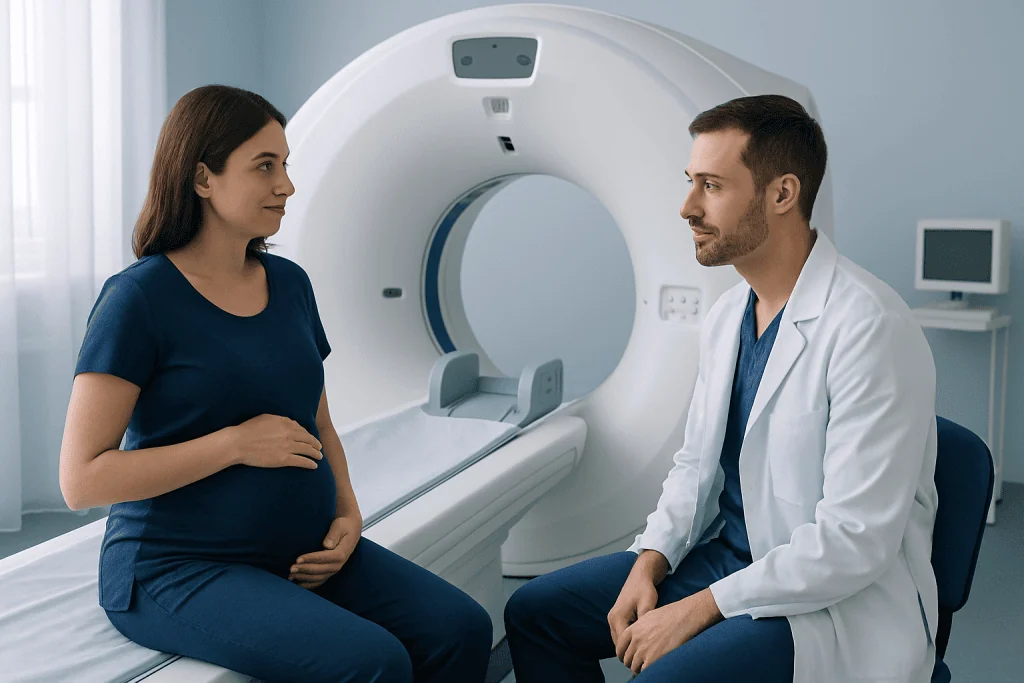
Before a CT scan, the imaging team reviews the reason for the examination and confirms whether pregnancy is known or possible. If pregnancy is confirmed, the radiologist may adjust the protocol, recommend another imaging test, limit the scanned area or proceed with CT if it is the safest way to answer an urgent medical question.
For a CT scan of a body area distant from the uterus, such as the head, neck, chest or an arm or leg, fetal exposure is usually from minimal scattered radiation rather than the direct X-ray beam. For abdominal and pelvic studies, the fetus may be in or near the imaging field, so the team pays particular attention to whether CT is necessary and how the scan can be optimised.
The purpose of these precautions is not to withhold needed care. Untreated conditions such as severe infection, internal injury or a blood clot can pose significant risks to both the pregnant person and the pregnancy. When a clinically necessary CT is recommended, it is generally because the expected information can guide safer, timely treatment.
If iodine-based contrast is considered, clinicians weigh the anticipated diagnostic value against the situation. It can cross the placenta, but it may be used when necessary. The care team can explain why contrast is or is not recommended and whether any follow-up is appropriate after birth.
What Happens if You Get a CT Scan and You Didn't Know You Were Pregnant?
Discovering a pregnancy after a CT scan can be worrying, but it is important not to assume that the scan has caused harm. In many situations, especially when the head, chest or extremities were scanned, fetal exposure is extremely low because the uterus was not directly imaged. A single diagnostic examination is generally very different from the high radiation exposures associated with known developmental effects.
The patient should contact the doctor or obstetric care provider and tell them the date of the scan, the body region examined and whether contrast was used. The radiology department can review the specific study and, when appropriate, estimate fetal exposure. This assessment helps the care team provide individualised reassurance and follow-up rather than relying on general assumptions.
Routine pregnancy care usually continues unless the treating clinicians advise otherwise. The value of reviewing the exposure is to document what happened, answer questions accurately and plan care based on the actual examination, stage of pregnancy and overall health.
Do They Give You a Pregnancy Test Before a CT Scan?
Imaging facilities commonly ask people of childbearing potential whether they are pregnant or could be pregnant before an examination that may expose the uterus or pelvis to radiation. This may be done verbally, through a written screening form or both. It is important to answer as accurately as possible, including if there is a missed period, unprotected intercourse, fertility treatment or uncertainty about pregnancy status.
A pregnancy test is not required before every CT scan. Whether testing is offered or requested depends on the planned body area, the urgency of the scan, local policies and the clinical situation. For example, a pregnancy test may be more relevant before a non-urgent abdominal or pelvic CT than before an urgent head CT.
In an emergency, medical teams may need to proceed with imaging before a test result is available if delaying diagnosis could be dangerous. They still use appropriate precautions and select the most suitable test for the patient’s condition. Patients should never avoid disclosing a possible pregnancy out of concern that care will be refused; the information helps the team make imaging safer.
Preparation, Step-by-Step Procedure and Recovery
Preparation depends on the type of CT. Patients should bring relevant medical information and tell the team about pregnancy, allergies, kidney problems, thyroid disease, diabetes medicines and previous reactions to contrast. If contrast is planned, the team may give instructions about eating or drinking beforehand; patients should follow the directions provided by their own imaging department.
During the examination, the patient lies on a narrow table that moves through the doughnut-shaped CT scanner. The radiographer positions the body carefully and may ask the patient to hold their breath briefly. The scanner itself does not touch the body, and the imaging portion commonly takes only a few minutes. If intravenous contrast is used, a temporary warm sensation or metallic taste can occur.
After a scan without contrast, most people can resume usual activities immediately. Following contrast, staff may briefly observe the patient, particularly if there is a history of allergies or other health concerns. Drinking fluids is often encouraged unless a doctor has advised fluid restriction. A radiologist interprets the images and sends a report to the referring clinician, who explains what the findings mean in the context of symptoms and pregnancy.
For complex imaging decisions, CT scanning may be coordinated by radiology, obstetrics, emergency medicine and other relevant specialties. Acibadem International’s multidisciplinary specialists and JCI-accredited hospitals support diagnosis and treatment planning for international patients when clinically appropriate.
Can You See a Baby on a CT Scan?
A fetus may be visible on a CT scan if the abdomen or pelvis is included in the images, but CT is not the preferred method for viewing or monitoring a baby. Ultrasound is the standard imaging method for evaluating fetal growth, heartbeat, anatomy, placental position and many pregnancy-related concerns because it does not use ionising radiation.
MRI may sometimes be used for additional evaluation of the fetus or placenta when ultrasound does not provide enough information. CT is reserved for maternal indications when it is needed to diagnose or manage the pregnant patient’s condition. If a fetus is incidentally seen on a CT study, the CT images should not replace dedicated prenatal assessment.
Anyone with questions about fetal development, bleeding, pain, reduced fetal movement later in pregnancy or other pregnancy symptoms should contact their maternity care team. Prompt assessment with the most appropriate pregnancy-focused test is more useful than relying on incidental CT findings.
Risks, Benefits and When to Seek Medical Care
The main potential risk of CT during pregnancy is radiation exposure to the developing fetus, particularly when the abdomen or pelvis is directly scanned. Risk depends on dose, body region and timing in pregnancy. Radiology professionals use modern protocols designed to obtain diagnostically useful images with the lowest practical radiation exposure. Shielding alone does not replace careful scan planning and is not suitable for every examination.
The benefit is the ability to diagnose time-sensitive conditions accurately. Missing or delaying treatment for serious injury, internal bleeding, appendicitis, severe infection or a possible blood clot can be more harmful than a justified imaging study. Decisions should be shared where possible, with clear discussion of why CT is recommended and what alternatives may be available.
When to seek medical care: urgent medical assessment is important for severe or worsening chest pain, sudden shortness of breath, coughing blood, fainting, significant injury, severe abdominal pain, heavy vaginal bleeding, high fever, confusion, weakness on one side of the body or a severe new headache. Pregnant people should contact their obstetric team or emergency services according to local guidance rather than delaying care because they are concerned about imaging.
For non-urgent questions, patients can ask their doctor whether ultrasound or MRI is suitable, what body region will be scanned, whether contrast is necessary and how the result may change treatment. Understanding the purpose of the test can make a necessary CT scan feel more manageable.
Frequently asked questions
Is a CT scan during pregnancy always dangerous?
No. CT uses radiation, so it is considered carefully during pregnancy, but a medically necessary scan should not automatically be avoided. The likely benefit of diagnosing an urgent condition may outweigh the potential risk, especially when the scan is outside the abdomen and pelvis.
Is MRI safer than CT in pregnancy?
MRI does not use ionising radiation and is often considered when it can answer the clinical question. However, CT may be quicker, more available or better suited to certain emergencies, injuries and vascular concerns. The best examination depends on the suspected condition and how urgently answers are needed.
Can a chest CT affect the baby?
In a chest CT, the fetus is usually outside the direct imaging field, so fetal exposure is generally very low and mainly due to scattered radiation. A chest CT may be needed to investigate serious conditions such as a suspected blood clot in the lungs. The radiology team can tailor the examination to the clinical need.
Should a pregnant patient refuse CT contrast?
Contrast decisions should be based on the reason for the scan. Iodine-based contrast can cross the placenta, but clinicians may use it when it is important for diagnosing a potentially serious maternal condition. The patient should ask why contrast is recommended and discuss any personal medical concerns with the care team.
How long does a CT scan take during pregnancy?
The actual image acquisition is often completed within minutes, although preparation, safety screening and positioning take longer. An examination involving intravenous contrast may require additional time. Most patients can return to normal activities shortly afterward unless their condition or contrast instructions require otherwise.
Will a CT scan show pregnancy complications?
CT is not the usual first test for pregnancy complications and is not a substitute for prenatal ultrasound. Ultrasound, and sometimes MRI, are generally better suited to assessing the fetus, placenta and many pelvic pregnancy concerns. CT may be used if doctors need to investigate an urgent maternal condition that other imaging cannot adequately assess.
References
- American College of Radiology
- American College of Obstetricians and Gynecologists
- Radiological Society of North America
- International Atomic Energy Agency
- U.S. Food and Drug Administration
This article is for general information only and is not a substitute for professional medical advice. Please consult a qualified doctor about your individual situation.
Explore treatments in Turkey — costs, top hospitals & a free quote
JCI-accredited · board-certified surgeons · reply within 24h
Add us as a Preferred Source to see more of our trusted health content across Google Search, AI Overviews and Discover.