How Gamma Radiation Is Produced: How It Works, Results and What to Expect

Gamma rays are high-energy electromagnetic waves released during nuclear changes and certain particle interactions. Medical gamma radiation is generated, measured and delivered under strict safety procedures.
Key Takeaways
- Gamma rays are high-energy electromagnetic waves released during nuclear changes and certain particle interactions.
- Medical gamma radiation is generated, measured and delivered under strict safety procedures.
- The health effect of gamma radiation depends on dose, duration, distance and the part of the body exposed.
- Focused gamma-ray treatment can target some tumors and brain conditions without an open surgical incision.
- Gamma rays do not remain in the body after external radiation treatment ends.
- People with possible radiation exposure or new concerning symptoms should seek prompt medical advice.
Gamma radiation is produced when an unstable atomic nucleus releases excess energy, or when certain high-energy particle interactions occur. In medicine, carefully controlled gamma rays may be used for imaging and for focused treatments such as stereotactic radiosurgery.
Overview: How Gamma Radiation Is Produced
How gamma radiation is produced depends on the setting. Most gamma rays are emitted when an unstable atomic nucleus changes into a more stable form and releases excess energy. This energy travels as a high-frequency electromagnetic wave called a gamma photon. Gamma rays may also arise from high-energy processes in space and from interactions involving particles and matter.
In healthcare, gamma radiation is produced in controlled equipment or from sealed radioactive sources. It can be used to create images of body processes or to deliver a planned dose of radiation to a precisely defined target. Medical teams select the source, dose and technique according to the purpose of the examination or treatment.
Gamma radiation is not automatically harmful or beneficial. Its effects depend on the amount of energy absorbed, the duration of exposure, the distance from the source and whether exposure is limited to a targeted area. Medical uses follow radiation-safety principles designed to keep exposure as low as reasonably achievable while obtaining the needed diagnostic or treatment benefit.
How Does Gamma Radiation Work?
Gamma rays are a form of ionizing radiation. This means they carry enough energy to remove electrons from atoms and molecules. When gamma rays pass through tissue, they can ionize molecules directly or create charged particles that cause ionization. This process can alter cell structures, including DNA.
For diagnostic nuclear medicine, a small amount of a radiopharmaceutical may be introduced into the body. The substance collects in particular tissues or follows a biological process, then emits gamma photons that are detected outside the body by specialized cameras. The resulting images can help clinicians assess organ function, blood flow or abnormal tissue activity.
For treatment, radiation is aimed at a tumor, vascular abnormality or other carefully selected target. Radiation can damage the DNA of targeted cells, limiting their ability to divide. Healthy cells can also be affected, which is why treatment planning seeks to concentrate the dose on the target and reduce exposure to surrounding tissue.
One form of highly focused treatment is Gamma Knife radiosurgery, which uses multiple precisely directed beams that meet at an intracranial target. Each individual beam has a relatively low effect along its path, while the combined dose is strongest where the beams converge.
Is Gamma Radiation Bad for Humans?

Gamma radiation can harm human tissue when exposure is sufficiently high or uncontrolled. Because gamma rays penetrate deeply, large doses may injure cells and increase the long-term risk of cancer. Very high whole-body exposures can cause radiation illness, but these circumstances are uncommon outside serious occupational, industrial or nuclear incidents.
Medical exposure is different from accidental exposure because it is justified by a clinical need, planned in advance and monitored. Imaging tests use the lowest practical amount of radiation, while radiation treatment deliberately uses higher localized doses to control or destroy abnormal cells. The expected benefit must outweigh the potential risk.
Patients should tell their care team if they are pregnant, could be pregnant, are breastfeeding or have had recent imaging or radiation treatment. The team can determine whether an examination should be adjusted, postponed or replaced by another approach. Questions about individual risk are best discussed with the doctor, radiologist, nuclear medicine physician or radiation oncologist involved in care.
Gamma-Ray Treatment: Candidacy and Planning
Gamma-ray-based treatment is not appropriate for every condition. Candidacy depends on the diagnosis, target location and size, prior treatment, general health and the potential effect on nearby organs. Focused cranial radiosurgery may be considered for selected brain tumors, certain benign tumors, arteriovenous malformations, trigeminal neuralgia and some other neurological conditions.
For example, some people with brain tumors may be assessed by a multidisciplinary team to determine whether surgery, conventional radiotherapy, systemic treatment, focused radiosurgery, observation or a combination is most suitable. The final plan is individualized and based on imaging, pathology when available, symptoms and treatment goals.
Before treatment, specialists review high-quality MRI, CT or other scans to identify the target and critical nearby structures. They use dedicated planning software to calculate beam directions and radiation dose. In many cases, a neurosurgeon, radiation oncologist, medical physicist and radiology team work together to prepare the plan.
- Focused gamma-ray treatment may be useful for small, well-defined targets.
- It may be considered when open surgery would carry substantial risk or is not preferred.
- Some larger, diffuse or rapidly progressing conditions need another treatment approach.
- Previous radiation and the sensitivity of surrounding tissue influence decisions.
What Happens During a Gamma Radiation Procedure?
The exact steps vary between diagnostic tests and radiation treatments. For a nuclear medicine scan, a radiopharmaceutical may be injected, swallowed or inhaled. There may be a waiting period while it distributes through the body, followed by imaging with a gamma camera. Most scans are painless, although remaining still can be important for image quality.
For Gamma Knife radiosurgery, patients usually attend a planning visit or have planning completed on the treatment day. A head frame or a custom immobilization mask helps keep the head in the same position throughout imaging and treatment. MRI, CT or angiography may be used to map the target accurately.
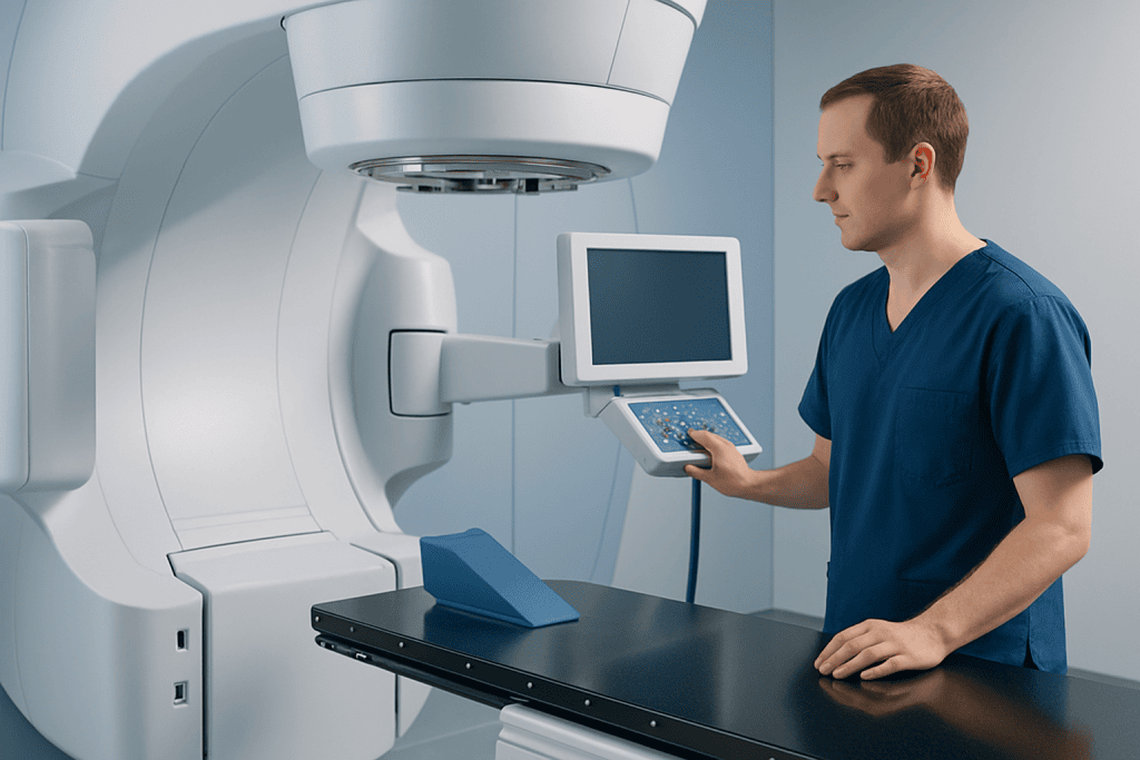
After the treatment plan is confirmed, the patient lies on a treatment couch while the machine delivers the programmed beams. The procedure itself is generally painless. The treatment team monitors the patient from outside the room and can communicate throughout. Depending on the treatment plan, the session may last from minutes to several hours.
Most focused gamma-ray procedures are outpatient treatments, but the care team will provide individual instructions. Patients may need a companion to take them home, especially if sedation, contrast material or medications were used. They should follow any restrictions related to driving, work and usual medicines.
Benefits, Risks and Recovery Timeline
The principal benefit of focused gamma-ray treatment is that it can deliver radiation accurately without an open surgical incision. This may reduce the need for general anesthesia and may allow a faster return to usual activities than traditional surgery for appropriately selected patients. However, it does not provide an immediate physical removal of a tumor or lesion.
Results often develop gradually. Depending on the condition, a treated tumor may stop growing, shrink over months, or show treatment-related changes on follow-up imaging. For vascular abnormalities, closure can take time. Symptoms may improve gradually, remain unchanged for a period or require additional treatment. Regular scans and clinical appointments are important to assess response.
Short-term effects can include tiredness, headache, temporary scalp tenderness, nausea or swelling near the treatment target. Side effects depend greatly on the treated area and the dose. Less common but potentially serious risks can include brain swelling, seizures, new neurological symptoms, injury to nearby nerves or tissue, radiation necrosis, and the need for further treatment.
Recovery after an uncomplicated outpatient radiosurgery session is commonly brief. Some people rest for the remainder of the day and resume many regular activities within one or two days, while others need longer based on symptoms and the condition being treated. The care team may prescribe medicines to manage swelling or symptoms and will explain the follow-up schedule.
Is Gamma the Deadliest Radiation?
Gamma radiation is among the more penetrating types of ionizing radiation, so it can travel through the body and affect internal organs when exposure is significant. However, describing one radiation type as the “deadliest” is not medically precise. Harm depends on the radiation dose, exposure time, distance from the source, shielding, route of exposure and the tissues involved.
Alpha particles, for example, do not penetrate intact skin well but can be very harmful if alpha-emitting material enters the body through breathing, swallowing or a wound. Beta radiation has different penetration and exposure characteristics. Gamma rays and X-rays can penetrate more deeply, so shielding and controlled use are especially important.
Radiation professionals measure and manage exposure using standardized safety practices. In clinical care, trained teams use equipment checks, treatment planning, quality assurance and protective procedures to limit unnecessary radiation to patients, staff and visitors.
How Long Do Gamma Rays Last?
Gamma rays themselves do not “last” in the way a substance remains in the body. A gamma photon travels at the speed of light until it interacts with matter, where it may be absorbed or scattered. After external-beam radiation or Gamma Knife treatment, the patient does not become radioactive and does not emit gamma rays to other people.
What may last is the radioactivity of a source or radiopharmaceutical. Radioactive materials decay over time according to their physical half-life, which differs for each isotope. In nuclear medicine, the selected radiopharmaceutical usually loses radioactivity over a predictable period and is also cleared from the body through normal biological processes.
After a nuclear medicine test or therapy involving radioactive material, the clinical team may give temporary instructions about hydration, toilet hygiene, close contact with young children or pregnant people, and travel. These instructions vary by isotope and procedure, so patients should follow the specific advice they receive rather than applying general rules.
When to Seek Medical Care
People should contact their medical team promptly after radiation treatment if they develop a severe or worsening headache, repeated vomiting, new weakness or numbness, confusion, vision changes, a seizure, fever, or difficulty speaking or walking. These symptoms do not always mean a serious complication, but they need timely assessment, particularly after treatment involving the brain.
Anyone who believes they have had an unplanned or substantial radiation exposure should move away from the source if it is safe to do so and contact local emergency services or public health authorities for instructions. They should not attempt to handle a suspected radioactive object or source.
For planned care, it is helpful to ask about the purpose of radiation, expected benefits, alternatives, possible side effects and the follow-up plan. Acibadem International’s multidisciplinary specialists and JCI-accredited hospitals assess and treat suitable international patients with coordinated radiation and neurological care.
Frequently asked questions
How gamma radiation is produced in medicine?
In medicine, gamma radiation may come from sealed radioactive sources or from radiopharmaceuticals used in nuclear medicine. The source is selected and controlled for a specific imaging or treatment purpose. Equipment, treatment planning and safety checks help ensure that radiation is delivered or measured appropriately.
How does gamma radiation work?
Gamma radiation carries energy that can ionize atoms and molecules as it passes through material. In imaging, emitted gamma photons are detected by specialized cameras to show body processes. In treatment, a planned dose can damage the DNA of targeted abnormal cells and reduce their ability to grow or divide.
Is gamma radiation bad for humans?
Uncontrolled or high-dose gamma radiation can damage living tissue and raise health risks. In contrast, medical use is carefully planned when the expected diagnostic or treatment benefit outweighs the risk. The individual risk depends on the procedure, dose, area treated and personal health circumstances.
Is gamma the deadliest radiation?
No single radiation type is always the most dangerous. Gamma rays are highly penetrating, but health effects depend on dose, exposure route, time, distance and shielding. Other forms of ionizing radiation can be very harmful in different circumstances, especially if radioactive material enters the body.
How long do gamma rays last in the body?
External gamma-ray treatment does not leave gamma rays in the body or make a person radioactive. For nuclear medicine procedures, the radioactive tracer remains only temporarily and decays and clears according to the specific material used. The care team will provide personalized safety instructions when needed.
Does Gamma Knife treatment involve an actual knife?
No. Gamma Knife radiosurgery does not use a surgical blade or make an incision. It is a form of stereotactic radiosurgery that directs many focused radiation beams toward a target in the brain. It is called radiosurgery because of its precision, not because it involves conventional surgery.
References
- International Atomic Energy Agency
- U.S. Food and Drug Administration
- National Cancer Institute
- Radiological Society of North America
- World Health Organization
This article is for general information only and is not a substitute for professional medical advice. Please consult a qualified doctor about your individual situation.
Explore treatments in Turkey — costs, top hospitals & a free quote
JCI-accredited · board-certified surgeons · reply within 24h
Add us as a Preferred Source to see more of our trusted health content across Google Search, AI Overviews and Discover.