Muga Scan — Explained by Medical Evidence, Not Myths

A muga scan measures how well the heart pumps blood, especially the left ventricle. It uses a small amount of radioactive tracer and a special camera to create moving images of the beating heart.
Key Takeaways
- A muga scan measures how well the heart pumps blood, especially the left ventricle.
- It uses a small amount of radioactive tracer and a special camera to create moving images of the beating heart.
- Doctors often use it to track heart function before, during, or after certain cancer treatments or in people with heart disease.
- The test is generally safe, noninvasive, and valued for providing a precise ejection fraction measurement.
- Results must be interpreted in context with symptoms, medical history, and other heart tests.
A muga scan is a nuclear medicine heart test that shows how effectively the heart’s chambers pump blood. It is most often used to measure ejection fraction and to monitor heart function over time, especially when a person is receiving treatments that may affect the heart.
What a Muga Scan Shows
A muga scan is a type of nuclear medicine test that evaluates how well the heart pumps blood. The name comes from “multiple gated acquisition,” which refers to the way the test synchronizes images with the heartbeat. In everyday terms, it helps doctors see the heart in motion and calculate the ejection fraction, which is the percentage of blood pumped out of a ventricle with each beat.
This test is especially useful when a very consistent measurement of pumping function is needed over time. For example, a doctor may order a muga scan before and during treatment with medicines that can affect the heart muscle. It can also support the evaluation of symptoms such as shortness of breath, fatigue, or reduced exercise tolerance when heart function is a concern.
Unlike a standard X-ray or CT scan, a muga scan does not focus on heart anatomy alone. Instead, it emphasizes function. That is why it is often considered alongside other heart tests such as an echocardiogram or, in selected situations, cardiac MRI. Each test answers slightly different questions, and the best choice depends on the person’s medical needs.
How the Test Works
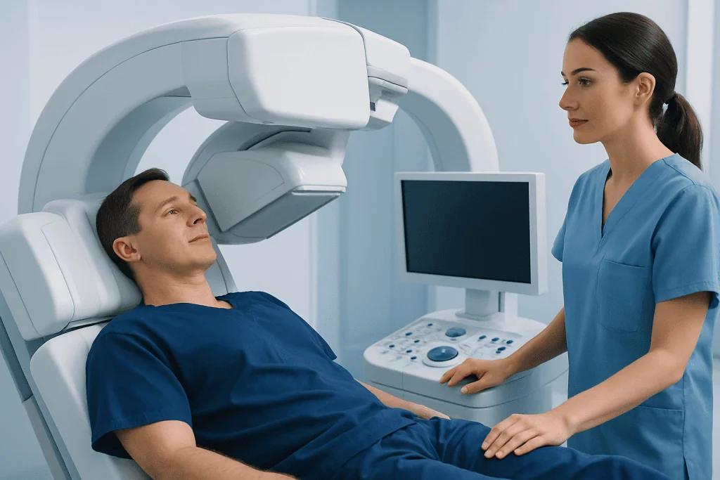
During a muga scan, a small amount of radioactive tracer is injected into a vein. The tracer attaches to red blood cells, allowing a special camera to track blood flow through the heart as it beats. Sticky patches called electrodes are placed on the chest to record the heartbeat, and the camera captures images timed to different points in the cardiac cycle.
The result is a moving set of images that shows how the ventricles fill and empty. From these images, the doctor can calculate the ejection fraction, most commonly for the left ventricle. This number helps indicate whether the heart’s pumping function is normal, mildly reduced, or more significantly impaired.
The amount of radiation used is low and is generally considered acceptable when the test is medically indicated. Even so, doctors usually order a muga scan only when the information is likely to guide care. If a person is pregnant, could be pregnant, or is breastfeeding, it is important to discuss this with the healthcare team before the test so the safest approach can be chosen.
Why Doctors Order a Muga Scan

The most common reason for a muga scan is to measure and follow ventricular function with a high degree of reproducibility. This is especially helpful in cancer care, where some chemotherapy or targeted therapies may affect the heart. In that setting, the test helps determine whether treatment can continue as planned or whether heart-protective steps are needed.
A doctor may also use a muga scan when there is a need to clarify or confirm findings from other tests. It can be useful in the assessment of heart failure, cardiomyopathy, prior heart damage, or unexplained symptoms suggestive of reduced pumping strength. People being evaluated for heart failure may have a muga scan if a precise ejection fraction measurement is important for decision-making.
In some cases, the test is ordered when an echocardiogram image is limited by body shape, lung disease, prior chest surgery, or other technical factors. Because muga scans are known for consistency when repeated, they can be helpful for tracking changes over months. Doctors may compare current results with prior studies to see whether heart function is stable, improving, or worsening.
What to Expect Before, During, and After
Preparation is usually simple. A person may be asked to avoid caffeine or certain medicines for a short time before the test, although instructions vary by center and by the reason the scan is being done. It is best to wear comfortable clothing and to follow any specific directions from the imaging team.
During the scan, the person typically lies still on an exam table while the gamma camera takes pictures from one or more angles. The test itself is painless, though the injection and electrode placement may cause minor temporary discomfort. It is important to remain still during image capture so the pictures are clear and accurate.
After the test, most people can return to normal activities right away unless told otherwise. Drinking fluids may be advised to help the body clear the tracer. Side effects are uncommon, but the healthcare team should be informed if there is redness at the injection site, unexpected symptoms, or concerns after going home.
- Bring a list of current medicines.
- Tell the team about pregnancy, possible pregnancy, or breastfeeding.
- Ask whether caffeine, exercise, or heart medications should be limited before the exam.
- Inform the staff if there is difficulty lying flat or staying still.
Understanding Muga Scan Results
The main value reported from a muga scan is the ejection fraction. In general, a higher number means the ventricle is pumping out a larger share of its blood with each beat, while a lower number suggests reduced pumping strength. However, the interpretation is not based on one number alone. Doctors also consider symptoms, blood pressure, medications, prior imaging, and the reason the scan was ordered.
A normal or near-normal result may reassure both patient and clinician that heart function is preserved at that time. A lower-than-expected result can suggest weakened heart muscle function and may prompt further evaluation or changes in treatment. For example, a person receiving therapies that affect the heart may need closer monitoring, a medication adjustment, or referral to a cardiologist.
Results do not diagnose every heart problem. A muga scan does not provide the same detail about heart valves, the pericardium, or coronary artery narrowing that other tests may provide. If doctors suspect conditions such as coronary artery disease or structural abnormalities, they may recommend additional evaluation, including tests such as echocardiography or other cardiac imaging.
Benefits, Limits, and Safety Considerations
One of the main strengths of a muga scan is accuracy and consistency in measuring ejection fraction over time. This makes it particularly useful for serial follow-up. The test is noninvasive, usually well tolerated, and does not require sedation in most adults.
At the same time, no single test is perfect. A muga scan focuses on pumping function and may not fully explain why the function is reduced. It also involves exposure to a small amount of ionizing radiation, so doctors balance the expected benefit of the information against the need to keep radiation exposure as low as reasonably possible.
Alternative tests may be chosen depending on the clinical question. An echocardiogram is widely used because it is convenient and does not use radiation. In more complex cases, doctors may consider advanced tests such as electrophysiology studies for rhythm-related concerns, though this is a different type of assessment and not a substitute for ventricular function imaging. The best testing plan is individualized.
When to Seek Medical Care
A muga scan is not an emergency treatment; it is a diagnostic test. Still, the symptoms that lead to the test can sometimes signal a significant heart problem. Medical care should be sought promptly if there is chest pain, severe shortness of breath, fainting, a new rapid or irregular heartbeat, or sudden swelling in the legs or abdomen.
People who already have known heart disease or are receiving treatments that may affect the heart should contact their doctor if they notice worsening fatigue, reduced exercise capacity, trouble breathing when lying flat, or unexplained weight gain from fluid retention. These changes may suggest that heart function needs reassessment.
Near the end of a patient’s evaluation, care is often coordinated between cardiology, oncology, and imaging specialists. Acibadem International’s multidisciplinary specialists in JCI-accredited hospitals diagnose and treat heart conditions for international patients when further assessment or follow-up is needed.
Frequently asked questions
What is a muga scan used for?
A muga scan is used to measure how well the heart pumps blood, especially the function of the left ventricle. It is often ordered to monitor heart function before, during, or after treatments that may affect the heart, or to help evaluate suspected heart weakness.
Is a muga scan the same as an echocardiogram?
No. Both tests can assess heart function, but they work in different ways. A muga scan uses a radioactive tracer and a special camera, while an echocardiogram uses ultrasound waves and can provide more detail about heart valves and structure.
How long does a muga scan take?
The full appointment often takes around one to two hours, depending on the imaging center and the exact protocol used. The imaging portion itself is usually shorter, but time is also needed for preparation, tracer injection, and positioning.
Is a muga scan painful or risky?
The test is generally not painful apart from the brief discomfort of a needle stick for the injection. Risks are usually low, but the scan does involve a small amount of radiation, so it is ordered when the expected medical benefit outweighs that exposure.
Can a muga scan detect blocked arteries?
Not directly. A muga scan shows how well the heart chambers pump but does not diagnose coronary artery blockages in the same way as stress testing, coronary CT, or angiography. If blocked arteries are suspected, other tests may be needed.
Do muga scan results always mean there is heart failure?
No. A reduced ejection fraction can occur for several reasons and does not automatically mean a person has heart failure symptoms or a specific diagnosis. Doctors interpret the result together with the person’s symptoms, medical history, examination, and other tests.
References
- American Heart Association
- National Heart, Lung, and Blood Institute
- Radiological Society of North America
- Society of Nuclear Medicine and Molecular Imaging
- European Society of Cardiology
This article is for general information only and is not a substitute for professional medical advice. Please consult a qualified doctor about your individual situation.
Explore treatments in Turkey — costs, top hospitals & a free quote
JCI-accredited · board-certified surgeons · reply within 24h