Optic Neuritis: Eye Pain, Vision Loss, and Neurology Evaluation

Optic neuritis commonly causes vision loss or blurred vision in one eye, often with pain that worsens when the eye moves. Many people recover a significant amount of vision, but the condition should be assessed promptly to confirm the diagnosis and check for related neurological conditions.
Key Takeaways
- Optic neuritis commonly causes vision loss or blurred vision in one eye, often with pain that worsens when the eye moves.
- Many people recover a significant amount of vision, but the condition should be assessed promptly to confirm the diagnosis and check for related neurological conditions.
- Magnetic resonance imaging, eye examination, visual field testing, and sometimes blood tests help identify the cause and guide care.
- Treatment may include observation, corticosteroids in selected cases, and management of an underlying condition such as multiple sclerosis, neuromyelitis optica, infection, or autoimmune disease.
- Sudden vision changes, severe eye pain, weakness, numbness, or symptoms in both eyes should be evaluated urgently by qualified specialists.
Optic neuritis is inflammation of the optic nerve, the pathway that carries visual information from the eye to the brain. It often causes eye pain, blurred or dim vision, and changes in color perception, and it may require both ophthalmology and neurology evaluation.
Overview
Optic neuritis is inflammation of the optic nerve, which connects the eye to the brain. When this nerve becomes inflamed, visual signals may not travel normally, leading to blurred, dim, or reduced vision. The condition can affect one eye or, less commonly, both eyes. It may appear suddenly over hours to days and is often noticed because vision changes are accompanied by discomfort around the eye.
Optic neuritis is important because it sits at the crossroads of eye health and nervous system health. Some cases occur after a viral illness or as part of an immune reaction. Others may be associated with neurological conditions such as multiple sclerosis, neuromyelitis optica spectrum disorder, or myelin oligodendrocyte glycoprotein antibody-associated disease. A careful evaluation helps doctors understand whether optic neuritis is an isolated event or part of a broader medical condition.
Although the symptoms can be unsettling, many patients experience substantial improvement over time, especially with appropriate diagnosis and follow-up. The key is not to ignore new vision loss or eye pain. Early assessment by an ophthalmologist, neuro-ophthalmologist, or neurologist can confirm the diagnosis, rule out other causes of vision loss, and identify the safest treatment plan.
Symptoms of Optic Neuritis
The most typical symptom of optic neuritis is vision loss in one eye that develops over a few days. Vision may become blurry, cloudy, dim, or less sharp. Some people describe a gray or dark area in the center of vision, while others notice that the entire visual field looks faded. Reading, recognizing faces, or seeing in low light may become more difficult.
Eye pain is also common, especially pain that worsens when the eye moves. This discomfort may feel like soreness behind the eye or pressure around the eye socket. Color vision can change as well; red objects may look washed out, dull, or less vivid in the affected eye. Some patients notice flashing lights, flickering, or brief visual disturbances with eye movement.
Symptoms can vary depending on the location and severity of inflammation. Common features include:
- Blurred or reduced vision, usually in one eye
- Pain with eye movement
- Reduced color brightness, especially red desaturation
- A central blind spot or patchy visual field loss
- Worsening vision with heat or exertion in some patients
- Occasional headache or tenderness around the eye
Optic neuritis usually does not cause a red eye, thick discharge, or severe surface irritation. Those signs may suggest other eye conditions. Because different eye and brain disorders can produce similar symptoms, a medical examination is needed rather than relying on symptoms alone.
Causes and Risk Factors

Optic neuritis is most often related to inflammation of myelin, the protective coating around nerve fibers. When myelin is disrupted, signals through the optic nerve slow down or become distorted. In many adults, optic neuritis may be associated with demyelinating disease, including multiple sclerosis. However, having optic neuritis does not automatically mean a person has multiple sclerosis; MRI findings, history, examination, and follow-up all help clarify risk.
Other immune-mediated conditions can also inflame the optic nerve. Neuromyelitis optica spectrum disorder and MOG antibody-associated disease may cause optic neuritis that is more severe, recurrent, or likely to involve both eyes. Autoimmune disorders such as sarcoidosis, lupus, or vasculitis can sometimes involve the optic nerve. Infections are less common but may include certain viral, bacterial, or inflammatory conditions depending on the patient’s medical history and geographic exposure.
Risk factors can differ by cause, but several patterns are recognized. Optic neuritis is more often diagnosed in young to middle-aged adults, and some forms are more common in women. A personal or family history of autoimmune disease, previous neurological symptoms, or recurrent episodes of vision loss may increase the need for broader evaluation. Certain medications, toxins, nutritional deficiencies, and inherited optic nerve disorders can mimic optic neuritis, so doctors consider these possibilities during diagnosis.
Because the causes are diverse, treatment is not one-size-fits-all. The medical team focuses on identifying whether the inflammation is typical, atypical, recurrent, infectious, autoimmune, or related to another neurological disorder. This distinction helps protect vision and guides long-term care.
Diagnosis and Neurology Evaluation
Diagnosis begins with a detailed history and eye examination. The doctor asks when symptoms started, whether pain occurs with eye movement, whether one or both eyes are affected, and whether there have been neurological symptoms such as numbness, weakness, imbalance, bladder changes, or unusual fatigue. A review of recent infections, autoimmune disease, medications, and previous episodes is also important.
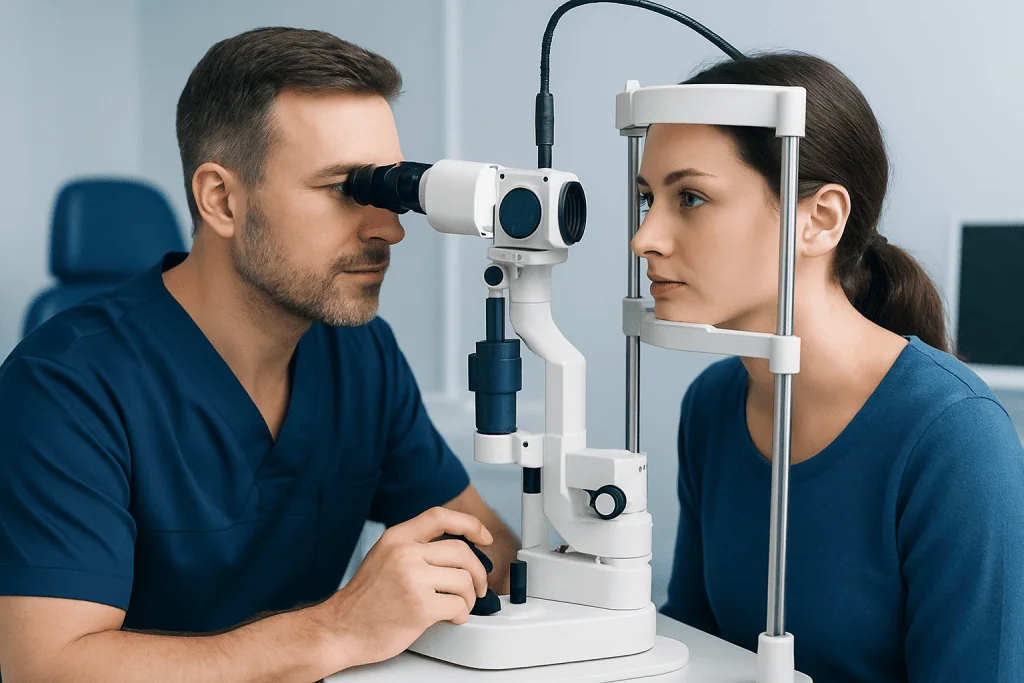
An ophthalmologist may check visual acuity, color vision, pupils, eye pressure, and the back of the eye. In optic neuritis, the pupil in the affected eye may respond less strongly to light, a finding called a relative afferent pupillary defect. The optic disc may look swollen in some patients, but it can also appear normal if inflammation is located behind the eye. Visual field testing can map areas of missing or reduced vision.
Magnetic resonance imaging of the brain and orbits with contrast is commonly used to look for optic nerve inflammation and to check for brain lesions that may suggest demyelinating disease. Optical coherence tomography can measure the retinal nerve fiber layer and help monitor optic nerve health over time. Visual evoked potentials may be used in selected cases to measure how quickly signals travel from the eye to the brain.
Blood tests may be recommended when symptoms are atypical, severe, recurrent, bilateral, or associated with systemic symptoms. Testing can look for infections, inflammatory markers, autoimmune antibodies, and specific antibodies linked to neuromyelitis optica spectrum disorder or MOG antibody-associated disease. In some cases, lumbar puncture is considered to examine cerebrospinal fluid. A neurology evaluation is especially useful when MRI findings, symptoms, or examination raise concern for a wider nervous system condition.
Treatment Options
Treatment depends on the type of optic neuritis, the severity of vision loss, and the suspected cause. Some typical cases improve without specific treatment, and the medical team may recommend close monitoring. In other cases, corticosteroids are used to reduce inflammation and speed visual recovery. Steroids may be given intravenously or by mouth depending on the clinical situation, but the decision should be individualized because risks and benefits vary.
For optic neuritis linked to multiple sclerosis or a high risk of future demyelinating events, neurologists may discuss disease-modifying therapies. These medications are not used simply to treat eye pain; they are considered when the overall neurological picture supports a diagnosis or elevated risk. The goal is to reduce future disease activity, not to guarantee a specific vision outcome. Patients should have a clear conversation about expected benefits, monitoring, and possible side effects.
If optic neuritis is related to neuromyelitis optica spectrum disorder, MOG antibody-associated disease, infection, or another autoimmune condition, treatment may be different from typical optic neuritis. Some patients need additional immune therapies, plasma exchange, antibiotics, antivirals, or longer-term preventive medication. This is why accurate diagnosis is essential before choosing therapy.
During recovery, vision may improve gradually over weeks to months. Some patients notice that colors, contrast, or fine detail take longer to recover than basic vision. Follow-up visits help document progress, watch for recurrence, and adjust treatment if symptoms do not improve as expected.
Recovery, Prevention, and Self-care
Recovery from optic neuritis is often gradual. Pain may improve before vision fully returns, and visual clarity can fluctuate during the healing period. Some people feel that vision worsens temporarily with heat, exercise, fever, or hot showers; this heat-related change usually reflects nerve signal sensitivity rather than new damage, but it should still be discussed with the doctor if persistent or severe.
Self-care cannot replace medical treatment, but it can support comfort and safety. Patients may benefit from resting the eyes during periods of visual strain, using good lighting for reading, and avoiding driving until vision is safe and legally adequate. Managing fever, staying hydrated, and pacing physical activity may help reduce temporary symptom worsening in some individuals.
There is no guaranteed way to prevent every case of optic neuritis. Prevention focuses on managing underlying conditions and reducing recurrence risk when a specific diagnosis is made. Patients with multiple sclerosis, neuromyelitis optica spectrum disorder, MOG antibody-associated disease, or systemic autoimmune disease should follow their specialist’s monitoring plan. Stopping prescribed immune or neurological medication without medical advice can increase the risk of relapse in some conditions.
Emotional support is also part of recovery. Sudden vision changes can affect work, reading, mobility, and confidence. Clear explanations, follow-up plans, and practical adjustments can make the recovery period less stressful. Patients should ask their healthcare team what changes are expected, what warning signs to watch for, and when to return for reassessment.
When to See a Doctor
Any new, unexplained vision loss should be assessed promptly, especially if it develops over hours or days, affects one eye, or is accompanied by pain with eye movement. A same-day or urgent evaluation is appropriate if vision loss is severe, both eyes are affected, symptoms are worsening quickly, or there are additional neurological signs such as weakness, numbness, trouble walking, double vision, or difficulty speaking.
Patients should also seek medical care if they have recurrent episodes of blurred vision, color fading, or eye pain, even if previous symptoms improved. Repeated attacks may indicate an underlying inflammatory or autoimmune condition that benefits from long-term management. People with known multiple sclerosis, neuromyelitis optica spectrum disorder, or other immune disorders should contact their specialist promptly when new visual symptoms occur.
Optic neuritis is best evaluated through collaboration between eye and nervous system specialists. Depending on the case, this may include an ophthalmologist, neuro-ophthalmologist, neurologist, radiologist, and immunology or infectious disease specialist. For international patients, Acibadem International’s multidisciplinary specialists and JCI-accredited hospitals can support diagnosis and treatment planning for optic neuritis and related neurological conditions.
Frequently asked questions
Is optic neuritis the same as multiple sclerosis?
No. Optic neuritis can occur in people with multiple sclerosis, but it can also happen as an isolated episode or due to other inflammatory, autoimmune, or infectious causes. MRI findings, neurological symptoms, examination results, and follow-up help determine whether multiple sclerosis is present or likely.
Does vision come back after optic neuritis?
Many people regain a significant amount of vision over weeks to months, especially in typical optic neuritis. Some may continue to notice subtle changes in color, contrast, or clarity. Recovery depends on the cause, severity, and whether there are recurrent attacks or other optic nerve conditions.
Why does optic neuritis cause pain when moving the eye?
The optic nerve is surrounded by tissues that can become inflamed and sensitive. When the eye moves, it can tug slightly on the inflamed area, causing discomfort behind or around the eye. Pain often improves as inflammation settles, sometimes before vision fully recovers.
What tests are commonly used for optic neuritis?
Doctors usually perform an eye examination, visual acuity testing, color vision testing, pupil assessment, and visual field testing. MRI of the brain and orbits is often recommended to confirm optic nerve inflammation and evaluate for demyelinating disease. Blood tests, optical coherence tomography, visual evoked potentials, or lumbar puncture may be used in selected cases.
Are steroids always needed for optic neuritis?
Not always. Some typical cases improve with observation and careful follow-up, while corticosteroids may be recommended to speed recovery or treat more severe inflammation. The choice depends on the clinical picture, MRI findings, underlying cause, and the patient’s overall health.
Can optic neuritis affect both eyes?
Yes, but typical optic neuritis more often affects one eye. Involvement of both eyes, very severe vision loss, recurrence, or poor recovery may suggest an atypical form and usually requires broader evaluation. Doctors may look for conditions such as neuromyelitis optica spectrum disorder, MOG antibody-associated disease, infection, or systemic autoimmune disease.
When is optic neuritis an emergency?
Urgent medical evaluation is needed for sudden or rapidly worsening vision loss, symptoms in both eyes, severe headache, new weakness or numbness, double vision, trouble walking, or difficulty speaking. These symptoms do not always mean a serious outcome, but they should be assessed quickly so the correct diagnosis and treatment can begin.
References
- American Academy of Ophthalmology
- National Institute of Neurological Disorders and Stroke
- Mayo Clinic
- Multiple Sclerosis International Federation
- The Optic Neuritis Study Group
This article is for general information only and is not a substitute for professional medical advice. Please consult a qualified doctor about your individual situation.
Explore treatments in Turkey — costs, top hospitals & a free quote
JCI-accredited · board-certified surgeons · reply within 24h