PET Scan Contrast: Preparation, Procedure and Results
PET imaging uses a radioactive tracer rather than conventional dye in most examinations. Some PET/CT scans also use iodine-based CT contrast to improve anatomic detail.
Key Takeaways
- PET imaging uses a radioactive tracer rather than conventional dye in most examinations.
- Some PET/CT scans also use iodine-based CT contrast to improve anatomic detail.
- Preparation commonly includes fasting, hydration and avoiding strenuous exercise, but instructions vary by scan type and health condition.
- Areas of increased tracer uptake are not automatically cancer; inflammation, infection and normal body activity can also appear active.
- Most people can resume usual activities soon after a PET scan, with simple precautions advised for a short period.
- People should tell the imaging team about pregnancy, breastfeeding, diabetes, kidney disease, allergies and all medicines before the appointment.
PET scan contrast usually means a small amount of radioactive tracer given before imaging; in PET/CT, an iodine-based CT contrast agent may also be used in some cases. The scan helps clinicians assess how tissues are functioning, but results always need interpretation alongside symptoms, medical history and other tests.
PET Scan Contrast: What It Means and How It Works
In everyday language, “PET scan contrast” may describe the substance used to make certain tissues easier to see. For most positron emission tomography (PET) scans, this is not a standard X-ray dye. It is a radiotracer: a tiny amount of a radioactive substance that is usually injected into a vein and then taken up by body tissues. The most commonly used tracer is fluorodeoxyglucose (FDG), a glucose-like substance.
Cells use energy at different rates. After FDG is given, the PET scanner detects signals from the tracer and creates images showing areas of higher or lower uptake. This can help clinicians evaluate cancer, inflammation, infection, heart muscle and some brain disorders. PET is frequently paired with computed tomography (CT) or magnetic resonance imaging (MRI) so functional PET information can be matched with detailed body anatomy.
A PET/CT examination may include a separate iodine-based CT contrast agent, given by mouth or vein, when clearer CT detail is needed. This is different from the PET radiotracer. The imaging team will explain which substances are planned and whether any additional preparation is required.
Who May Need a PET Scan and When Contrast Is Used
A PET scan may be recommended when a clinician needs more information about tissue activity than a standard CT, MRI or X-ray can provide. In oncology, it can help assess the extent of a known cancer, check response to treatment, investigate a concerning finding or look for possible recurrence. It may also be used in selected heart and neurological assessments.
Whether CT contrast is used depends on the clinical question, the body area being imaged and a person’s health history. Intravenous CT contrast can make blood vessels and organs more distinct, while oral contrast may help outline the digestive tract. Some PET/CT examinations do not require either type of CT contrast.
Before the scan, patients should inform the team about pregnancy or possible pregnancy, breastfeeding, diabetes, kidney problems, asthma, previous contrast reactions and medicines or supplements. These details help the clinician select the safest imaging protocol. A PET scan is one part of an assessment and does not independently confirm a diagnosis.
- FDG PET is often used to assess glucose use in tissues.
- Other tracers may be selected for prostate cancer, neuroendocrine tumors, heart conditions or brain conditions.
- CT contrast is considered separately from the PET tracer and is not appropriate for everyone.
Preparing for a PET Scan

Preparation supports accurate results because food, exercise, blood glucose and some medicines can affect tracer uptake. The imaging center will provide individual instructions, and those directions should take priority. Many FDG PET scans require fasting for several hours before the appointment, with plain water usually encouraged unless the care team says otherwise.
People with diabetes need personalized instructions because fasting and insulin or other glucose-lowering medicines must be managed safely. They should not stop prescribed medication without advice from the clinician coordinating the scan. It is also important to report recent infections, vaccinations, surgeries or biopsies, since these may temporarily increase activity in lymph nodes or healing tissues.
Wear comfortable clothing without metal where possible, and bring prior imaging reports or discs if requested. The team may check blood glucose before the tracer is administered. If glucose is outside the range needed for reliable imaging, the scan may need to be delayed or adapted.
What should you not do before a PET scan? Unless the imaging team gives different directions, patients should avoid eating during the specified fasting window, drinking sugary beverages, chewing gum or using products with sugar. They should also avoid strenuous activity for at least a day before many FDG PET scans, because recently exercised muscles can take up more tracer. Smoking, alcohol and caffeine instructions vary, so patients should follow the instructions for their particular scan.
What foods should I avoid 24 hours before a PET scan? For many FDG PET scans, clinicians may advise limiting carbohydrates and sugars before fasting, particularly for certain cardiac or specialized scans. Foods that can raise blood glucose include sweets, sweetened drinks, pastries, white bread, rice, pasta and some breakfast cereals. However, dietary plans differ by PET tracer and medical condition, so patients should not begin a restrictive diet unless their imaging provider has specifically recommended it.
Why do you have to rest the day before a PET scan? Resting reduces extra glucose use by skeletal muscles. Vigorous exercise, heavy lifting and prolonged physical exertion may produce increased muscle tracer uptake that can make images harder to interpret. Gentle normal activity is often acceptable, but the imaging center can clarify what level of activity is suitable.
Step by Step: What Happens During the Procedure
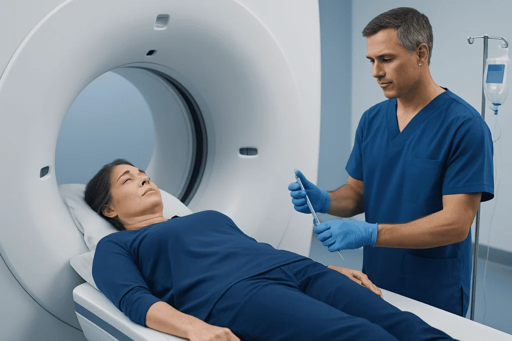
At check-in, staff confirm medical history, medications, allergies and preparation. A small blood sample may be taken to measure glucose. The radiotracer is generally injected through a vein in the arm or hand. The injection is similar to a routine blood draw and the tracer usually causes no noticeable sensation.
After the injection, the person rests quietly in a comfortable room while the tracer distributes through the body. This uptake period is often around 30 to 60 minutes, although timing varies by tracer and scan type. Keeping warm, relaxed and relatively still is helpful. Talking, chewing, walking around or exercising can affect tracer distribution in some studies.
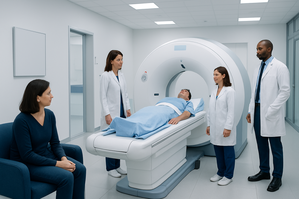
For imaging, the person lies on a narrow table that moves slowly through the scanner. PET/CT scanners are usually short, open rings rather than a long tunnel. The scan itself is painless, though lying still may be uncomfortable for people with pain, stiffness or anxiety. The complete visit commonly lasts a few hours because it includes preparation, uptake time and scanning.
If intravenous CT contrast is used, some people briefly notice warmth, a metallic taste or a feeling of needing to urinate. These sensations generally pass quickly. Staff monitor patients and are prepared to manage uncommon reactions.
Understanding PET Scan Images and Results
A radiologist or nuclear medicine physician reviews the PET images together with CT or MRI findings, prior scans and the reason for testing. The report is sent to the clinician who requested the study. Results may be available within several days, depending on the center and whether comparison with previous examinations is needed.
What color does cancer show on a PET scan? Cancer does not have one universal color on a PET scan. Image colors are chosen by the viewing software, and areas with higher tracer uptake may appear bright or “hot” in colors such as yellow, orange or red. A bright area is not automatically cancer: normal organs, muscles, inflammation, infection and healing tissue can also show increased uptake.
Likewise, a cancer may not always show strong uptake. Some cancers are less metabolically active, very small or better evaluated with a tracer other than FDG. For these reasons, the report describes the location and pattern of uptake rather than relying on color alone. A clinician may recommend blood tests, follow-up imaging or a biopsy when further clarification is needed.
In cancer care, PET findings can contribute to staging and treatment planning, but they are interpreted with pathology and other tests. Patients who are undergoing evaluation or treatment for cancer should discuss what their individual scan result means with their oncology team.
Benefits, Risks and Recovery Timeline
PET imaging can identify changes in tissue activity that may occur before structural changes are visible on other scans. When combined with CT or MRI, it can provide a more complete view of both function and anatomy. This information may help clinicians choose further tests, plan treatment or monitor response over time.
The radiation exposure from a PET scan comes from the radiotracer and, when included, the CT portion. The amount is carefully planned for the examination, and the tracer naturally loses radioactivity over time and leaves the body mainly through urine. Imaging is ordered when the expected clinical value outweighs the small radiation-related risk.
Reactions to the PET radiotracer are very uncommon. If iodine-based CT contrast is used, mild effects such as temporary warmth or nausea can occur, while allergic-type reactions are uncommon but possible. Kidney function may need review before certain contrast-enhanced CT studies. Anyone who has had a past contrast reaction should tell the team in advance.
Most people can return to usual activities immediately after the scan. Drinking extra water, if medically appropriate, and emptying the bladder regularly can help clear the tracer. Centers may advise avoiding prolonged close contact with infants or pregnant people for several hours; the exact guidance depends on the tracer used. Breastfeeding instructions should always be obtained directly from the nuclear medicine team.
When to Seek Medical Care
Patients should contact the imaging center before their appointment if they become unwell, develop an infection, receive a vaccination, are unable to follow fasting instructions or discover that they may be pregnant. The team can decide whether the scan should proceed, be modified or be rescheduled. They should also be contacted promptly if diabetes management is making fasting difficult.
After an examination with CT contrast, urgent medical care is appropriate for symptoms of a serious allergic reaction, such as trouble breathing, swelling of the face or throat, widespread hives, severe dizziness or fainting. Delayed rash, persistent vomiting or significant changes in urination should also be reported to a clinician, especially in people with kidney disease.
A PET scan itself is not usually an emergency test. However, symptoms such as sudden chest pain, severe shortness of breath, new weakness on one side of the body, confusion or fainting need urgent medical assessment rather than waiting for a scheduled imaging appointment.
Acibadem International’s multidisciplinary specialists and JCI-accredited hospitals support international patients who need diagnostic imaging and coordinated care, including PET-CT scanning when clinically appropriate.
Frequently asked questions
Is PET scan contrast the same as CT contrast?
No. PET imaging usually uses a radiotracer that emits signals detected by the scanner, while CT contrast is commonly an iodine-based substance used to improve detail on CT images. A PET/CT scan may use the PET tracer alone or may include CT contrast depending on the purpose of the examination.
Does the PET tracer make people feel unwell?
Most people do not feel the PET radiotracer after injection. Allergic reactions to commonly used PET tracers are very rare. If CT contrast is also given, temporary warmth or a metallic taste may occur.
Can a person drink water before a PET scan?
Plain water is commonly allowed and may be encouraged, but instructions vary by study. Drinks containing sugar, milk, juice or flavored additives are often restricted before an FDG PET scan. The imaging center’s directions should always be followed.
Can diabetes affect a PET scan?
Yes. High or rapidly changing blood glucose can alter how FDG is distributed and may reduce image quality. People with diabetes should receive individualized food and medication instructions from the imaging team or their treating clinician.
How long does it take for PET scan contrast to leave the body?
The radiotracer loses radioactivity naturally and is cleared mainly in urine over the following hours. Drinking fluids, when safe for the person’s health, and urinating regularly may help. The nuclear medicine team can provide tracer-specific guidance about close contact and breastfeeding.
Does a bright spot on a PET scan always mean cancer?
No. Bright or high-uptake areas can reflect cancer, but they can also be caused by inflammation, infection, normal organ activity, recent exercise or healing after treatment. The interpreting specialist considers the pattern, location, CT or MRI findings and medical history before drawing conclusions.
References
- Radiological Society of North America
- Society of Nuclear Medicine and Molecular Imaging
- American Cancer Society
- National Cancer Institute
- U.S. Food and Drug Administration
This article is for general information only and is not a substitute for professional medical advice. Please consult a qualified doctor about your individual situation.
Explore treatments in Turkey — costs, top hospitals & a free quote
JCI-accredited · board-certified surgeons · reply within 24h
Add us as a Preferred Source to see more of our trusted health content across Google Search, AI Overviews and Discover.