Enamel Hypoplasia: Diagnosis, Outlook, and Modern Treatment Approaches

Enamel hypoplasia happens when enamel does not develop fully before a tooth erupts. It may cause pits, grooves, discoloration, tooth sensitivity, and a higher risk of decay.
Key Takeaways
- Enamel hypoplasia happens when enamel does not develop fully before a tooth erupts.
- It may cause pits, grooves, discoloration, tooth sensitivity, and a higher risk of decay.
- Causes can include genetic conditions, illness, nutritional problems, trauma, or disruptions during tooth development.
- Treatment depends on severity and may include fluoride, bonding, fillings, crowns, or other restorative dental care.
- Regular dental follow-up is important to protect affected teeth and monitor long-term oral health.
Enamel hypoplasia is a developmental defect in which tooth enamel does not form completely, leaving teeth thinner, softer, or uneven in certain areas. With early diagnosis and modern dental care, symptoms can often be managed well and teeth can usually be protected and restored.
Overview
Enamel hypoplasia is a condition in which the outer protective layer of the tooth, called enamel, does not develop to its usual thickness. This can leave one tooth or several teeth with visible defects such as pits, grooves, rough surfaces, or missing areas of enamel. Because enamel cannot grow back once it has formed, the main goals of care are early recognition, protection of the tooth, and restoration when needed.
This condition can affect baby teeth, permanent teeth, or both. Some people notice it as white, yellow, or brown patches, while others first become aware of it because of tooth sensitivity or repeated cavities in the same area. The appearance and severity vary widely, from minor cosmetic changes to more significant structural weakness.
Enamel hypoplasia is different from enamel demineralization that happens after teeth erupt. In hypoplasia, the problem begins during tooth development, before the tooth is visible in the mouth. A dentist may also distinguish it from related enamel defects, such as amelogenesis imperfecta, which is usually inherited and often affects many teeth more extensively.
How enamel hypoplasia looks and feels
The signs of enamel hypoplasia can be subtle or obvious. Some teeth may appear smaller than expected in one area, look rough or irregular, or show thin spots where the enamel did not fully form. Color changes can also happen because thinner enamel allows the underlying dentin to show through more clearly.
Common features include:
- Pits or grooves in the tooth surface
- White, cream, yellow, or brown patches
- Rough or uneven enamel texture
- Tooth sensitivity to cold, heat, sweets, or brushing
- Teeth that chip more easily or wear down faster
- A higher tendency to develop cavities
Children and adults may also feel self-conscious if front teeth are affected. Even mild defects can trap plaque more easily, which may increase the likelihood of decay or gum irritation. When the enamel loss is deeper, chewing discomfort or rapid breakdown of the tooth surface may occur.
Causes and risk factors

Enamel forms while teeth are developing under the gums, so anything that disrupts this process can contribute to enamel hypoplasia. In some people, the cause is genetic. In others, it is linked to health problems, nutritional deficiencies, medications, infections, or trauma that occurred during pregnancy, infancy, or early childhood.
Possible causes and risk factors include:
- Inherited enamel disorders
- Premature birth or low birth weight
- Severe illness or high fever during early childhood
- Nutritional deficiencies, especially during key growth periods
- Maternal smoking, certain illnesses, or poor nutrition during pregnancy
- Trauma or infection affecting a developing tooth
- Exposure to some medications or environmental factors during tooth formation
A local injury to a baby tooth can sometimes affect the developing permanent tooth beneath it. This may lead to a single patch or line of defective enamel on the permanent tooth. In other cases, several teeth are involved, which can suggest a more widespread developmental disturbance.
Not every enamel defect has a clearly identifiable cause. A dentist may review medical history, birth history, childhood illnesses, and family history to better understand why the enamel formed this way and whether related conditions should be considered.
Diagnosis and dental assessment
Diagnosis usually begins with a clinical dental examination. A dentist looks at the shape, texture, thickness, and color of the enamel and checks whether the affected areas are more likely to collect plaque, develop decay, or trigger sensitivity. The pattern of involvement often gives important clues about the underlying cause.
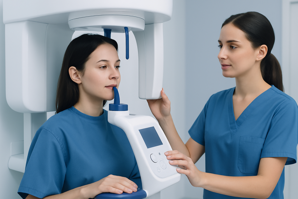
Dental X-rays may be used to evaluate the tooth structure more fully and to see whether the enamel defect is limited to the surface or associated with deeper problems. The dentist may also assess bite alignment, tooth wear, and whether the pulp or inner part of the tooth could be at risk.
Because enamel hypoplasia can resemble other conditions, careful diagnosis matters. A dentist may distinguish it from dental fluorosis, early decay, acid erosion, or generalized inherited enamel conditions. If symptoms are severe, the person may be referred for pediatric dental care in children or specialized restorative care in adults.
In some situations, the assessment includes broader medical questions. If the enamel defects are widespread, recurrent, or linked with other developmental concerns, a clinician may recommend further evaluation to look for nutritional, metabolic, or genetic factors contributing to the problem.
Modern treatment approaches
Treatment for enamel hypoplasia is individualized. The best option depends on how many teeth are involved, which teeth are affected, whether the main concern is sensitivity, appearance, structural weakness, or active decay, and the age of the patient. Many people benefit from a combination of protective and restorative approaches over time.
For mild cases, treatment may focus on reducing sensitivity and lowering cavity risk. This can include fluoride applications, remineralizing products, sealants, and close preventive follow-up. If defects trap plaque or are likely to decay, small restorations may be placed to protect the tooth surface.
When the enamel defect is more visible or structurally significant, restorative dentistry may be recommended. Depending on the tooth and the amount of damage, options may include tooth-colored bonding, fillings, inlays, onlays, or dental crowns to strengthen and cover the tooth. In selected cosmetic cases, dental veneers may improve shape and appearance once the tooth is healthy and stable.
If a tooth is severely damaged and cannot be preserved, replacement options may be discussed, including dental implant treatment in appropriate adult patients. The aim is not only to improve appearance, but also to restore comfort, chewing function, and long-term protection of the mouth.
Outlook and long-term dental health
The outlook for enamel hypoplasia is often good when it is recognized early and monitored regularly. Although lost or underdeveloped enamel cannot regenerate, affected teeth can frequently be protected successfully for many years with good preventive care and timely treatment. The long-term result depends mainly on the severity of the defect and how well the teeth are maintained.
Mild cases may need only observation and preventive support. More extensive defects may require repeated restorative care over time, especially in children whose mouths are still growing. Because enamel-deficient areas are more vulnerable, even small changes in sensitivity, wear, or discoloration should be reviewed promptly.
People with enamel hypoplasia often benefit from a long-term care plan rather than a single treatment step. This may include regular hygiene visits, monitoring of restorations, cavity prevention, and periodic reassessment as the teeth and bite change with age. With this approach, many patients maintain comfortable function and an acceptable appearance.
Prevention and self-care
Not all cases of enamel hypoplasia can be prevented, especially when genetics play a role. However, good prenatal care, support for infant and child nutrition, prompt treatment of childhood illnesses, and injury prevention may reduce some risks during tooth development. Once the condition is present, daily home care becomes very important.
Helpful self-care steps include:
- Brushing gently twice a day with fluoride toothpaste
- Cleaning between teeth daily with floss or another recommended tool
- Limiting frequent sugary snacks and acidic drinks
- Using a soft-bristled toothbrush if teeth are sensitive
- Keeping regular dental checkups and cleanings
- Following the dentist’s advice about fluoride, sealants, or sensitivity products
People should avoid abrasive whitening methods or harsh brushing, as these can aggravate sensitivity and wear already-thin enamel. If there is grinding or clenching, a dentist may recommend protective measures because mechanical stress can worsen enamel breakdown. At the later stages of care, Acibadem International’s multidisciplinary specialists and JCI-accredited hospitals diagnose and treat dental conditions for international patients when coordinated evaluation is needed.
When to seek medical care
A dental appointment is advisable if a child or adult has teeth with pits, grooves, unusual discoloration, rough enamel, or persistent sensitivity. Early evaluation is especially helpful when the problem involves front teeth, several teeth at once, or repeated cavities in the same area.
More urgent dental review may be needed if there is tooth pain, rapid chipping, swelling, difficulty eating, or signs of infection. Parents should also seek assessment if a child’s newly erupted teeth look abnormal, as early protection can reduce future damage.
If enamel defects are accompanied by broader health concerns, such as developmental issues, nutritional concerns, or a family history of inherited enamel disorders, a doctor or dentist may suggest additional medical evaluation. Prompt professional advice helps clarify the cause and supports the most appropriate treatment plan.
Frequently asked questions
Is enamel hypoplasia the same as cavities?
No. Enamel hypoplasia is a developmental enamel defect that happens while the tooth is forming, whereas cavities are areas of tooth decay caused by acids and bacteria after the tooth erupts. However, teeth with enamel hypoplasia are more likely to develop cavities because their protective surface is weaker.
Can enamel hypoplasia be reversed?
The missing or underdeveloped enamel itself cannot grow back. Even so, dentists can often protect the tooth, reduce sensitivity, and restore shape and strength with preventive and restorative treatments. Early care usually improves long-term outcomes.
Does enamel hypoplasia affect children only?
It often becomes noticeable in childhood because the defect begins during tooth development, but adults can also have it. Some people are diagnosed later when sensitivity, cosmetic concerns, or repeated dental problems lead to examination.
What does enamel hypoplasia look like?
It may appear as pits, grooves, thin spots, rough patches, or areas of white, yellow, or brown discoloration. In some cases, the affected teeth look chipped or uneven. The exact appearance depends on how much enamel was affected during development.
Is enamel hypoplasia painful?
It can be painless, especially when the defect is mild. But if the enamel is thin or missing, the tooth may become sensitive to temperature changes, sweets, or brushing. Pain is more likely if decay, cracking, or nerve irritation develops.
How is enamel hypoplasia treated in children?
Treatment in children depends on which teeth are affected and how severe the defects are. A dentist may use fluoride, sealants, bonding, fillings, or protective crowns, while also monitoring growth and oral hygiene closely. The aim is to preserve the teeth, keep the child comfortable, and support normal eating and development.
References
- American Dental Association
- National Institute of Dental and Craniofacial Research
- American Academy of Pediatric Dentistry
- Mayo Clinic
- World Health Organization
This article is for general information only and is not a substitute for professional medical advice. Please consult a qualified doctor about your individual situation.
Explore treatments in Turkey — costs, top hospitals & a free quote
JCI-accredited · board-certified surgeons · reply within 24h

More from the Health Library



Related Specialists

Dr. Ümran Karabulut Doğan
Gynecology & Obstetrics
Dr. Mustafa Tanıdır
General Surgery
Prof. Dr. Osman Güven
Orthopedic Surgery & Traumatology
Prof. Dr. Moşe Benhabib
Gynecology & Obstetrics