Male vs Female Skeleton: Key Differences and How Doctors Tell Them Apart

The pelvis is usually the most reliable area for distinguishing a male vs female skeleton. The skull can provide helpful clues, but it is generally less accurate than the pelvis on its own.
Key Takeaways
- The pelvis is usually the most reliable area for distinguishing a male vs female skeleton.
- The skull can provide helpful clues, but it is generally less accurate than the pelvis on its own.
- Doctors do not rely on a single feature because bone traits overlap between individuals.
- Age, ancestry, hormones, growth, and medical conditions can affect skeletal appearance.
- Imaging and specialist assessment are used when skeletal differences matter in medical care.
Male vs female skeleton differences are real, but they are usually patterns rather than absolute rules. Doctors and forensic specialists look at several bones together—especially the pelvis and skull—to estimate biological sex as accurately as possible.
Male vs Female Skeleton at a Glance
In a male vs female skeleton comparison, the clearest differences are usually seen in the pelvis, skull, and overall bone robustness. In general, male skeletons tend to have larger, denser, and more rugged-looking bones, while female skeletons more often show a broader pelvis and finer muscle attachment markings. However, these are tendencies, not fixed rules, and there is considerable overlap.
Clinicians, radiologists, anatomists, and forensic experts therefore use a pattern-based approach. Rather than asking whether one feature alone is “male” or “female,” they consider multiple skeletal traits together, along with age, medical history, and imaging findings.
Below is a simple side-by-side summary of common differences:
- Pelvis: Female pelvis is usually wider and shaped to support childbirth; male pelvis is often narrower and taller.
- Pelvic inlet: Often more oval or round in females, more heart-shaped in males.
- Subpubic angle: Usually wider in females, narrower in males.
- Skull: Male skulls often show a more prominent brow ridge, squarer jaw, and larger mastoid processes.
- Forehead and chin: Female skulls often appear smoother, with a more vertical forehead and narrower chin.
- Long bones: Male bones are often longer and thicker overall, though size alone is not diagnostic.
- Muscle markings: Male skeletons may show stronger muscle attachment sites, especially with higher lifelong muscle mass.
These comparisons are most useful when the skeleton is fully developed. In children and adolescents, differences are much less pronounced because many sex-related skeletal features become clearer only after puberty.
How Clinicians Tell Them Apart
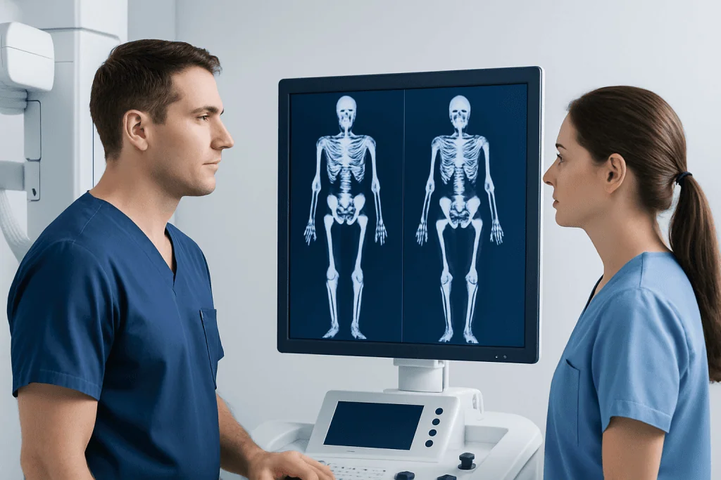
When doctors or other specialists need to estimate skeletal sex, they usually start with the pelvis. This is because the pelvic bones are the most consistently different between biological males and females. The width of the pelvic outlet, the shape of the pubic arch, the sciatic notch, and the contour of the sacrum all provide important clues.
The skull is often the next most informative area. A clinician may assess the brow ridge, eye socket margins, jaw shape, mastoid process, and the back of the skull where neck muscles attach. A more pronounced brow ridge and squarer chin may suggest a male pattern, while smoother contours and finer features may suggest a female pattern.
Long bones can also help, especially when measured carefully. The length and diameter of the femur, humerus, and other large bones may support an overall estimate, but these measurements are influenced by height, ancestry, nutrition, physical activity, and health conditions. For that reason, they are interpreted with caution.
In medical practice, experts often combine physical examination of bones with imaging such as X-rays, CT, or MRI when appropriate. If bone anatomy is being assessed because of trauma, congenital variation, or suspected disease, imaging may also help rule out problems such as fractures, deformity, or osteoporosis.
Why the Pelvis Matters Most

The pelvis is usually the strongest clue in a male vs female skeleton assessment because it reflects functional differences related to body structure and childbirth. In females, the pelvic cavity is typically broader, the iliac blades are more flared, and the subpubic angle is wider. The sacrum is often shorter and wider, helping create a roomier birth canal.
In males, the pelvis is often narrower, with a deeper and more funnel-shaped appearance. The subpubic angle tends to be more acute, and the pelvic inlet may look more heart-shaped. These differences are often easier to recognize in adults than in younger people whose bones are still developing.
Even so, the pelvis is not perfectly “male” or “female” in every person. Natural human variation is common. Pregnancy history, developmental differences, prior injury, arthritis, or surgery can also change pelvic appearance and make interpretation less straightforward.
If pelvic symptoms such as pain, instability, or limited movement are present, doctors focus first on the patient’s health rather than on sex estimation. In that setting, evaluation may include a physical examination and orthopedic rehabilitation planning if recovery support is needed after injury or surgery.
What the Skull and Other Bones Can Show
The skull can offer useful supporting evidence, especially when the pelvis is unavailable or cannot be assessed clearly. Male-pattern skulls often have a more sloping forehead, larger brow ridge, stronger jawline, larger mastoid process behind the ear, and more visible muscle attachment areas. Female-pattern skulls more often have smoother contours, sharper orbital margins, and a rounder or narrower chin.
Ribs, vertebrae, and the shoulder girdle may also show broader trends. For example, male skeletons often have relatively broader shoulders and more robust upper-limb bones, while female skeletons may have proportionally different trunk and pelvic relationships. Still, these bones are less reliable than the pelvis and should not be used alone for a firm conclusion.
Hormones influence how bones develop over time. Testosterone tends to increase bone size and muscle attachment prominence, while estrogen plays a major role in skeletal maturation and bone maintenance. Conditions that alter hormone levels, delayed puberty, or certain endocrine disorders can affect the expected pattern of skeletal traits.
In some situations, a doctor may order imaging to look more closely at the head, spine, or joints—not to label a skeleton as male or female, but to understand symptoms, injury, or disease. Depending on the concern, this might involve MRI or other diagnostic imaging methods.
Limits, Overlap, and Special Situations
A key point for patients and families is that skeletal sex estimation is not the same as identity, and it is not always exact. Biology is complex, and many bone features overlap. A smaller male skeleton may share traits often seen in females, and a larger female skeleton may show more robust traits often associated with males.
Accuracy also depends on the age of the person and the condition of the bones. In children, sex-related skeletal differences are subtle and difficult to assess. In older adults, bone loss, arthritis, past fractures, and degenerative changes may alter landmarks. This is one reason doctors avoid overconfidence when interpreting skeletal features.
Ancestry and population background also influence average bone shape and size. Methods developed in one population may not transfer perfectly to another. Specialists therefore use validated tools carefully and interpret them within clinical context rather than relying on a one-size-fits-all rule.
There are also medical situations where a patient’s skeleton does not fit typical male or female patterns. Differences in sexual development, prior hormonal treatment, congenital bone conditions, or skeletal disorders can all affect bone appearance. If there are concerns about bone strength or structure, doctors may also assess related problems such as scoliosis or metabolic bone disease.
What to Do in Medical and Everyday Situations
Most people do not need a skeleton-based sex assessment in daily life. These evaluations are usually relevant in medicine, surgery planning, radiology, anthropology, or forensic work. If a patient is having scans or treatment for a bone or joint problem, the practical priority is understanding the anatomy accurately so care can be tailored to the individual.
For someone with bone pain, injury, posture changes, or concerns about bone density, the best next step is a medical evaluation rather than trying to interpret anatomy alone. A clinician may ask about symptoms, growth history, menstrual or hormonal history, prior fractures, family history, and physical activity. Imaging and bone health tests can then be chosen if needed.
Treatment depends on the issue found. Fractures may need immobilization or surgery; chronic joint or spine problems may need physical therapy, medication, or targeted procedures. If bone loss is suspected, doctors may investigate nutrition, hormone balance, and risk factors and may recommend physical therapy and rehabilitation as part of recovery and mobility support.
Near the end of a patient’s diagnostic pathway, multidisciplinary teams may help connect imaging findings with symptoms and function. Acibadem International’s multidisciplinary specialists and JCI-accredited hospitals diagnose and treat bone, joint, and spine conditions for international patients when expert assessment is needed.
When to Seek Medical Care
Medical attention is important if a person has persistent bone or joint pain, repeated fractures, visible deformity, difficulty walking, new back curvature, or unexplained loss of height. These symptoms may point to an underlying bone, spine, or metabolic problem that needs assessment.
Urgent care is advisable after major trauma, if there is severe pain, inability to bear weight, numbness, weakness, or sudden loss of bladder or bowel control. These signs can suggest a serious injury or nerve-related problem that should not wait.
It is also reasonable to see a doctor for questions about bone development, delayed puberty, asymmetry, or concerns raised during imaging. A clinician can explain whether a variation is a normal anatomical difference or part of a condition that requires follow-up.
Patients should remember that online comparisons of a male vs female skeleton are educational, but they cannot replace professional diagnosis. If symptoms are present, a qualified doctor can recommend the most appropriate examination, imaging, and treatment plan.
Frequently asked questions
What is the biggest difference between a male vs female skeleton?
The pelvis is usually the most important difference. Female pelves are generally broader and shaped to support childbirth, while male pelves are often narrower and deeper. Doctors still look at several features together because individuals vary.
Can doctors tell sex from a skull alone?
A skull can provide useful clues, but it is usually less reliable than the pelvis by itself. Features such as the brow ridge, jaw shape, and mastoid process may suggest a male or female pattern. Even so, overlap is common, so experts prefer to assess multiple bones.
Are male bones always larger than female bones?
Not always. On average, male bones tend to be larger and more robust, but there is wide natural variation. Height, ancestry, hormones, nutrition, and physical activity all influence bone size.
At what age do skeletal differences become clear?
Many differences become easier to recognize after puberty. Before then, children’s skeletons often do not show strong sex-related features, which makes assessment more difficult. This is why age matters in skeletal interpretation.
Can disease change how a skeleton looks?
Yes. Conditions such as osteoporosis, arthritis, endocrine disorders, congenital differences, and old fractures can alter bone shape or density. These changes can affect how easily skeletal traits are interpreted and may also require treatment.
Is skeletal sex estimation the same as gender identity?
No. Skeletal sex estimation is a biological assessment based on bone traits, usually for medical or scientific purposes. Gender identity is a person’s own internal sense of gender and is not determined by bones.
References
- Cleveland Clinic
- MedlinePlus
- National Institute of Arthritis and Musculoskeletal and Skin Diseases
- Radiological Society of North America
- World Health Organization
This article is for general information only and is not a substitute for professional medical advice. Please consult a qualified doctor about your individual situation.
Explore treatments in Turkey — costs, top hospitals & a free quote
JCI-accredited · board-certified surgeons · reply within 24h

More from the Health Library



Related Specialists

Dr. Zeynep Kuzu
Pediatrics
Assoc. Prof. Dr. Burak Elmas
Vitro Fertilization and Reproductive Medicine Center
Dr. Hatem Doğu
Anesthesiology
Asst. Prof. Dr. Hilal Kurtoğlu Gümüşel
Cardiology