Sonohysterogram: A Complete Medical Overview

A sonohysterogram is an ultrasound-based test that gives a clearer view of the uterine cavity than a standard pelvic ultrasound alone. It is often recommended for abnormal uterine bleeding, infertility evaluation, suspected polyps, fibroids, or scar tissue inside the uterus.
Key Takeaways
- A sonohysterogram is an ultrasound-based test that gives a clearer view of the uterine cavity than a standard pelvic ultrasound alone.
- It is often recommended for abnormal uterine bleeding, infertility evaluation, suspected polyps, fibroids, or scar tissue inside the uterus.
- The procedure is usually brief and done without general anesthesia, though mild cramping can occur.
- Timing matters: it is often scheduled after menstruation but before ovulation in people who are still having periods.
- Results can help guide next steps, including observation, medication, or procedures such as hysteroscopy.
A sonohysterogram is a specialized pelvic ultrasound that uses sterile saline to gently expand the uterus so its inner lining can be seen more clearly. It is commonly used to investigate abnormal bleeding, fertility concerns, suspected polyps or fibroids, and some recurrent pregnancy problems.
What a sonohysterogram is and why it is used
A sonohysterogram, also called saline infusion sonography, is a pelvic ultrasound test that uses a small amount of sterile saline placed inside the uterus during imaging. The fluid gently opens the uterine cavity, making the outline of the endometrium easier to see than on a standard ultrasound alone. This can help doctors identify structural causes of symptoms that might otherwise be difficult to distinguish.
It is most often used to evaluate abnormal uterine bleeding, infertility, repeated implantation failure, and suspected problems inside the uterine cavity. Common findings include endometrial polyps, submucosal fibroids, adhesions, congenital uterine shape differences, and areas of endometrial thickening. In some situations, it may also be used as part of planning for procedures or surgery.
Compared with more invasive testing, a sonohysterogram is usually done in an outpatient setting and does not require incisions. It offers more detail than a routine transvaginal ultrasound for the inner contour of the uterus, while remaining less invasive than hysteroscopy. That makes it a useful next step when symptoms or previous test results suggest that a closer look is needed.
Who may need a sonohysterogram
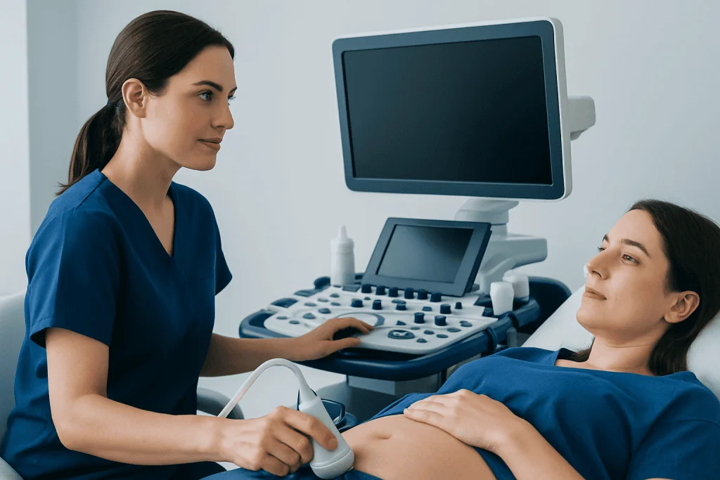
A doctor may recommend a sonohysterogram for people with heavy menstrual bleeding, bleeding between periods, bleeding after menopause, or a persistently thickened endometrium seen on ultrasound. It may also be part of an infertility workup, especially when there is concern about the uterine cavity or when previous imaging has raised questions that need clarification.
The test can be helpful when a standard ultrasound suggests a possible growth or irregularity but cannot reliably show whether it is inside the cavity, in the uterine wall, or pressing on the cavity from outside. This distinction matters because treatment options differ. For example, a submucosal fibroid may affect bleeding and fertility differently from a fibroid located deeper in the uterine muscle.
It is not the right test for every situation. People with a known or possible pregnancy, active pelvic infection, or significant unexplained pelvic pain may need a different approach or further evaluation first. A clinician will review symptoms, medical history, and timing in the menstrual cycle before deciding whether the test is appropriate.
- Abnormal uterine bleeding
- Suspected endometrial polyps
- Possible uterine fibroids affecting the cavity
- Infertility or recurrent pregnancy loss evaluation
- Assessment of scar tissue or uterine shape differences
How the procedure is performed

A sonohysterogram is usually performed in a gynecology or imaging clinic. The test often begins with a transvaginal ultrasound to assess the uterus and ovaries. Then, a speculum is inserted into the vagina, the cervix is cleaned, and a thin catheter is gently passed through the cervix into the uterus. After the speculum is removed, sterile saline is slowly infused while ultrasound images are taken.
As the saline fills the uterine cavity, the walls separate slightly, allowing the doctor or sonographer to see the lining more clearly. The imaging portion usually takes only a few minutes. The entire appointment may last longer because of preparation, discussion, and review of comfort during the test.
Most people feel pressure or menstrual-like cramping rather than sharp pain. Light spotting afterward can happen. In many cases, the test is scheduled shortly after the menstrual period ends and before ovulation, because this timing offers the clearest view of the uterine lining and helps avoid performing the test during an early pregnancy.
What the results can show
The main strength of a sonohysterogram is its ability to outline the inside of the uterus. It can show whether the cavity is smooth and normal or whether something is projecting into it. A rounded growth attached to the lining may suggest a polyp, while a broader bulge from the uterine muscle may suggest a submucosal fibroid. Areas where the walls appear stuck together may indicate adhesions.
The test may also help identify congenital differences in uterine shape, such as a septum, and may clarify whether endometrial thickening looks diffuse or focal. This distinction can help determine whether a person needs follow-up imaging, biopsy, hysteroscopy, or no additional testing. When fertility is the concern, a normal result can also be useful because it helps rule out cavity problems that could interfere with implantation.
Although highly informative, a sonohysterogram does not answer every question. It may suggest a diagnosis, but tissue sampling or direct visualization can still be needed. If the findings point to structural changes such as uterine fibroids or a uterine cavity lesion, the next step may include further gynecologic evaluation and treatment planning.
Preparation, risks, and recovery
Preparation is usually simple. A doctor may ask about the date of the last menstrual period, the chance of pregnancy, history of pelvic infection, current symptoms, and medicines being taken. Some people are advised to take an over-the-counter pain reliever before the appointment if they commonly experience menstrual cramps, but this should be done only according to a clinician’s guidance.
Risks are generally low, but no procedure is completely risk-free. Mild cramping, temporary dizziness, or light spotting can occur. Infection is uncommon but possible, which is why active pelvic infection is an important reason to postpone the test. Rarely, the procedure may not be completed if the cervix is difficult to pass or if discomfort is significant.
Most people can return to normal daily activities right away. It is common to have a small amount of watery discharge afterward because the saline drains out. Patients are usually told to contact their doctor if they develop fever, increasing pelvic pain, foul-smelling discharge, or heavy bleeding after the test.
How sonohysterogram compares with other tests
A standard transvaginal ultrasound is often the first imaging test because it is widely available and useful for evaluating the uterus, ovaries, and pelvis overall. However, it may not always clearly define the inner uterine cavity. A sonohysterogram adds detail specifically for the endometrial cavity and can help decide whether a finding is likely to be clinically important.
Hysteroscopy allows a doctor to look directly inside the uterus with a thin camera and, in some cases, treat a problem during the same session. It is more invasive than a sonohysterogram but may be the preferred next step when there is a strong suspicion of a cavity lesion that needs confirmation or removal. MRI may be used in selected situations when broader pelvic anatomy or more complex uterine conditions need further characterization.
If the test shows a lesion that appears removable or a structural issue requiring treatment, a doctor may discuss options such as hysteroscopy or surgery depending on symptoms, fertility goals, and the type of finding. When fibroids are involved, management may also include monitoring, medication, or procedures such as myomectomy in carefully selected patients.
Treatment after a sonohysterogram and when to seek medical care
A sonohysterogram itself is a diagnostic test, not a treatment. What happens next depends on the result and the reason the test was ordered. Some people need no further care if the uterine cavity looks normal. Others may need additional testing, an endometrial biopsy, medical treatment for bleeding, or a procedure to remove or correct a structural problem.
When a focal lesion such as a polyp is suspected, treatment may involve removal if it is causing symptoms, affecting fertility, or raising concern on follow-up evaluation. If heavy bleeding is related to a broader uterine condition, management may be individualized and could include medications or, in some cases, endometrial ablation for those who are not planning future pregnancy. A gynecologist will help weigh the benefits and limitations of each option.
Medical care should be sought promptly after the procedure if there is fever, worsening pelvic pain, fainting, heavy bleeding, or discharge with a strong odor. People should also consult a doctor if abnormal bleeding persists, fertility concerns continue, or symptoms return after treatment. Near the end of the care pathway, Acibadem International’s multidisciplinary specialists in JCI-accredited hospitals diagnose and treat uterine conditions for international patients when further evaluation or management is needed.
Frequently asked questions
Is a sonohysterogram painful?
Most people describe a sonohysterogram as uncomfortable rather than very painful. Mild to moderate cramping can happen when the catheter is placed or when the saline enters the uterus, but the discomfort usually passes quickly after the test.
How long does a sonohysterogram take?
The imaging portion is often brief, usually only several minutes. The full visit may take longer because of check-in, preparation, explanation of the procedure, and post-test instructions.
When is the best time in the menstrual cycle to have a sonohysterogram?
It is commonly scheduled after menstruation ends but before ovulation. This timing often provides the clearest view of the uterine lining and reduces the chance of performing the test during an early pregnancy.
Can a sonohysterogram detect fibroids or polyps?
Yes, it can be very helpful for showing whether a polyp or fibroid projects into the uterine cavity. It may not replace all other tests, but it often helps clarify findings seen on a routine ultrasound.
Is a sonohysterogram the same as hysteroscopy?
No. A sonohysterogram uses ultrasound and saline to outline the inside of the uterus, while hysteroscopy uses a thin camera to look directly into the cavity. Hysteroscopy is more invasive but can also allow treatment during the same procedure in some cases.
Are there any risks after a sonohysterogram?
Light spotting, watery discharge, and temporary cramping are relatively common and usually settle quickly. Patients should contact a doctor if they develop fever, increasing pain, heavy bleeding, or unusual discharge afterward.
References
- American College of Obstetricians and Gynecologists
- American Institute of Ultrasound in Medicine
- Royal College of Obstetricians and Gynaecologists
- National Institute for Health and Care Excellence
- Radiological Society of North America
This article is for general information only and is not a substitute for professional medical advice. Please consult a qualified doctor about your individual situation.
IVF & fertility treatment in Turkey — success rates and costs
JCI-accredited · board-certified surgeons · reply within 24h
Check your numbers in seconds
BMI, calories, due date, blood pressure and 30+ more clinical calculators — free, instant, doctor-reviewed ranges.