Elastography: A Complete Medical Overview
Elastography measures tissue stiffness, which can provide clues about disease. It is commonly used for the liver, breast, thyroid, prostate, and other soft tissues.
Key Takeaways
- Elastography measures tissue stiffness, which can provide clues about disease.
- It is commonly used for the liver, breast, thyroid, prostate, and other soft tissues.
- The test is usually quick, painless, and done with ultrasound or MRI.
- Results do not stand alone and must be interpreted with symptoms, exam findings, and other tests.
- Elastography can help monitor chronic conditions over time and may reduce unnecessary biopsies in some cases.
Elastography is a noninvasive imaging method that measures how stiff or soft body tissues are, helping doctors assess organs and lumps without surgery. It is most often used alongside ultrasound or MRI to support diagnosis, guide follow-up, and sometimes reduce the need for biopsy.
Overview: what elastography is and why it is used
Elastography is a medical imaging technique that shows how stiff or elastic a tissue is. In general, healthy and diseased tissues can feel different: some conditions make tissue harder, while others change its structure in more subtle ways. By measuring these differences, elastography can give doctors added information that a standard scan may not show clearly on its own.
Most commonly, elastography is performed as part of an ultrasound exam, though it can also be done with MRI. It does not usually replace other imaging tests. Instead, it adds a functional layer of information, helping clinicians understand whether a liver is becoming scarred, whether a thyroid nodule appears more suspicious, or whether a breast lump needs closer assessment.
One important point for patients is that elastography is a test, not a diagnosis by itself. A stiff area does not automatically mean cancer, and a soft area does not always mean everything is normal. Doctors interpret the findings together with symptoms, blood tests, physical examination, and sometimes biopsy results when needed.
How elastography works
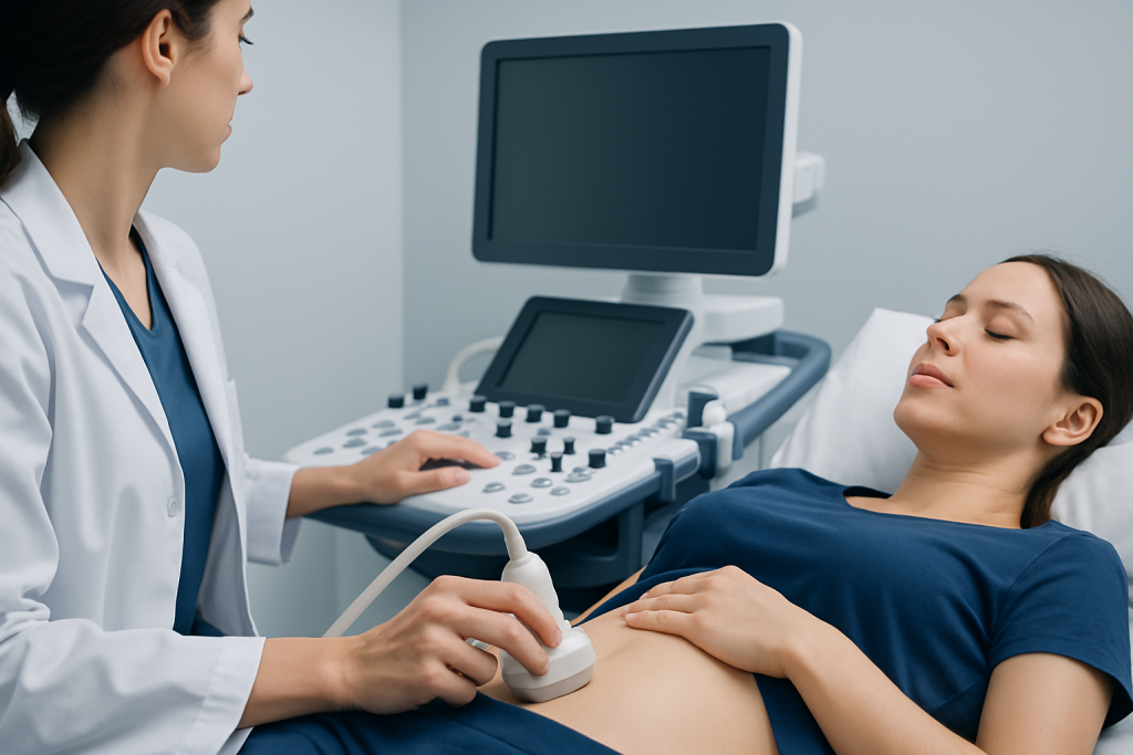
Elastography works by applying or tracking tiny mechanical waves through tissue and measuring how quickly the tissue responds. In simple terms, stiffer tissue tends to transmit these waves differently from softer tissue. The imaging system then converts this information into numbers, color maps, or both, giving the doctor a visual and measurable estimate of tissue stiffness.
There are two main approaches. Ultrasound elastography includes techniques such as transient elastography, strain elastography, and shear wave elastography. MRI elastography uses magnetic resonance imaging to map tissue stiffness, often in deeper organs such as the liver. Which method is chosen depends on the clinical question, the body area being examined, and the equipment available.
Patients often ask whether the test involves radiation. Standard ultrasound elastography does not use ionizing radiation, and MRI elastography also avoids radiation exposure. This is one reason elastography can be useful for repeated monitoring in long-term conditions, especially chronic liver disease.
- Ultrasound elastography: widely available, quick, and commonly used in liver, thyroid, breast, and soft tissue assessment.
- MRI elastography: helpful when a broader or deeper tissue assessment is needed, particularly in liver evaluation.
- Quantitative results: some methods provide measured stiffness values that can be tracked over time.
Common uses of elastography in medicine
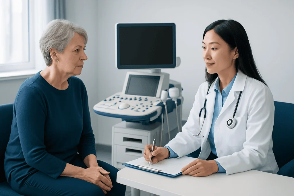
Elastography is used in several areas of medicine, but the liver is one of its best-known applications. In chronic liver disease, repeated injury can lead to fibrosis, or scarring. Elastography can help estimate the degree of fibrosis without needing an invasive procedure in every patient. It is often part of the evaluation for liver conditions, fatty liver disease, hepatitis, and cirrhosis follow-up.
In breast imaging, elastography may help characterize a lump found on examination or mammography. Some benign lesions are soft, while some suspicious lesions are firmer. Even so, elastography is not used alone to confirm or rule out breast cancer. It is one piece of the overall assessment, sometimes used alongside targeted imaging and tissue sampling such as biopsy.
Doctors may also use elastography for thyroid nodules, prostate evaluation, musculoskeletal soft tissues, lymph nodes, and certain pancreatic or abdominal findings. Depending on the clinical setting, it may help decide whether a lesion can simply be observed, whether repeat imaging is appropriate, or whether further testing is needed.
A practical advantage is that elastography may reduce uncertainty when standard imaging findings are borderline. In the right context, it can support earlier recognition of disease, more tailored follow-up, and better planning for treatment options such as MRI or ultrasound-guided reassessment.
What to expect before, during, and after the test
The preparation for elastography depends on the organ being examined. For liver elastography, patients may be asked not to eat for a few hours beforehand because food intake can affect blood flow and stiffness readings. For breast, thyroid, or superficial soft tissue elastography, there is often little or no special preparation.
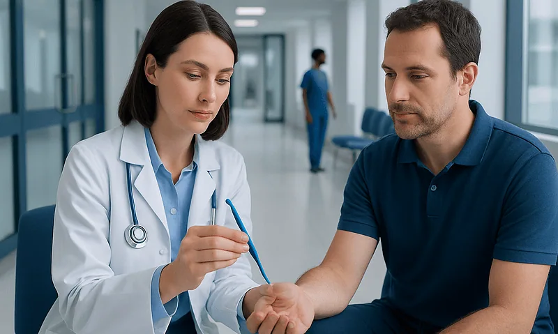
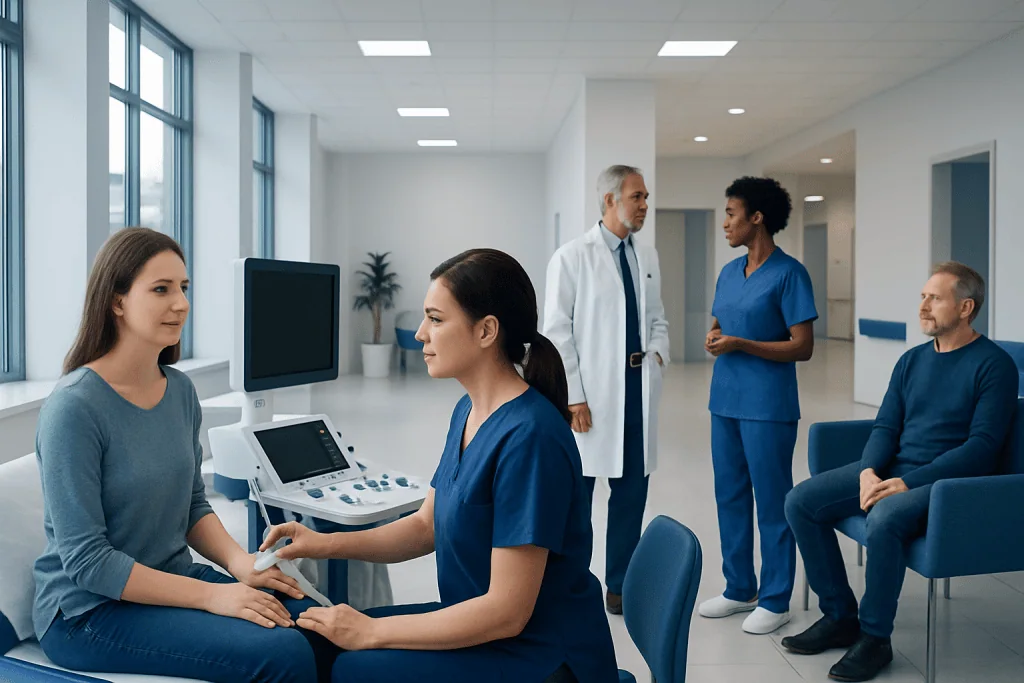
During an ultrasound elastography exam, the patient usually lies on an examination table while gel is applied to the skin. The clinician places a probe over the area being studied. The test is typically painless, though mild pressure may be felt. A liver study often takes only a few minutes once positioning is complete. MRI elastography takes longer and may involve lying still inside the MRI scanner.
After the test, most patients can return to normal activities right away. There is generally no recovery period. Results may be available quickly, but a final interpretation often depends on the full radiology report and review by the treating doctor. If the findings are unclear, the next step may include blood tests, repeat imaging, or another diagnostic procedure rather than immediate treatment.
Understanding results and their limits
Elastography results are often reported as a stiffness measurement or as an image showing areas of relative softness and hardness. In liver care, higher stiffness may suggest more fibrosis, but values can be influenced by more than scarring alone. Inflammation, congestion, recent meals, obesity, and technical factors may sometimes affect the reading.
For breast, thyroid, or other focal lesions, a stiff area may be more concerning than a soft one, but this is not absolute. Benign conditions such as scar tissue, calcification, inflammation, or certain noncancerous nodules can also appear firm. That is why doctors do not rely on elastography in isolation when making major decisions.
It is also important to understand that elastography is not equally useful for every body type or every condition. Some examinations may be technically limited by body habitus, deep lesions, motion, or difficult probe positioning. When results do not match the clinical picture, doctors may recommend a different imaging method or tissue diagnosis to clarify what is happening.
Used well, elastography is valuable because it adds context. It can help monitor progression over time, compare changes between visits, and support more personalized care. The clearest benefit often comes not from a single number, but from how that number fits into the broader medical picture.
Benefits, risks, and possible alternatives
The main benefits of elastography are that it is noninvasive, usually painless, and often quick to perform. For many patients, especially those with chronic liver disease, it offers a way to monitor tissue changes over time without repeated invasive sampling. In selected situations, it may help reduce unnecessary procedures or guide doctors toward the most appropriate next test.
The risks are generally low. Ultrasound elastography is considered very safe and does not expose the patient to ionizing radiation. MRI elastography is also noninvasive, though standard MRI safety rules still apply, including screening for certain implanted devices or metal fragments. Some people may find MRI uncomfortable because of noise or enclosed space.
Alternatives depend on the medical question. Standard ultrasound, CT, MRI, blood tests, mammography, and biopsy may all play a role. In liver disease, biopsy remains important in selected cases, especially when test results conflict or when a precise tissue diagnosis is needed. The best choice depends on the suspected condition, the patient’s history, and what information is required for treatment decisions.
When to seek medical care
Medical care should be sought when symptoms suggest a condition that may need imaging or further evaluation. Examples include a new breast lump, persistent neck swelling, unexplained abdominal pain, jaundice, abnormal liver blood tests, ongoing fatigue with known liver disease, or a mass noticed anywhere in the body. Elastography may be one of several tests used to understand the cause.
People with chronic hepatitis, fatty liver disease, heavy alcohol use, obesity, diabetes, or a family history of liver disease may also benefit from medical follow-up even if they feel well. Likewise, any lump that is enlarging, painful, firm, or associated with skin changes should be assessed by a qualified doctor rather than monitored at home without advice.
Urgent assessment is appropriate for severe abdominal pain, rapidly worsening jaundice, significant swelling, unexplained weight loss, or symptoms that are progressing quickly. A clinician can decide whether elastography is useful or whether other tests are more appropriate first.
For patients who need coordinated evaluation, Acibadem International’s multidisciplinary specialists and JCI-accredited hospitals diagnose and treat conditions in which elastography may be helpful, as part of a broader imaging and clinical assessment.
Frequently asked questions
Is elastography the same as an ultrasound?
Not exactly. Elastography is a special technique that is often added to a standard ultrasound exam to measure tissue stiffness. It can also be performed with MRI in some cases.
What diseases can elastography help detect?
Elastography can support the evaluation of liver fibrosis, breast lumps, thyroid nodules, prostate abnormalities, and some soft tissue or lymph node findings. It helps doctors estimate stiffness, which may provide clues about scarring, inflammation, or malignancy, but it does not confirm a diagnosis on its own.
Is elastography painful?
Most patients find elastography comfortable or only mildly uncomfortable. Ultrasound elastography usually feels like a routine scan with light pressure on the skin. MRI elastography is also noninvasive, though lying still in the scanner may be inconvenient for some people.
Can elastography replace a biopsy?
Sometimes it can reduce the need for biopsy, but it does not replace biopsy in every situation. If imaging results are unclear, conflicting, or strongly suspicious, a tissue sample may still be the safest way to make a definite diagnosis.
How should a patient prepare for elastography?
Preparation depends on the body area being examined. For liver elastography, fasting for several hours may be recommended, while many other elastography exams require little or no special preparation. The imaging team usually provides instructions in advance.
How accurate is elastography?
Elastography can be very helpful, especially when used for the right clinical question and interpreted by experienced clinicians. However, its accuracy can be affected by factors such as inflammation, body habitus, lesion depth, and technical limitations. It is most reliable when combined with other medical information.
References
- World Health Organization
- Radiological Society of North America
- American College of Radiology
- European Association for the Study of the Liver
- National Institute of Diabetes and Digestive and Kidney Diseases
This article is for general information only and is not a substitute for professional medical advice. Please consult a qualified doctor about your individual situation.
Oncology care in Turkey — second opinion and treatment plan
JCI-accredited · board-certified surgeons · reply within 24h