Lymphoscintigraphy: What Patients Need to Know

Lymphoscintigraphy uses a small amount of radioactive tracer to map lymphatic flow. It is often ordered to assess lymphedema or identify sentinel lymph nodes before cancer surgery.
Key Takeaways
- Lymphoscintigraphy uses a small amount of radioactive tracer to map lymphatic flow.
- It is often ordered to assess lymphedema or identify sentinel lymph nodes before cancer surgery.
- The test is generally safe, minimally invasive, and usually involves only small injections and imaging over time.
- Results help doctors understand whether lymph drainage is normal, delayed, blocked, or redirected.
- Preparation is usually simple, but patients should follow instructions about clothing, pregnancy, breastfeeding, and prior procedures.
Lymphoscintigraphy is a specialized imaging test that shows how lymph fluid moves through the body. It is commonly used to evaluate lymphedema, lymphatic blockage, and sentinel lymph node pathways, helping doctors plan diagnosis and treatment with more precision.
Overview: what lymphoscintigraphy shows
Lymphoscintigraphy is an imaging test that helps doctors see how lymph fluid drains through the lymphatic system. To perform the test, a very small amount of radioactive tracer is injected into the skin or tissue, usually in an arm, leg, breast area, or near the site being evaluated. A special camera then takes images as the tracer travels through lymph vessels and lymph nodes.
This test is different from standard X-rays, CT, or ultrasound because it focuses on lymphatic function rather than just structure. In other words, it shows how well lymph is moving, whether drainage is delayed, and where flow may be blocked or rerouted. That functional information can be especially useful when swelling, prior surgery, radiation, or cancer raises questions about lymphatic health.
Lymphoscintigraphy is most often used when a doctor suspects lymphedema, wants to investigate unexplained swelling, or needs to map sentinel lymph nodes before certain cancer operations. It may also be considered when lymphatic leakage or congenital lymphatic abnormalities are suspected. For patients, the test is usually outpatient, relatively straightforward, and often completed without sedation.
Why doctors order this test
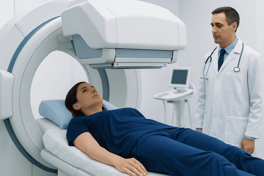
Doctors request lymphoscintigraphy when they need a clearer picture of lymph drainage. One of the most common reasons is the assessment of lymphedema, a condition in which lymph fluid builds up and causes persistent swelling, typically in an arm or leg. The test can help distinguish true lymphatic dysfunction from swelling related to vein problems, inflammation, injury, or other causes.
Another important use is sentinel lymph node mapping. In some cancers, such as certain breast cancers or melanoma, surgeons may need to identify the first lymph node or nodes that drain the tumor area. Lymphoscintigraphy helps show that pathway so surgery can be better targeted. In that context, the test may be used alongside procedures related to breast cancer care or skin cancer evaluation.
Less commonly, lymphoscintigraphy may help investigate:
- Unexplained chronic limb swelling
- Possible lymphatic obstruction after surgery or radiation
- Suspected primary lymphatic disorders present from birth or early life
- Lymph leakage, such as chylous complications in selected cases
- Treatment planning for lymphatic rehabilitation or surgery
Because it provides functional information, the test can complement physical examination and other imaging rather than replace them. The final interpretation is always considered together with symptoms, medical history, and other test results.
What the procedure is like
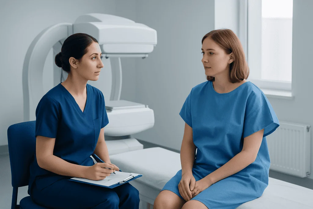
Before the scan begins, a healthcare professional injects a small amount of tracer just under the skin. The exact injection site depends on the reason for the study. For limb swelling, injections are often placed between the fingers or toes. For sentinel node mapping, the tracer is injected near the tumor or biopsy site. The injections may cause brief stinging, but they are usually quick.
After injection, images are taken with a gamma camera at set intervals. Some scans begin immediately, while others include delayed images over one or more hours to see how the tracer progresses. Patients may be asked to move the limb, walk, massage the area gently, or perform simple exercises to encourage lymph flow, depending on the protocol used.
The test itself is painless once the injections are over. During imaging, the patient lies or sits still while the camera records the tracer’s movement. The camera does not enclose the entire body like an MRI machine, so many people find it easier to tolerate. Total appointment time varies, but it is often longer than routine imaging because pictures may need to be repeated over time.
In many cases, patients can go home the same day and return to usual activities shortly afterward. The medical team will explain any special instructions, including hydration, skin care, or whether close contact precautions are needed, although the amount of radioactivity used is very low.
Preparing for lymphoscintigraphy and understanding safety
Preparation is usually simple. Patients are often advised to wear comfortable clothing and avoid tight garments that may compress the area being studied unless specifically instructed otherwise. It is helpful to bring prior imaging reports, a list of medications, and details of any past surgeries, radiation treatment, infections, or long-standing swelling, since these can affect interpretation.
Patients should tell the care team if they are pregnant, might be pregnant, or are breastfeeding. Although the radiation dose is typically low, these situations may require special precautions or a different plan. Allergic reactions to the tracer are uncommon, and complications are rare, but all imaging procedures should still be individualized.
Lymphoscintigraphy is generally considered safe. The tracer dose is small, and the procedure is minimally invasive. The most common issues are mild discomfort at the injection site, temporary redness, or minor bruising. Infection after the injection is unusual. For many people, the benefit of understanding lymphatic function outweighs the limited risks, especially when the result could guide treatment decisions.
If a patient has had recent surgery, active skin infection, severe inflammation, or another nuclear medicine test, the doctor may adjust the timing. Asking in advance about medications, skincare products, compression garments, and recent procedures can help the appointment go smoothly.
How results are interpreted
A nuclear medicine specialist reviews how quickly and how completely the tracer moves through lymphatic vessels and into lymph nodes. In a normal study, the tracer follows expected pathways and reaches regional lymph nodes within a typical time frame. In an abnormal study, transport may be delayed, absent, asymmetric, or diverted through collateral channels.
In patients with suspected lymphedema, lymphoscintigraphy can show reduced lymphatic transport, backflow of tracer into the skin, interrupted channels, or poor visualization of expected nodes. These patterns help support the diagnosis and may also suggest severity or whether one side is more affected than the other. However, the report is only one part of diagnosis; symptoms and examination remain very important.
For sentinel lymph node mapping, the scan identifies the node or nodes most likely to receive drainage from the tumor area first. That information can help surgeons plan a more focused procedure, often as part of breast cancer treatment or other cancer-related operations. In some settings, the findings are combined with same-day surgical localization techniques.
Results may also influence rehabilitation planning. If the scan confirms impaired lymph drainage, the doctor may recommend compression, exercise, skin protection, or referral for specialized lymphedema treatment. If the study is normal, the team may investigate other causes of swelling, such as venous disease, medication effects, or inflammatory conditions.
Treatment and next steps after the scan
Lymphoscintigraphy itself is a diagnostic test, not a treatment. What happens next depends on why it was ordered and what it shows. If the test supports lymphedema, treatment usually focuses on symptom control, preserving function, and reducing the risk of complications. This may include compression garments, manual lymphatic drainage, exercise programs, weight management, and careful skin care.
Some patients benefit from structured rehabilitation, especially when swelling affects daily activity or follows cancer treatment. In selected situations, doctors may discuss surgical options, but these are not appropriate for everyone and usually follow a detailed evaluation. If swelling is related to cancer or a mass blocking lymph flow, treatment is directed at the underlying cause rather than the lymphatic symptom alone.
When lymphoscintigraphy is used before an operation, the scan helps guide the surgical plan. For example, identifying a sentinel node can make surgery more precise and may reduce the need for wider node removal in suitable patients. Depending on the case, this may fit into broader care such as surgical oncology or multidisciplinary cancer planning.
Near the end of the diagnostic process, patients may be referred to specialists in nuclear medicine, oncology, surgery, physical medicine, or vascular and lymphatic care. At Acibadem International, multidisciplinary specialists in JCI-accredited hospitals diagnose and treat conditions related to lymphatic imaging for international patients, using individualized treatment planning based on clinical findings and test results.
Self-care and when to seek medical care
For patients being evaluated for lymphatic problems, self-care often begins even before a final diagnosis. Protecting the skin, keeping the affected area clean and moisturized, avoiding injury, and following advice on movement or compression can help reduce irritation and support comfort. If swelling is present, patients should avoid self-treating with very tight bandaging unless guided by a trained professional, since incorrect compression can worsen symptoms.
General measures that may help include staying active within comfort limits, elevating a swollen limb when appropriate, maintaining a stable body weight, and reporting changes promptly. People with persistent swelling after cancer treatment should not assume it is normal or unavoidable. Early assessment can be useful, especially if symptoms are increasing over time.
Medical care should be sought if there is new or rapidly worsening swelling, redness, warmth, fever, skin pain, leakage of fluid, shortness of breath, or swelling that develops suddenly on one side. These symptoms may point to infection, a blood clot, or another condition that needs prompt evaluation. Patients should also contact their doctor if swelling interferes with daily activities, if a known lymphedema treatment plan stops working, or if there are concerns after recent surgery or radiation.
If there is concern about a related underlying condition, doctors may also investigate problems such as lymphedema or other causes of abnormal lymph drainage. A qualified clinician can explain whether lymphoscintigraphy is the right next step and what additional tests, if any, are needed.
Frequently asked questions
Is lymphoscintigraphy painful?
Most people tolerate lymphoscintigraphy well. The main discomfort is usually the small tracer injections at the beginning of the test, which may sting briefly. After that, the imaging portion is generally painless.
How long does a lymphoscintigraphy test take?
The appointment length varies depending on the reason for the study and how quickly the tracer moves through the lymphatic system. Some scans are completed within a couple of hours, while others require delayed images later the same day. The care team usually explains the schedule in advance.
What does lymphoscintigraphy diagnose?
Lymphoscintigraphy helps evaluate how lymph fluid drains. It is commonly used to support the diagnosis of lymphedema, investigate lymphatic blockage, and map sentinel lymph nodes before certain cancer surgeries. It may also help assess some less common lymphatic disorders.
Is the radiation from lymphoscintigraphy safe?
The test uses a very small amount of radioactive tracer, and it is generally considered safe when medically appropriate. The radiation exposure is usually low, but patients who are pregnant or breastfeeding should tell their doctor before the test. The imaging team can then decide on the safest approach.
Do patients need to fast before lymphoscintigraphy?
Many patients do not need special fasting, but instructions can vary by center and by the part of the body being studied. It is best to follow the preparation guidance given by the imaging department. Asking ahead about food, medications, and compression garments can help avoid delays.
Can a normal lymphoscintigraphy result rule out all causes of swelling?
No. A normal result may suggest that lymphatic drainage is preserved, but swelling can still come from many other causes, such as vein problems, inflammation, heart or kidney conditions, or medications. Doctors interpret the scan together with symptoms, examination, and other tests.
References
- National Cancer Institute
- Radiological Society of North America
- Society of Nuclear Medicine and Molecular Imaging
- National Institute of Diabetes and Digestive and Kidney Diseases
- American Cancer Society
This article is for general information only and is not a substitute for professional medical advice. Please consult a qualified doctor about your individual situation.
Explore treatments in Turkey — costs, top hospitals & a free quote
JCI-accredited · board-certified surgeons · reply within 24h
Add us as a Preferred Source to see more of our trusted health content across Google Search, AI Overviews and Discover.

More from the Health Library



Related Specialists

Assoc. Prof. Dr. Mustafa Emir Tavşanlı
Neurology
Dr. Parvana Mikailova
Clinical Laboratory
Assoc. Prof. Dr. Çağlar Baysal
Gastroenterology
Dr. A. Ezgi Sancaklı
Gynecology & Obstetrics