Exophytic — Explained by Medical Evidence, Not Myths

Exophytic describes the shape or direction of a growth, not whether it is benign or cancerous. An exophytic finding can appear on imaging, endoscopy, skin examination, or pathology reports.
Key Takeaways
- Exophytic describes the shape or direction of a growth, not whether it is benign or cancerous.
- An exophytic finding can appear on imaging, endoscopy, skin examination, or pathology reports.
- Symptoms depend on the growth's location, size, and whether it causes bleeding, pain, pressure, or blockage.
- Doctors usually evaluate exophytic lesions with examination, imaging, and sometimes biopsy.
- Treatment ranges from observation to medication, endoscopic removal, surgery, or cancer care, depending on the cause.
- Prompt medical review is important for persistent symptoms, unexplained bleeding, rapid growth, or weight loss.
Exophytic is a descriptive medical term, not a diagnosis. It means a tissue growth projects outward from the surface of an organ, skin, or lining, and the next step is to learn what the growth is, where it is, and whether it needs treatment.
What exophytic means
Exophytic means a growth extends outward from the surface of a tissue. Doctors use this word to describe appearance on a scan, during an examination, or in a pathology report. It does not by itself mean cancer, infection, or any single disease.
The term can be used in many parts of the body. A radiologist may describe an exophytic kidney mass that bulges outward from the kidney. A gastroenterologist may note an exophytic lesion in the stomach or colon during endoscopy. A dermatologist or gynecologist may use the word for a visible outward-growing lesion on the skin or cervix.
This distinction matters because patients often read the word in a report and assume the worst. In reality, exophytic is similar to terms like “solid,” “cystic,” or “polypoid” in that it helps describe form. The clinical meaning comes from the whole picture: location, symptoms, age, imaging features, lab results, and if needed, a tissue sample.
Where exophytic growths can occur
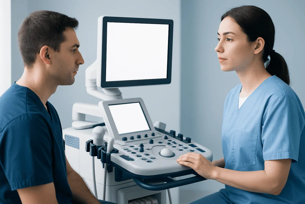
Exophytic growths can arise in organs, mucosal linings, or on the skin. Common examples include kidney lesions seen on ultrasound or CT, uterine fibroids that project outward from the uterus, gastrointestinal polyps or masses that protrude into a hollow organ, and skin growths that rise above the surrounding surface.
Some exophytic findings are discovered by chance during tests done for another reason. For example, an imaging scan may report an exophytic renal lesion, which could represent a benign cyst, a solid tumor, or another type of kidney abnormality. In the digestive tract, a doctor may describe a protruding or exophytic lesion before knowing whether it is inflammatory, benign, or malignant.
Because the same word applies to many body systems, context is essential. A small exophytic skin lesion and an exophytic pancreatic mass do not carry the same implications or need the same tests. That is why a report should always be interpreted by the clinician who knows the patient’s history and symptoms.
When the finding involves an organ commonly assessed for tumors, doctors may arrange more targeted imaging or referral. Depending on the body area, this can include MRI scanning or CT scanning to define size, borders, internal structure, and relation to nearby tissues.
Common symptoms and why some exophytic lesions cause none

Many exophytic lesions cause no symptoms at all, especially when they are small and found incidentally. Symptoms usually appear when the growth becomes large enough to irritate nearby tissue, bleed, become infected, or press on an organ, nerve, or blood vessel.
The exact symptoms depend on where the lesion is located. In the digestive tract, an exophytic growth may cause bleeding, changes in bowel habits, abdominal discomfort, fullness, or anemia. In the urinary system, symptoms can include flank pain, blood in the urine, or urinary obstruction. Skin or gynecologic lesions may be noticed because of visible growth, pain, discharge, itching, or contact bleeding.
General features that deserve attention include unexplained weight loss, loss of appetite, persistent fatigue, a rapidly enlarging mass, or recurrent bleeding. These signs do not prove cancer, but they do suggest the need for proper assessment rather than watchful waiting alone.
It is also common for a report to mention an exophytic lesion when the person’s symptoms are unrelated. In those situations, a doctor determines whether the finding is clinically important or simply an incidental change that can be monitored safely over time.
Causes and risk factors
An exophytic appearance can result from several different processes. Benign causes include cysts, polyps, fibroids, papillomas, inflammatory growths, scar tissue, and some infections. Other exophytic lesions are premalignant or malignant, meaning they contain abnormal cells that can invade or spread if untreated.
Risk factors vary by organ system. Age, smoking, chronic inflammation, family history, hormonal influences, certain viral infections, prior radiation, and long-standing irritation can all play a role in specific diseases. For example, outward-growing lesions in the digestive tract may relate to polyp formation, while some kidney masses reflect cystic or solid tumors.
Doctors look beyond the word exophytic and assess the lesion’s detailed features. Helpful clues include whether it is smooth or irregular, fluid-filled or solid, single or multiple, and stable or growing over time. Associated findings such as enlarged lymph nodes, ulceration, or invasion into surrounding tissue may increase concern and guide the next step.
When an exophytic lesion is suspected to be a tumor, specialists may evaluate for related conditions, including kidney cancer or colon cancer, depending on the site and imaging findings. This does not mean the diagnosis is already confirmed; it means the lesion needs a structured workup.
How doctors diagnose an exophytic lesion
Diagnosis starts with a careful history and physical examination. A clinician asks when symptoms began, whether the lesion has changed, and whether there is pain, bleeding, fever, bowel or urinary changes, or weight loss. The examination may include inspection, palpation, pelvic examination, skin assessment, or endoscopy depending on the body area.
Imaging is often the next step. Ultrasound can help distinguish cystic from solid lesions. CT and MRI offer more detail about size, shape, blood supply, and involvement of adjacent structures. In some situations, doctors compare older scans to see whether the finding is new or has remained unchanged over time.
Endoscopy or direct visualization may be useful for lesions in the gastrointestinal, airway, urinary, or gynecologic tract. Doctors can sometimes remove a small lesion during the same procedure or take a sample for analysis. If cancer is a possibility, tissue diagnosis is often the most reliable way to determine exactly what the lesion is.
A biopsy is not always needed, but when it is, pathology can show whether the lesion is benign, inflammatory, precancerous, or malignant. In digestive-tract lesions, endoscopy may allow both evaluation and biopsy. In some cancer workups, biopsy provides the information needed to plan treatment safely and accurately.
Treatment options depend on the cause, not the word itself
There is no single treatment for an exophytic lesion because exophytic is only a description. Management depends on the diagnosis, location, symptoms, size, growth pattern, and overall health of the patient. Some lesions only need monitoring, while others require removal or more specialized care.
Benign and asymptomatic lesions may be observed with repeat examination or interval imaging. If the lesion is causing pain, bleeding, pressure, or cosmetic concern, treatment may involve medications, minimally invasive removal, endoscopic therapy, or surgery. Infections or inflammatory causes are treated according to the underlying condition.
If testing shows a precancerous or cancerous growth, treatment is tailored by a multidisciplinary team. This may include surgery, endoscopic resection, chemotherapy, radiation therapy, or a combination approach. The goal is to remove or control the lesion while preserving normal function whenever possible.
Near the end of the diagnostic process, some patients benefit from care at centers that combine imaging, pathology, surgery, oncology, and organ-specific specialists. Acibadem International’s multidisciplinary specialists and JCI-accredited hospitals diagnose and treat exophytic lesions for international patients when expert evaluation is needed.
Self-care, follow-up, and questions to ask
Self-care begins with understanding the report and avoiding assumptions. Patients should ask where the lesion is, what it may represent, whether it has suspicious features, and what test or follow-up is recommended. Keeping copies of imaging and pathology reports can make future comparisons easier.
It is sensible to monitor for new symptoms such as bleeding, faster growth, worsening pain, changes in bowel or urinary habits, fever, or unexplained weight loss. People should not start supplements or home treatments intended to “shrink” a mass without medical advice, because these approaches can delay diagnosis or interfere with care.
Healthy habits still matter, even though they do not treat the lesion directly. Avoiding tobacco, limiting alcohol, eating a balanced diet, maintaining physical activity, and attending routine screening tests can support overall health and help detect certain conditions earlier. If the lesion is being observed, follow-up appointments should be kept as scheduled.
Questions that often help include:
- Is the lesion definitely benign, or is more testing needed?
- Should it be monitored, biopsied, or removed?
- What symptoms should prompt earlier review?
- When should repeat imaging or examination be done?
When to seek medical care
Medical care should be sought if an exophytic lesion is newly discovered and its cause is unclear, especially when there are symptoms. Prompt evaluation is particularly important for bleeding, blood in the stool or urine, a rapidly enlarging lump, persistent pain, trouble swallowing, bowel obstruction symptoms, or a lesion that keeps returning after treatment.
Urgent assessment is also needed for severe abdominal pain, vomiting with inability to keep fluids down, fainting, heavy bleeding, or signs of infection such as fever with redness, swelling, or discharge. These symptoms do not always indicate a serious disease, but they should not be ignored.
Even without urgent symptoms, a patient should arrange follow-up if a scan or report mentions an exophytic mass, lesion, or nodule and no one has explained the result clearly. Early clarification can reduce anxiety and help ensure that benign findings are not overtreated and important conditions are not missed.
Frequently asked questions
Does exophytic mean cancer?
No. Exophytic only describes the way a growth extends outward from a surface. Some exophytic lesions are benign, some are inflammatory, and some are cancerous, so the diagnosis depends on further evaluation.
What is the difference between exophytic and endophytic?
Exophytic means growing outward from a tissue surface. Endophytic generally means growing inward or within the tissue or organ. These terms describe growth pattern, which can help doctors interpret imaging or examination findings.
Can an exophytic lesion go away on its own?
Some benign or inflammatory lesions may shrink or resolve, depending on the cause. Others remain stable for years, and some grow over time. A doctor can advise whether observation is reasonable or whether testing is needed.
Is a biopsy always required for an exophytic mass?
Not always. If imaging clearly shows a simple benign lesion, a biopsy may not be needed. When the nature of the growth is uncertain or suspicious, biopsy can provide a more definite diagnosis.
What tests are usually done after a report says exophytic?
The next tests depend on the location of the finding. Doctors may recommend ultrasound, CT, MRI, endoscopy, lab tests, or a biopsy. Sometimes the best next step is simply repeat imaging after a defined interval.
Should a patient worry about an incidental exophytic finding?
An incidental finding should be taken seriously enough to review with a clinician, but it should not automatically cause alarm. Many incidental exophytic lesions are benign. The key is to understand what organ is involved and whether the report recommends more evaluation.
References
- National Cancer Institute
- American College of Radiology
- Mayo Clinic
- Merck Manual Consumer Version
- National Health Service
This article is for general information only and is not a substitute for professional medical advice. Please consult a qualified doctor about your individual situation.
Persistent digestive symptoms? Get evaluated in Turkey
JCI-accredited · board-certified surgeons · reply within 24h
Add us as a Preferred Source to see more of our trusted health content across Google Search, AI Overviews and Discover.