Pterional Craniotomy: What Patients Need to Know

A pterional craniotomy accesses the brain through the temple region, usually behind the hairline. It may be used for aneurysms, tumors, and other conditions affecting the front or side of the brain and skull base.
Key Takeaways
- A pterional craniotomy accesses the brain through the temple region, usually behind the hairline.
- It may be used for aneurysms, tumors, and other conditions affecting the front or side of the brain and skull base.
- Preoperative imaging and careful surgical planning help determine whether this approach is appropriate.
- Recovery varies according to the underlying condition, the extent of surgery, and a person’s overall health.
- New or worsening neurological symptoms after surgery require prompt medical assessment.
A pterional craniotomy is a neurosurgical approach that creates an opening behind the hairline at the temple to reach selected areas of the brain, skull base, and nearby blood vessels. It is commonly planned for conditions such as certain brain tumors, aneurysms, and other lesions, with the exact procedure tailored to the person’s diagnosis and anatomy.
What is a pterional craniotomy?
A pterional craniotomy is a type of brain surgery in which a neurosurgeon temporarily removes a small section of skull near the temple, in an area called the pterion. The pterion is where several skull bones meet. Through this opening, the surgeon can reach structures at the front and side of the brain, including areas near important arteries, nerves, and the skull base.
The procedure is also called a pterional approach or frontotemporal craniotomy. It is an established route in neurosurgery because it can provide access along natural spaces around the brain rather than requiring the surgeon to pass directly through healthy brain tissue. At the end of the operation, the bone flap is usually put back and secured with small plates or screws.
A pterional craniotomy is not a diagnosis in itself. It is the surgical route selected when its view and working corridor may be suitable for a particular problem. The reason for surgery, possible alternatives, and expected benefits should be discussed individually with the neurosurgical team.
Why might this approach be recommended?
The pterional approach may be used to treat conditions located around the frontal and temporal regions of the brain, the skull base, or the circle of Willis, a network of arteries at the base of the brain. The approach is often considered when the surgeon needs access to a lesion while protecting surrounding tissue as much as possible.
Examples include selected brain tumors, including some brain tumors, certain meningiomas, and pituitary-region or skull-base lesions. It may also be used in surgery for some cerebral aneurysms, including aneurysms that may need clipping rather than endovascular treatment. The best treatment depends on the size, location, shape, and urgency of the aneurysm, as well as the patient’s health.
Less commonly, this route may be part of surgery for vascular malformations, trauma-related bleeding, infection, or pressure caused by swelling. Not every condition in these areas requires open surgery. Some people may be better suited to monitoring, medication, radiation-based treatment, minimally invasive procedures, or endovascular embolization, depending on the diagnosis.
How surgeons plan and perform the operation
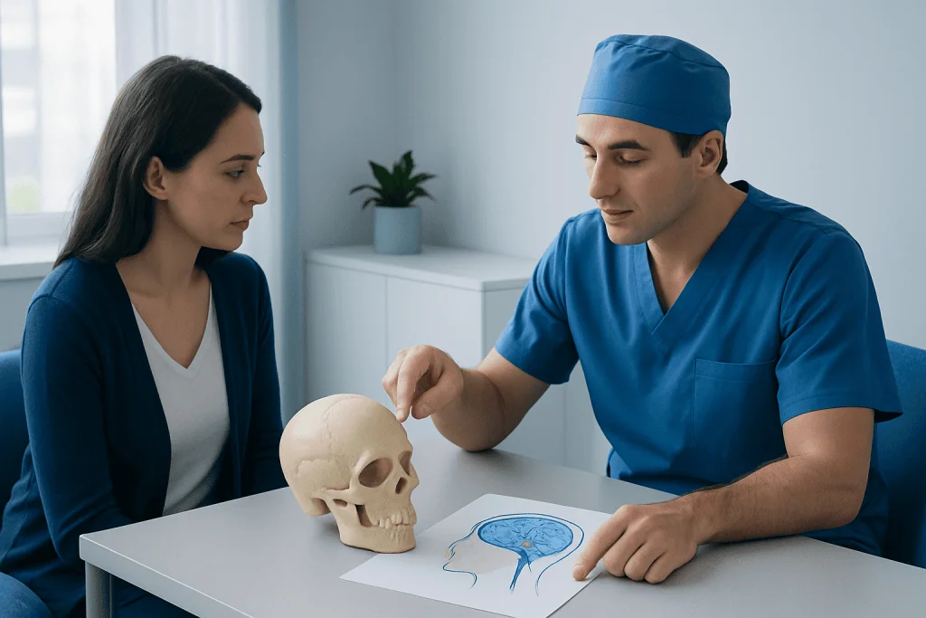
Planning begins with a detailed review of brain imaging, typically MRI, CT, CT angiography, or catheter angiography when blood vessels are involved. These scans help the team understand the exact location of the problem and its relationship to arteries, veins, nerves, and normal brain structures. Blood tests, medication review, and assessment of general fitness for anesthesia are also important.
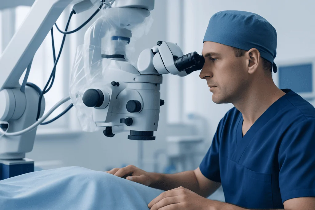
The operation is performed under general anesthesia. The head is carefully positioned, and an incision is usually made behind the hairline, starting in front of the ear and curving toward the forehead. The surgeon temporarily moves the scalp and muscle tissue, creates the bone opening, and uses magnification and specialized instruments to reach the planned target.
Whenever possible, the surgical team works through spaces between brain structures and uses monitoring techniques to help protect neurological function. After treating or removing the targeted lesion, the surgeon closes the protective layers, replaces the bone flap when appropriate, and closes the skin incision. The length and complexity of surgery vary considerably according to the condition being treated.
In some circumstances, the exact plan may need to change during surgery for safety reasons. The neurosurgeon should explain the planned procedure, realistic alternatives, and potential need for additional treatment before consent is given.
Benefits, limitations, and possible risks
The main potential benefit of a pterional craniotomy is access. Its angle may allow the surgeon to see and work around key structures at the front of the skull base and near major cerebral arteries. For an appropriate condition, this can support removal of a tumor, repair or clipping of an aneurysm, relief of pressure, or collection of tissue for diagnosis.
However, brain surgery always carries risks. These can include bleeding, infection, blood clots, seizures, fluid leakage, stroke, swelling, and reactions to anesthesia. Depending on the location of the condition and structures involved, there can also be changes in speech, movement, sensation, vision, memory, concentration, behavior, or hormone function. Some complications are temporary, while others may be longer lasting.
There may be numbness, tenderness, or weakness of the chewing muscle around the temple while healing. A visible scar is usually placed within or close to the hairline, but hair changes, contour changes at the temple, and jaw discomfort can occur. The individual likelihood of each risk depends more on the underlying problem and the planned extent of surgery than on the incision alone.
The care team balances these risks against the risks of leaving the condition untreated and against other available approaches. Asking the surgeon why this route is recommended in a specific case can help patients make an informed decision.
What to expect during recovery
After a pterional craniotomy, patients are usually monitored closely in a recovery area or intensive care setting. Staff regularly check alertness, pupils, speech, strength, sensation, blood pressure, and pain. A CT or MRI scan may be performed after surgery to assess the surgical area and establish a postoperative baseline.
Headache, fatigue, scalp tenderness, facial swelling, and bruising around the eye can occur in the early days after surgery. These effects often improve gradually, although recovery timelines differ. The hospital stay may range from a few days to longer, especially when surgery was performed after a rupture, stroke, major tumor surgery, or another serious neurological event.
At home, the team may advise rest balanced with gentle walking, incision care, avoiding heavy lifting or strenuous activity for a period, and taking prescribed medicines as directed. Driving, returning to work, and resuming exercise should be discussed with the treating team. People should not stop anti-seizure medicines, steroids, blood thinners, or other prescribed medications without medical advice.
Follow-up appointments are essential. They allow the team to review wound healing, pathology results when tissue was removed, neurological recovery, rehabilitation needs, and any further treatment. Depending on the diagnosis, follow-up may include imaging, radiation treatment, medication, or brain tumor surgery planning as part of broader care.
Preparing for surgery and supporting recovery
Preparation is individualized, but patients are commonly asked to provide a full medication and supplement list, including blood thinners, diabetes medicines, herbal products, and medicines for seizures. The surgical team will explain which medicines to continue, adjust, or pause before surgery. It is important not to make medication changes independently.
Stopping smoking and avoiding nicotine before and after surgery can support wound healing and reduce respiratory complications. Good nutrition, hydration, sleep, and management of long-term conditions such as high blood pressure or diabetes may also help with surgical readiness. Family members or trusted friends can assist with transport, meals, medication reminders, and follow-up visits after discharge.
Rehabilitation may include physical therapy, occupational therapy, speech and language therapy, or neuropsychological support. These services are offered according to a person’s symptoms and goals. Recovery can involve physical healing as well as adjustment to fatigue, emotional changes, concentration difficulties, or temporary limits in independence.
For international patients, Acibadem International’s multidisciplinary specialists and JCI-accredited hospitals can assess and treat neurosurgical conditions, coordinating care across neurosurgery, neurology, imaging, rehabilitation, and related specialties.
When to seek medical care
Anyone with a known brain lesion, aneurysm, or progressive neurological symptoms should seek timely assessment from a qualified clinician. Symptoms that merit medical review include persistent or worsening headaches, new seizures, changes in vision, weakness, numbness, speech difficulty, confusion, balance problems, or significant personality or cognitive changes. These symptoms have many possible causes, but they should not be ignored.
After a craniotomy, patients should contact their surgical team promptly for increasing redness, drainage, worsening swelling, fever, severe or escalating headache, persistent vomiting, or concerns about the incision. New confusion, weakness, loss of consciousness, a seizure, sudden vision changes, or difficulty speaking should be treated as urgent symptoms.
Emergency services should be contacted immediately for symptoms suggestive of stroke, such as sudden facial drooping, arm weakness, speech difficulty, severe sudden headache, or loss of consciousness. Early evaluation is important because neurological emergencies need rapid assessment and treatment.
Frequently asked questions
How serious is a pterional craniotomy?
A pterional craniotomy is major surgery because it involves opening the skull to treat a brain or blood-vessel condition. Its level of risk and expected recovery depend on why it is being performed, the location of the lesion, and the person’s health. The neurosurgical team can explain the individual risk profile in detail.
How long does recovery take after a pterional craniotomy?
Initial physical recovery often takes several weeks, but full recovery may take months. Some people recover more quickly, while others need rehabilitation or further treatment for the underlying condition. Fatigue and concentration changes can persist for a time even when the incision has healed.
Will hair be shaved for a pterional craniotomy?
Hair preparation varies by hospital and surgical technique. Often, only a small area near the incision is trimmed, and the incision is planned behind the hairline when possible. The surgical team can explain what to expect for an individual operation.
Can a pterional craniotomy affect speech or memory?
It can, particularly when surgery is near brain areas involved in language, memory, or attention. Whether this is likely depends on the side of the brain, the lesion, the extent of surgery, and individual anatomy. Preoperative assessment and intraoperative monitoring may be used when appropriate to help reduce risk.
Is a pterional craniotomy used for brain aneurysms?
Yes, it is a traditional open surgical approach for selected brain aneurysms, especially those near certain major arteries at the base of the brain. Some aneurysms are instead treated with minimally invasive endovascular techniques. A neurosurgeon and neurointerventional team can help determine the most suitable option.
What should patients ask before a pterional craniotomy?
Useful questions include why this approach is recommended, what alternatives are available, and what outcome is expected from surgery. Patients may also ask about major risks, likely hospital stay, follow-up imaging, rehabilitation, activity restrictions, and signs that require urgent medical attention.
References
- American Association of Neurological Surgeons
- National Institute of Neurological Disorders and Stroke
- Mayo Clinic
- National Health Service
- World Health Organization
This article is for general information only and is not a substitute for professional medical advice. Please consult a qualified doctor about your individual situation.
Hair transplant in Turkey — costs, graft counts & a free assessment
JCI-accredited · board-certified surgeons · reply within 24h