

Robotic Thoracic Surgery
Robotic thoracic surgery is a minimally invasive approach for selected chest and lung procedures, using robotic instruments for precision, smaller incisions, and faster recovery.
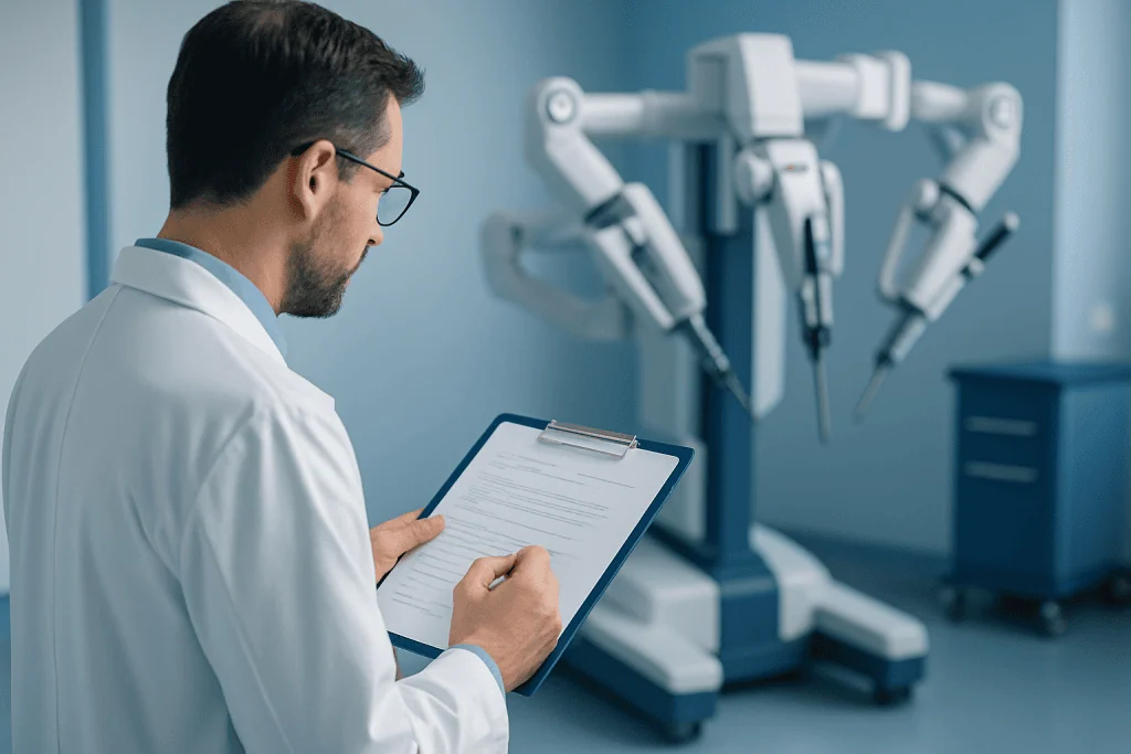
Quick answer
Robotic thoracic surgery is a minimally invasive way to perform selected chest and lung operations using robotic instruments controlled by a surgeon for greater precision through small incisions. At Acibadem in Turkey, it is used for carefully chosen thoracic procedures with preoperative assessment, image-guided planning, and postoperative care focused on pain control, breathing support, and recovery.
Robotic Thoracic Surgery: A Less Invasive Path for Complex Chest Care
Being told you may need chest or lung surgery can raise immediate and understandable concerns. Many patients worry about pain, breathing after surgery, time away from family or work, and whether a procedure will affect their long-term quality of life. For international patients, there are additional questions: How will the diagnosis be confirmed? Who will coordinate the care? Is the hospital experienced with complex thoracic procedures? What happens after returning home?
Robotic thoracic surgery is one of the minimally invasive approaches used for selected conditions of the lungs, chest cavity, mediastinum, esophagus, pleura, and thymus. Instead of making a large incision between the ribs, surgeons work through several small incisions using highly controlled robotic instruments and magnified three-dimensional visualization. For appropriately selected patients, this can mean less surgical trauma, a shorter hospital stay, reduced postoperative discomfort, and a faster return to daily life compared with traditional open thoracic surgery.
At the same time, robotic thoracic surgery is still major surgery. It requires careful diagnosis, experienced thoracic surgeons, modern anesthesia and intensive care support, precise imaging, and a team that can make the right decision if a minimally invasive plan needs to be modified for safety. The goal is not simply to use advanced technology, but to choose the most appropriate operation for the patient’s disease, anatomy, overall health, and treatment goals.
For many people, especially those being evaluated for lung nodules, early-stage lung cancer, thymic tumors, mediastinal masses, or selected benign chest conditions, robotic surgery may offer a refined balance: cancer-quality or disease-focused surgery performed through smaller access points, with a recovery designed to help patients breathe, move, and regain independence as early as medically appropriate.
What Robotic Thoracic Surgery Is
Robotic thoracic surgery is a form of minimally invasive chest surgery in which a thoracic surgeon operates using robotic instruments introduced through small incisions in the chest wall. The surgeon remains in full control throughout the procedure. The robotic platform does not operate independently; it translates the surgeon’s hand movements into precise movements of slender instruments inside the chest.
The approach is used in selected procedures involving the lungs, lymph nodes, mediastinum, pleura, thymus, diaphragm, and, in some centers and cases, the esophagus. It is part of a broader family of minimally invasive thoracic surgery, which also includes video-assisted thoracoscopic surgery. The main difference is that robotic systems may provide enhanced depth perception, stable magnified visualization, wristed instrument movement, and improved access in narrow or delicate areas of the chest.
During robotic thoracic surgery, the surgeon views the operative field on a high-definition, three-dimensional monitor. Small instruments are positioned through ports between the ribs. These instruments can bend and rotate in ways that help the surgeon dissect tissue, seal blood vessels, remove tumors or diseased lung sections, and sample or remove lymph nodes with high precision. An assistant surgeon and operating room team remain at the patient’s side to support instrument exchange, suction, stapling, specimen retrieval, and safety monitoring.
Robotic thoracic surgery may be used for diagnostic procedures, such as obtaining tissue from a suspicious lung nodule or mediastinal mass, as well as therapeutic operations, such as removing a lung segment, lung lobe, thymus gland, or selected chest tumors. In cancer care, the operation is planned according to evidence-based protocols, staging results, pulmonary function, tumor location, and multidisciplinary recommendations.
Not every patient is a candidate. Prior chest surgery, extensive adhesions, advanced disease, tumor invasion into major structures, severe cardiopulmonary risk, or emergency situations may make an open operation safer or more appropriate. A skilled thoracic team evaluates both the technical possibility of robotic surgery and whether it is the best option for the patient’s outcome.
Who May Need Robotic Thoracic Surgery
Patients may be considered for robotic thoracic surgery after symptoms, imaging findings, or diagnostic tests suggest a condition in the chest that requires biopsy or surgical treatment. Some patients feel well and discover a lung nodule or mediastinal mass during imaging performed for another reason. Others seek care because of persistent symptoms that need investigation.
Common symptoms that may lead to thoracic evaluation include a cough that does not resolve, coughing up blood, shortness of breath, recurrent chest infections, unexplained chest discomfort, hoarseness, difficulty swallowing, fatigue, or unintentional weight loss. Some thymic and mediastinal conditions may be associated with muscle weakness or autoimmune conditions, while pleural disease may cause fluid accumulation around the lung and breathlessness.
Diagnosis usually begins with a careful medical history and physical examination, followed by imaging. A chest X-ray may detect an abnormality, but more detailed evaluation often requires computed tomography of the chest. Depending on the situation, doctors may recommend PET imaging, magnetic resonance imaging, bronchoscopy, endobronchial ultrasound, pulmonary function testing, cardiac assessment, blood tests, or image-guided biopsy. For suspected cancer, staging is essential because treatment decisions depend on whether disease is localized, regionally advanced, or metastatic.
Patients who may be considered for robotic thoracic surgery include those with early-stage lung cancer suitable for surgical removal, suspicious lung nodules that require diagnosis or treatment, benign lung lesions that are growing or symptomatic, selected mediastinal tumors, thymoma or thymic hyperplasia, pleural disorders, recurrent pneumothorax, or certain chest wall and diaphragm conditions. The decision is individualized and takes into account disease biology, imaging results, surgical risk, lung reserve, age, general fitness, and patient preferences.
For international patients, a second opinion is often valuable before committing to surgery. A review of imaging, pathology reports, pulmonary tests, and prior treatments can confirm whether robotic surgery is feasible, whether additional diagnostic steps are needed, or whether another treatment such as open surgery, radiation therapy, systemic therapy, or active surveillance would be more appropriate.
Conditions and Indications Addressed by Robotic Thoracic Surgery
Robotic thoracic surgery may be used for a range of chest conditions when the location of the disease and the patient’s health allow minimally invasive access. In lung surgery, it can be used for wedge resection, segmentectomy, lobectomy, and, in carefully selected cases, more complex resections. These procedures may be performed for lung cancer, suspicious nodules, metastatic lesions limited to the lung, or benign lesions that require removal.
For lung cancer, surgery is most often considered when disease appears localized and the patient has adequate lung and cardiac function. The surgical plan may include removal of a lung lobe or smaller anatomic lung segment, along with lymph node evaluation. The extent of resection depends on tumor size, position, lung function, tissue diagnosis, and staging. Robotic surgery can support precise dissection around blood vessels, airways, and lymph node stations, which is important for both treatment and staging.
Robotic techniques may also be used for mediastinal masses, including selected thymomas, thymic cysts, bronchogenic cysts, neurogenic tumors, and other lesions located between the lungs. For patients with myasthenia gravis associated with thymic disease or thymic enlargement, removal of the thymus may be part of the treatment plan when recommended by the neurology and thoracic surgery teams.
Pleural conditions may also be treated using minimally invasive thoracic surgery. These include recurrent pneumothorax, pleural biopsies, pleural cysts, or selected cases requiring removal of abnormal pleural tissue. Some patients with recurrent fluid around the lung may require diagnostic or therapeutic procedures to improve breathing and obtain tissue for diagnosis.
Other indications can include selected esophageal or diaphragmatic procedures, chest infections requiring drainage or decortication in appropriate stages, and removal of certain benign chest tumors. However, robotic surgery is not defined by the diagnosis alone. It is selected when the surgeon believes the disease can be managed safely, completely, and effectively through small incisions without compromising the medical objective of the operation.
How Robotic Thoracic Surgery Is Performed
Preparation and Evaluation Before Surgery
Preparation begins with confirming the diagnosis and understanding the patient’s overall condition. The thoracic surgery team reviews imaging studies, pathology if available, medical history, medications, allergies, smoking history, prior operations, and current symptoms. For patients with suspected or confirmed cancer, cases are commonly discussed in a multidisciplinary tumor board or specialist meeting that may include thoracic surgeons, pulmonologists, medical oncologists, radiation oncologists, radiologists, nuclear medicine physicians, pathologists, anesthesiologists, and intensive care specialists.
Pulmonary function testing is often used to estimate how well the lungs will tolerate surgery, especially if part of the lung must be removed. Cardiac evaluation may be recommended for patients with heart disease, advanced age, diabetes, reduced exercise tolerance, or abnormal findings. Blood tests, electrocardiography, and anesthesia assessment help identify risks that should be optimized before the procedure.
Patients are usually advised to stop smoking before surgery because smoking increases the risk of pneumonia, poor wound healing, air leaks, and breathing complications. Certain blood-thinning medications may need to be paused or adjusted under medical supervision. The team provides instructions about fasting, medication use, arrival time, expected hospital stay, and breathing exercises. International patients may also receive guidance on travel timing, medical documentation, translation support, and post-discharge follow-up planning.
The Procedure Itself
Robotic thoracic surgery is typically performed under general anesthesia. A specialized breathing tube allows the anesthesiology team to ventilate one lung while the surgeon operates on the other side of the chest. This creates the working space needed for safe visualization and instrument movement. The patient is positioned carefully to protect nerves, joints, and pressure points during the operation.
The surgeon makes several small incisions between the ribs. Through these openings, a camera and robotic instruments are introduced into the chest. Carbon dioxide may be used in selected cases to improve working space. The surgeon controls the instruments from a console in the operating room, while the bedside surgical team assists with instrument exchange, suction, stapling, specimen handling, and emergency readiness.
The specific operation depends on the condition. For a lung nodule, the surgeon may remove a wedge-shaped piece of lung tissue or an anatomic segment. For lung cancer, a lobectomy or segmentectomy with lymph node assessment may be performed when appropriate. For thymic or mediastinal surgery, the surgeon carefully separates the lesion from surrounding structures such as the heart, great vessels, nerves, and lungs. For pleural procedures, abnormal tissue can be biopsied or removed, and techniques may be used to reduce recurrence of pneumothorax or fluid accumulation.
Once the target tissue is removed, it is placed in a protective retrieval bag and taken out through one of the small incisions, which may be slightly enlarged if needed. A chest tube is commonly placed to drain air and fluid from the chest and help the lung re-expand. The incisions are then closed, and the patient is transferred to the recovery area or intensive care unit depending on the procedure and medical condition.
Technology Used During Robotic Thoracic Surgery
Robotic thoracic surgery relies on several categories of technology that work together to improve visualization, precision, safety, and perioperative monitoring. High-definition three-dimensional imaging allows the surgeon to see fine structures in the chest with depth perception and magnification. Wristed robotic instruments provide controlled movement in narrow spaces, which may be especially helpful around blood vessels, airways, lymph nodes, and mediastinal structures.
Modern operating rooms also use advanced anesthesia monitoring, lung isolation techniques, energy devices for tissue sealing, surgical stapling systems, imaging review tools, and specimen localization methods when required. In some cases, preoperative image-guided marking or bronchoscopic localization may help identify small or deep lung nodules. Pathology support may be used during surgery for selected cases to help guide the extent of resection.
The value of technology depends on how it is integrated into clinical decision-making. A robotic system can enhance the surgeon’s capability, but good outcomes rely on accurate diagnosis, appropriate patient selection, disciplined surgical technique, experienced anesthesia care, and a team prepared to convert to an open procedure if that becomes the safest choice.
Typical Duration and Hospital Stay
The length of robotic thoracic surgery varies according to the procedure, disease location, prior surgery, anatomy, and whether lymph node assessment or additional steps are needed. A diagnostic biopsy or wedge resection may be shorter, while lobectomy, segmentectomy, thymectomy, or complex mediastinal surgery may take longer. Patients should view any estimated time as a guide rather than a fixed expectation.
After surgery, the hospital stay depends on pain control, breathing ability, chest tube drainage, lung expansion, mobility, and the patient’s overall health. Many patients undergoing minimally invasive thoracic surgery are able to leave the hospital earlier than they might after open surgery, but discharge occurs only when the team confirms that it is medically safe. Some patients need additional observation, especially if they have underlying lung disease, persistent air leak, infection risk, or other medical conditions.
Recovery Process After Surgery
Recovery begins immediately after the operation. Nurses and respiratory therapists help patients cough, breathe deeply, use breathing exercise devices, sit up, and walk as early as appropriate. Early movement helps reduce the risk of blood clots, pneumonia, and muscle deconditioning. Pain is managed with a tailored plan that may include oral medications, regional anesthesia techniques, and non-opioid options when suitable.
The chest tube is removed when air leak and fluid drainage are acceptable and imaging confirms adequate lung expansion. Patients receive instructions about wound care, showering, activity limits, breathing exercises, medication use, and warning signs such as fever, worsening shortness of breath, increasing chest pain, redness or drainage from incisions, or leg swelling.
Most patients gradually resume light daily activities within days to a few weeks, depending on the procedure and their baseline health. More strenuous exercise, heavy lifting, long flights, and return to work should be discussed with the surgeon. For cancer patients, final pathology results guide whether additional treatment such as chemotherapy, immunotherapy, targeted therapy, or radiation therapy is recommended. Follow-up may include clinic visits, imaging surveillance, pulmonary rehabilitation, or coordination with physicians in the patient’s home country.
Why Acting Early Matters and the Risks of Delay
Early evaluation is important because many thoracic conditions are more treatable when identified before they progress. A small lung cancer, for example, may be suitable for surgical removal with curative intent, while a more advanced tumor may require a different and more complex treatment strategy. A growing mediastinal mass may eventually press on nearby structures, and recurrent pleural problems can lead to repeated infections, scarring, or reduced lung expansion.
Delaying care can also make diagnosis less straightforward. Symptoms such as chronic cough, shortness of breath, or chest discomfort may be attributed to infection, smoking history, asthma, reflux, or aging, but persistent or unexplained symptoms deserve appropriate imaging and specialist review. In patients with suspicious nodules, timing of follow-up or biopsy should be guided by imaging characteristics, risk factors, and international recommendations rather than anxiety or avoidance.
For patients who already have a diagnosis, delay can affect treatment choices. Tumors may grow, lymph nodes may become involved, lung function may decline, and the opportunity for a less invasive operation may narrow. In benign disease, repeated inflammation or infection can create scar tissue that makes surgery more difficult and recovery more demanding.
Acting early does not always mean rushing into an operation. It means obtaining a timely, accurate diagnosis and a clear treatment plan from an experienced team. In some cases, careful surveillance is the right choice. In others, surgery should proceed without unnecessary delay. The key is to make the decision with complete information and appropriate specialist input.
Benefits of Robotic Thoracic Surgery
For selected patients, robotic thoracic surgery may offer several practical and clinical advantages compared with traditional open chest surgery.
| Benefit | What It Means for You |
|---|---|
| Smaller incisions | Because the operation is performed through small access points, patients may experience less tissue disruption and a more discreet scar pattern than with a large thoracotomy incision. |
| Less postoperative discomfort for many patients | A minimally invasive approach may reduce rib spreading and muscle trauma, which can help with breathing exercises, walking, and earlier return to daily activities. |
| Enhanced visualization and precision | Magnified three-dimensional imaging and controlled instrument movement can help the surgeon work carefully around blood vessels, airways, lymph nodes, and delicate chest structures. |
| Shorter hospital stay in appropriate cases | Many patients recover in the hospital for a shorter period than is typical after open surgery, although the actual discharge date depends on safety, chest tube status, and overall recovery. |
| Earlier mobility and breathing recovery | Reduced surgical trauma may make it easier to walk, cough, breathe deeply, and participate in respiratory recovery exercises after surgery. |
| Strong role in multidisciplinary cancer care | When used for lung cancer or other thoracic tumors, robotic surgery can be integrated with staging, pathology review, systemic therapy, radiation therapy, and structured follow-up. |
Recovery Timeline After Robotic Thoracic Surgery
Recovery varies by procedure and individual health, but the following timeline reflects what many patients can generally expect after robotic thoracic surgery.
| Time Period | What Patients Can Expect |
|---|---|
| Day 1 | Patients are monitored closely after anesthesia. Breathing exercises, pain control, early sitting, and walking often begin as soon as medically appropriate. A chest tube may be in place. |
| First Week | Many patients focus on walking more each day, using breathing exercises, managing incision care, and transitioning to oral pain medication. Some patients are discharged during this period if recovery is stable. |
| First Month | Energy gradually improves. Light daily activities usually become easier, while heavy lifting and strenuous exercise may still be restricted. Pathology results and any additional treatment plan are reviewed. |
| Longer Term | Patients continue rebuilding stamina and lung capacity. Cancer patients may enter a surveillance program or begin additional therapy if recommended. Follow-up imaging and specialist visits are scheduled according to the diagnosis. |
Factors That Influence Outcomes and a Good Result
Outcomes after robotic thoracic surgery depend on a combination of disease-related, patient-related, and treatment-related factors. One of the most important is accurate diagnosis. A suspicious lung nodule, for example, must be evaluated in the context of imaging features, growth pattern, smoking history, prior cancer history, and overall risk. When cancer is suspected or confirmed, staging helps determine whether surgery alone is appropriate or whether treatment should involve chemotherapy, immunotherapy, targeted therapy, or radiation therapy.
The type and extent of surgery also matter. Removing too little tissue may fail to address the disease adequately, while removing more lung than necessary may affect breathing capacity. For this reason, thoracic surgeons balance oncologic principles with preservation of lung function. In selected early lung cancers, segmentectomy may be considered; in other cases, lobectomy remains the more appropriate operation. Lymph node evaluation is important for accurate staging and future treatment decisions.
Patient health strongly influences recovery. Lung function, heart disease, diabetes, kidney function, nutrition, smoking status, weight, physical conditioning, and frailty can all affect surgical risk. Patients with chronic obstructive pulmonary disease or reduced respiratory reserve may still be candidates for surgery, but they require careful assessment and optimization. Smoking cessation, pulmonary rehabilitation, medication adjustment, and improved nutrition can all support better recovery.
The surgeon’s experience with minimally invasive thoracic techniques is another important factor. Robotic surgery involves a learning curve and requires coordinated teamwork. Good results depend not only on the surgeon at the console, but also on anesthesia expertise, nursing care, respiratory therapy, intensive care support, pathology, imaging, and postoperative monitoring. A hospital that routinely manages complex thoracic patients is better equipped to anticipate issues such as air leaks, arrhythmias, pneumonia, bleeding, or pain management challenges.
Finally, follow-up is essential. Surgery is not complete when the incision closes. Patients need clear pathology interpretation, wound assessment, imaging surveillance, medication guidance, and a plan for returning to activity and travel. For international patients, coordination with physicians at home can help ensure continuity after returning to their country.
Why International Patients Choose Acibadem for Robotic Thoracic Surgery
International patients considering robotic thoracic surgery often look for more than an operation. They need a reliable diagnostic pathway, specialists who can interpret complex findings, hospital systems that meet international quality standards, and a care team that understands the practical realities of traveling for treatment. Acibadem Hospitals in Turkey provide thoracic care within JCI-accredited hospitals, supported by multidisciplinary collaboration, advanced diagnostic resources, and dedicated international patient services.
At Acibadem, patients with suspected or confirmed thoracic disease are evaluated through a structured process. Imaging, pathology, pulmonary function results, previous medical records, and treatment history are reviewed carefully. When cancer or complex disease is involved, cases may be discussed by multidisciplinary tumor boards or specialist boards. This helps ensure that surgery is recommended in the correct clinical context and that alternatives such as surveillance, systemic therapy, radiation therapy, interventional pulmonology, or open surgery are considered when appropriate.
Experienced thoracic surgeons at Acibadem use minimally invasive approaches, including robotic surgery, for selected chest and lung procedures. The decision to use robotic surgery is individualized. It depends on the diagnosis, tumor or lesion location, anatomy, lung function, previous treatments, and the patient’s overall health. The emphasis is on choosing the approach that can treat the condition effectively while supporting recovery and safety.
Modern diagnostic and surgical technology supports this process. High-resolution imaging helps define the size, location, and relationship of chest lesions to nearby structures. PET imaging, bronchoscopy, endobronchial ultrasound, image-guided biopsy, pulmonary function testing, and advanced pathology techniques may be used when clinically indicated. In the operating room, three-dimensional visualization, robotic instrumentation, refined anesthesia monitoring, and minimally invasive surgical tools assist the team in performing precise procedures through smaller incisions.
For international patients, communication and coordination are central to the experience. Acibadem International provides support in more than 20 languages, helping patients prepare medical records, schedule consultations, understand treatment plans, coordinate hospital appointments, and plan travel-related details. This support is particularly important for thoracic surgery patients, who may need preoperative testing, inpatient recovery, pathology review, and guidance about when it is safe to fly home.
Personalized treatment planning is especially important in thoracic disease because two patients with similar imaging findings may need very different care. A small lung nodule may require surveillance, biopsy, wedge resection, segmentectomy, or lobectomy depending on its features. A mediastinal mass may need surgical removal, tissue diagnosis first, or treatment by another specialty. A patient with lung cancer may need surgery alone or a combination of therapies. Acibadem’s multidisciplinary structure is designed to align these decisions with international, evidence-based protocols while taking into account the patient’s personal priorities and medical circumstances.
Patients also benefit from the presence of comprehensive hospital services under one system. Thoracic surgery may involve pulmonology, oncology, radiology, pathology, nuclear medicine, anesthesia, intensive care, pain management, physiotherapy, and rehabilitation. When these disciplines work together, patients receive clearer recommendations and more coordinated care before, during, and after surgery.
Choosing care abroad is a significant decision. Patients and families often want to know that their questions will be answered, their records will be reviewed thoughtfully, and their treatment plan will be explained in language they understand. The aim is to provide medically rigorous care while supporting the emotional and practical needs of people who have traveled far from home for treatment.
Moving Forward With Confidence and Clarity
Robotic thoracic surgery can be an important option for selected patients who need chest or lung surgery and wish to understand whether a minimally invasive approach is appropriate. It may offer smaller incisions, careful precision, and a recovery pathway that helps many patients return to normal activities sooner than traditional open surgery. However, the best operation is always the one that fits the diagnosis, stage of disease, anatomy, lung function, and overall health of the individual patient.
If you have been diagnosed with a lung nodule, lung cancer, mediastinal mass, thymic disease, recurrent pneumothorax, pleural disorder, or another chest condition, a specialist review can help clarify your options. A second opinion may be particularly valuable if you have received different recommendations, are unsure whether surgery is necessary, or want to know whether robotic thoracic surgery is possible in your case.
Acibadem’s thoracic surgery teams and international patient services can review your medical information, explain the diagnostic steps that may be needed, and help you understand the expected procedure, recovery, and follow-up plan. Taking the next step begins with an accurate evaluation and a conversation with the right specialists.
This information is general in nature and is not a substitute for professional medical advice, diagnosis, or treatment. Decisions about robotic thoracic surgery should be made after consultation with qualified physicians who can evaluate your individual medical condition.
Preparation
- Evaluation usually includes chest imaging, pulmonary function tests, blood tests, anesthesia assessment, and review of current medications. Patients may be asked to stop smoking, adjust blood thinners, and fast before surgery. The surgical team explains the planned approach, risks, and expected recovery steps.
Aftercare
- After surgery, patients are monitored for breathing, pain control, and chest tube drainage if used. Early walking, breathing exercises, and wound care help reduce complications and support recovery. Follow-up visits review pathology results when relevant and guide return to normal activities.
Turkey vs UK, Germany & USA
Robotic thoracic surgery may be considered for selected lung, mediastinal and chest procedures when a minimally invasive approach is clinically appropriate. Costs and patient experience vary by country, hospital setting, surgeon expertise, procedure complexity and what is included in the care package.
The comparison below highlights cost and experience factors for international patients considering robotic thoracic surgery in different healthcare systems.
| Factor | Turkey | UK | Germany | USA |
|---|---|---|---|---|
| Price drivers | Often offered as bundled international patient packages; final cost depends on procedure type, robotic system use, hospital stay and pathology needs. | Private care may vary by hospital and consultant; public pathways may involve eligibility and referral processes. | Costs depend on hospital category, specialist fees, diagnostics, length of stay and rehabilitation planning. | Costs can vary widely due to facility fees, surgeon fees, anaesthesia, imaging, pathology and insurance arrangements. |
| Hospital and surgeon factors | Experience with minimally invasive thoracic surgery, robotic platform availability and multidisciplinary tumour board access can influence planning. | Choice of consultant, private hospital access and availability of robotic thoracic programmes affect timing and pathway. | Specialist thoracic centres may provide structured diagnostics, surgery and follow-up, with costs linked to institutional protocols. | High variation between academic centres, private hospitals and insurance networks; surgeon and facility selection can strongly affect billing. |
| Accreditation and quality | International patients may choose JCI-accredited hospitals such as Acibadem, with coordinated quality and safety processes. | Quality is monitored through national and institutional standards; private providers may have additional accreditations. | Hospitals follow national quality requirements; specialist certification and centre experience may be relevant. | Accreditation, hospital network status and specialty programme reputation may affect both care pathway and cost. |
| Waiting times | Private international scheduling may allow coordinated appointments after records are reviewed. | Public pathways may involve waiting lists; private care may shorten access depending on consultant availability. | Scheduling depends on referral pathway, diagnostics and specialist availability. | Access can be quick in some private settings but depends on insurance authorisation and network rules. |
| Travel and language logistics | International patient teams may assist with translation, airport transfers, accommodation guidance and appointment coordination. | Less travel complexity for local residents; international patients may need to arrange accommodation and post-discharge support. | International patients may require translation support and coordination across diagnostics, surgery and follow-up. | Travel, accommodation, insurance communication and out-of-network billing may require detailed planning. |
| Typical package scope | Packages may include specialist consultation, preoperative tests, surgery, anaesthesia, hospital stay, standard medications and basic translation support. | Private packages may separate consultation, diagnostics, hospital fees and surgeon fees. | Packages may be itemised by diagnostic work-up, surgery, inpatient care and follow-up needs. | Billing is often itemised across multiple providers, including hospital, surgeon, anaesthesia, pathology and imaging. |
What affects your final cost:
- Type of thoracic procedure, such as wedge resection, segmentectomy, lobectomy, thymectomy or mediastinal mass surgery.
- Whether robotic surgery is suitable or another approach is safer.
- Complexity of the condition, tumour location, previous chest surgery and overall lung function.
- Preoperative imaging, bronchoscopy, biopsy, pathology and molecular testing needs.
- Length of hospital stay, intensive care requirements and recovery progress.
- Surgeon, anaesthesiology, operating room, robotic system and hospital facility fees.
- Translation, airport transfer, accommodation, companion support and follow-up arrangements.
Compare your options
Robotic thoracic surgery is one option within a broader range of thoracic treatment approaches. Suitability is decided by a specialist after reviewing diagnosis, imaging, lung function, medical history and treatment goals.
| Option | What it is | Typical use | Key considerations |
|---|---|---|---|
| Robotic-assisted thoracic surgery | A minimally invasive approach using robotic instruments controlled by the surgeon through small incisions. | Selected lung resections, thymectomy, mediastinal procedures and some chest wall or pleural operations. | May support precision and smaller incisions, but requires appropriate anatomy, experienced teams and access to a robotic platform. |
| Video-assisted thoracoscopic surgery | A minimally invasive technique using a camera and long instruments without robotic arms. | Many lung, pleural and mediastinal procedures when minimally invasive surgery is appropriate. | May offer similar recovery advantages in selected cases; choice depends on surgeon expertise and procedure complexity. |
| Open thoracic surgery | A traditional approach through a larger chest incision to provide direct access to the thoracic cavity. | Complex tumours, extensive adhesions, major bleeding risk or cases where minimally invasive surgery is not suitable. | May be necessary for safety or completeness of surgery, but can involve a longer recovery pathway. |
| Diagnostic thoracic procedures | Procedures such as bronchoscopy, mediastinal sampling or pleural biopsy to clarify diagnosis before treatment. | Staging, tissue diagnosis, infection evaluation or assessment of lymph nodes and mediastinal lesions. | Results may determine whether surgery, oncology treatment or another pathway is recommended. |
| Non-surgical or combined treatment | Treatments such as radiotherapy, chemotherapy, immunotherapy, targeted therapy or active surveillance when appropriate. | Cases where surgery is not recommended, or where treatment is combined before or after surgery. | Decision-making usually involves a multidisciplinary team and depends on diagnosis, stage and patient fitness. |
Trusted care for international patients
General information only — not medical or financial advice. Final costs depend on the factors above and your individual case; request a free, personalised quote.
Medical Units
Available at These Hospitals





Guides for This Treatment
Offered at These Centers
Frequently Asked Questions
What affects the cost of robotic thoracic surgery?
The final cost depends on the exact procedure, complexity of the case, robotic system use, surgeon and hospital fees, anaesthesia, imaging, pathology, hospital stay, intensive care needs and follow-up plan. Travel, translation and accommodation support may also affect the overall budget.
How can I get a personalised quote?
A personalised quote usually requires medical records, recent imaging, biopsy or pathology results if available, lung function tests and a summary of previous treatments. Acibadem can review your documents through a free consultation and provide guidance on the likely treatment plan and package scope.
What is usually included in an international patient package?
Package content varies, but it may include specialist consultation, standard preoperative tests, surgery, anaesthesia, hospital stay, routine medications, nursing care and translation support. Items such as extra diagnostics, extended stay, intensive care, advanced pathology or additional treatments may be quoted separately.
Is robotic thoracic surgery always more suitable than other approaches?
No. Robotic surgery is suitable only for selected patients and conditions. A thoracic surgeon decides the safest approach after assessing the diagnosis, tumour or lesion location, lung function, previous surgery, general health and expected treatment benefit.
Can international patients plan surgery without long waiting times?
Scheduling depends on medical urgency, the need for additional tests, surgeon availability and operating room planning. International patient teams can help coordinate record review, appointments, translation and travel logistics once the case is assessed.
Is this information medical or financial advice?
No. This is general educational information and does not replace consultation with a qualified specialist or a formal written quote. For an individual assessment, a free consultation is recommended.